Anemia sel sabit dudu siji tes, siji gejala, utawa siji tingkat urgensi. Panuntun sing ngutamake pasien iki misahake status panyandang, penyakit sing dikonfirmasi, pemantauan rutin, lan kahanan sing mbutuhake perawatan darurat.


- Sifat sel sabit tegese siji gen HbS; biasane ora nyebabake anemia kronis utawa krisis nyeri sing kambuh.
- Skrining bayi anyar lair ngenali sebagian besar bayi sing kena sadurunge gejala diwiwiti, ngidini penisilin lan tindak lanjut spesialis diwiwiti luwih dhisik.
- Hemoglobin electrophoresis misahake jinis hemoglobin lan mbantu ngesahake HbSS, HbSC, HbS-beta thalassemia, utawa status panyandang.
- Loro ing dada, sesak napas, demam 38,5°C utawa luwih, bingung, utawa lemes anyar mbutuhake evaluasi darurat sing cepet ing wong kanthi penyakit sel sabit.
- Sindrom dada akut bisa diwiwiti kanthi watuk, demam, loro ing dada, utawa oksigen sing kurang lan bisa saya parah sajrone jam tinimbang dina.
- Mudhun hemoglobin sing dumadakan 2 g/dL utawa luwih saka tingkat lumrahe wong bisa ngisyarati krisis aplastik, sekuèstrasi, getihen, utawa hemolisis sing saya cepet.
- Cacah retikulosit mbantu mbedakake respons sumsum sing aktif saka krisis aplastik; jumlah sing sithik nalika anemia saya parah minangka pola bebaya.
- Hidroksiurea minangka obat sing ngganti penyakit, dudu mung obat nyeri; pemantauan biasane kalebu tes getih lengkap saben 4 minggu nalika nyetel dosis.
Apa tegese anemia sel sabit—lan apa sing ora dingerteni
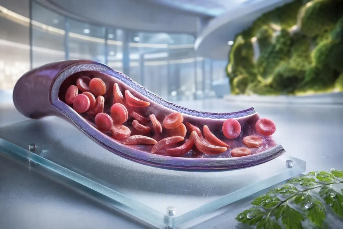
Anemia sel sabit biasane nuduhake penyakit HbSS, ing ngendi wong ngwarisake rong gen sing ngasilake hemoglobin S; sel abang bisa dadi kaku, rusak luwih dhisik, lan nyumbat pembuluh getih cilik. Trait sel sabit iku béda: siji gen HbS biasane cukup kanggo diwarisake sacara genetik nanging ora cukup kanggo nyebabake anemia kronis lan episode vasooklusif sing bola-bali katon ing HbSS.
Penyakit sel sabit minangka istilah umum sing kalebu HbSS, HbSC, HbS-beta-nol thalassemia, lan HbS-beta-plus thalassemia. HbSS asring nyebabake anemia sing paling kenthel, nanging tingkat keparahan beda-beda banget sanajan ing kulawarga sing padha; hemoglobin dhasar 7,5 g/dL bisa stabil kanggo wong diwasa lan mbebayani kanggo wong liya yen wis mudhun kanthi cepet.
Mutasi hemoglobin S ngganti siji asam amino ing beta-globin: valin ngganti asam glutamat ing posisi 6. Ing oksigen rendah, dehidrasi, asidosis, utawa demam, molekul HbS bisa polimerisasi ing njero sel. Owah-owahan fisik kasebut njlentrehake kenapa krisis bisa kedadeyan sawise penerbangan dawa, penyakit gastrointestinal, utawa wengi kurang cairan.
Kantesti AI minangka Analisa tes getih AI sing nempatake CBC, bilirubin, LDH, retikulosit, kreatinin, lan asil sadurunge menyang siji tampilan longitudinal; ora nggawe diagnosis penyakit sel sabit saka CBC wae. Kanggo nyegerake praktis babagan laporan hemoglobin, delengen apa tegese HGB.
Wiwit tanggal 4 September 2026, aku isih nemoni pasien sing salah ngandhani yen trait lan penyakit bisa diganti. Ora. Aturan klinis Dr. Thomas Klein iku prasaja: njaluk genotipe hemoglobin sing tepat, dudu pernyataan samar yen tes kasebut “positif.”
Napa genotipe ngganti perawatan
HbSC bisa duwe hemoglobin luwih dhuwur tinimbang HbSS nanging isih nyebabake komplikasi retina, nyeri, utawa nekrosis avaskular. HbS-beta thalassemia bisa meh padha karo trait utawa HbSS gumantung saka apa produksi beta-globin sebagian ana, mula tes molekuler kadhangkala ngrampungake pola elektroforesis sing ambigu.
Gejala penyakit sel sabit miturut umur lan garis dasar
Gejala penyakit sel sabit umume kalebu nyeri balung utawa dhadha episodik, kesel, penyakit kuning, sesak napas, pertumbuhan sing telat, lan infeksi sing kerep, nanging ora ana wong loro sing ngalami pola sing padha. Gejala biasane muncul sawise umur 4 nganti 6 wulan amarga hemoglobin janin nalika dilindhungi bayi saka polimerisasi HbS.
Daktilitis—pembengkakan tangan utawa sikil sing lara—bisa dadi tandha katon pisanan ing bayi. Ing bocah-bocah umur sekolah, nyeri weteng mbutuhake perawatan khusus amarga konstipasi, penyakit kandung empedu, pembesaran limpa, lan vasooklusi bisa krasa padha banget ing omah.
Penyakit kuning ing penyakit sel sabit nuduhake kerusakan sel abang sing terus-terusan lan paningkatan bilirubin ora langsung; ora ateges hepatitis kanthi otomatis. Urine peteng, tinja pucet, gatal, demam, utawa nyeri weteng tengen ndhuwur ngganti evaluasi kasebut lan mbutuhake evaluasi panyebab ati lan biliary; pandhuan kita kanggo pola bilirubin langsung nerangake bedane.
Wong diwasa bisa ngnormalake nyeri sing saya kerep. Ing pengalamanku, owah-owahan saka rong episode sing diatur ing omah saben taun dadi rong episode saben wulan sacara klinis signifikan sanajan kunjungan darurat durung mundhak. Gangguan turu, ora mlebu kerja, gejala ereksi, swasana ati sing kurang, lan toleransi olahraga sing suda kudu didokumentasikan amarga asring sadurunge owah-owahan formal ing perawatan.
Konsentrasi hemoglobin 6 nganti 9 g/dL bisa dadi kisaran stabil sing umum ing HbSS, nalika nilai ing ngisor 10 g/dL bakal ora dikarepake ing umume wong tanpa penyakit. Angka kasebut penting, nanging owah-owahan saka dhasar pribadine luwih penting.
Sapa sing kudu nimbang skrining panyandang sel sabit
Penyaringan sel sabit iku lumrah sadurunge meteng utawa ing wiwitan meteng kanggo wong sing status pembawane durung dingerteni, ora preduli saka penampilan, jeneng kulawarga, utawa leluhur sing diasumsi. Status pembawa diwarisake lan asring ora dingerteni nganti skrining bayi utawa tes hemoglobin sing ora dikarepke.
Yen wong tuwa kandung loro nggawa varian sing ana gandhengane karo HbS utawa beta-talasemia, saben meteng bisa duwe kasempatan 25% kanggo mewarisi kondisi hemoglobin sing signifikan sacara klinis, gumantung saka varian sing terlibat. Pembawa bisa duwe energi normal, asil CBC normal, lan ora ana riwayat episode nyeri.
Skrining utamané migunani nalika siji mitra duwe HbAS, sipat HbC, sipat beta-talasemia, mikrositosis sing ora bisa diterangake, utawa riwayat kulawarga penyakit sel sabit. Volume korpuskular rata-rata sing sithik ora kudu disalahake amarga kekurangan zat besi tanpa mriksa ferritin lan nimbang talasemia; mbandhingake Wacan MCV lan MCH.
Laporan global Organisasi Kesehatan Dunia 2022 ngira-ngira yen kelainan hemoglobin tetep dadi beban penyakit warisan utama, utamane ing ngendi akses skrining bayi anyar ora rata. Pendekatan sing paling ngajeni yaiku akses universal menyang skrining sing diinformasikake tinimbang nggunakake etnis minangka penjaga gerbang.
Kantesti iku sawijining platform interpretasi hasil tes getih AI sing bisa njlentrehake asil HbS sing dilaporake ing basa umum, nanging konfirmasi pembawa lan konseling reproduksi mbutuhake laboratorium lan klinisi sing akreditasi. Yen pasangan ngrencanakake meteng, takon apa tes mitra lan konseling genetik bisa ditindakake sadurunge konsepsi tinimbang sawise asil dadi sensitif wektu.
Carane elektroforesis hemoglobin ngesahake diagnosis
Elektroforesis hemoglobin misahake fraksi hemoglobin lan minangka tes inti kanggo ngonfirmasi anemia sel sabit utawa sipat, asring bebarengan karo kromatografi cair kinerja dhuwur lan, nalika dibutuhake, tes genetik. CBC rutin ora bisa ngonfirmasi HbSS utawa sipat amarga kekurangan zat besi lan talasemia bisa ngasilake indeks sel abang sing tumpang tindih.
Ing HbSS sing durung diobati, elektroforesis biasane nuduhake HbS utamane tanpa HbA, sanajan persentase HbF beda-beda. HbAS biasane nuduhake HbA lan HbS, asring kanthi HbA luwih dhuwur tinimbang HbS; persentase sing tepat bisa owah kanthi alpha-talasemia sing diwarisake bebarengan utawa transfusi anyar.
Transfusi anyar bisa nggawe asil elektroforesis mblusuk kira-kira 3 wulan amarga HbA donor isih ngalir. Iki uga sebabe HbA1c bisa nyuda glukosa rata-rata sawise transfusi utawa ing hemolysis turnover dhuwur; delengen nalika HbA1c ngapusi.
Goresan perifer bisa nuduhake sel target, polikromasia, lan bentuk sabit, nanging mikroskop ndhukung tinimbang ngganti tes fraksi. Dr. Klein wis weruh goresan darurat sing “normal” ngundurake klarifikasi nalika pasien wis ditransfusikake pirang-pirang minggu sadurunge-tansah ngandhani laboratorium lan tim hematologi babagan transfusi.
Metode skrining bayi anyar nemtokake HbS sadurunge penyakit klinis, nanging asil kasebut isih mbutuhake tes konfirmasi sing pas wektune. Pedoman bukti taun 2014 dening Yawn et al. nyaranake keterlibatan spesialis ing wiwitan urip, kanthi langkah-langkah pencegahan diwiwiti sadurunge komplikasi utama pisanan (Yawn et al., 2014).
Pemantauan getih rutin: apa sing ditindakake klinisi
Pemantauan rutin ing penyakit sel sabit biasane ngetren hemoglobin, retikulosit, sel putih, platelet, bilirubin, LDH, fungsi ginjel, lan protein urin—dudu siji asil sing kapisah. Perbandingan sing paling aman yaiku karo basis pasien dhewe sing stabil, idealne dijupuk nalika dheweke sehat lan ora bubar ditransfusi.
Hemolysis asring ngasilake LDH, bilirubin ora langsung, lan retikulosit sing mundhak kanthi haptoglobin sing sithik. Haptoglobin bisa kurang amarga alesan lian, kalebu penyakit ati, mula pola gabungan luwih kuwat tinimbang siji pananda; kita pandhuan interpretasi haptoglobin nuduhake sebabe.
Persentase retikulosit dhewe bisa ngluwihi tanggapan sumsum ing anemia sing signifikan. Dokter asring ngitung jumlah retikulosit absolut utawa tanggapan retikulosit sing dikoreksi; jumlah absolut sing mudhun nalika anemia saya parah nimbulake keprihatinan kanggo krisis aplastik sing ana gandhengane karo parvovirus B19.
Kantesti iku sawijining Piranti analisis tes getih berbasis AI sing mbandhingake asil hematologi sing diulang tinimbang nganggep LDH sing dhuwur minangka diagnosis mandiri. Sampel sing ditandhani minangka hemolyzed ing tabung bisa nambah kalium lan LDH kanthi palsu, mula asil sing ngagetake bisa mbutuhake gambar ulang; mriksa tes ulang sawise hemolysis.
Hydroxyurea biasane nambah MCV lan HbF saka wektu, nalika mudhunake neutrofil kanthi moderat ing dosis sing efektif. Owah-owahan kasebut bisa dikarepake, nanging jumlah neutrofil absolut ing ngisor ambang perawatan individu utawa jumlah platelet sing mudhun cepet mbutuhake review resep tinimbang ngatur obat dhewe.
Kapan nyeri minangka episode vas-oklusif—lan kapan ora
Nyeri vaso-oklusif asring jero, abot, lan dumunung ing punggung, dada, pinggul, anggota awak, utawa weteng, nanging nyeri anyar ora kudu otomatis dilabel minangka krisis sabit. Demam, bengkak fokal, ciloko, njaga weteng, utawa gejala neurologis siji-sijine nuduhake masalah sing mbutuhake panaliten lian.
Rencana nyeri ing omah biasane kalebu hidrasi awal, kehangatan, analgesia sing diresepake, istirahat, lan ambang eskalasi pribadi sing disepakati karo tim sabit. Ngombe volume sing akeh banget ora luwih aman: wong sing nandhang masalah ginjel utawa penyakit jantung bisa ngalami kelebihan cairan, utamane sajrone penyakit akut.
Nyeri sing tetep ora bisa dikontrol sawise rencana penyelamat sing biasa, nyegah cairan utawa obat-obatan tetep mudhun, utawa beda kualitas utawa lokasi kudu nyebabake saran klinis sing cepet. Sendi sing bengkak lan panas bisa nuduhake radhang sendi septik utawa infeksi balung tinimbang oklusi vaso-sing ora rumit; kita pandhuan tes nyeri sing ora bisa diterangake ngrancang investigasi pertama sing migunani.
Ing perawatan darurat, analgesia pas wektune lan penilaian ulang luwih penting tinimbang mbuktekake tingkat nyeri liwat nomer laboratorium. Pandhuan NICE babagan episode nyeri akut sel sabit nyaranake penilaian analgesia sing cepet lan review sing asring, amarga undertreatment dhewe nyumbang kanggo tekanan sing saya suwe lan pemulihan sing ditundha (NICE, 2012).
Aja nggunakake kompres adhem langsung ing area nyeri vaso-oklusif kajaba tim sampeyan kanthi khusus menehi saran; adhem bisa nyempitake pembuluh getih periferal. Rincian praktis kasebut katon cilik, nanging akeh pasien sing ngandhani aku ora ana sing nerangake nganti pirang-pirang episode sing angel.
Sindrom dada akut: darurat sing akeh wong kliwat
Sindrom dada akut minangka infiltrat paru-paru anyar ing pencitraan dada ditambah gejala pernapasan, demam, nyeri dada, utawa oksigen sing sithik ing wong sing duwe penyakit sel sabit, lan iku darurat. Iku bisa saya parah kanthi cepet, kadhangkala sawise mlebu rumah sakit amarga episode nyeri tinimbang nalika gejala dada diwiwiti.
Hubungi layanan darurat utawa lunga menyang perawatan darurat kanggo sesak ambegan anyar, nyeri dada, lambe biru-abu-abu, pingsan, watuk terus-terusan nganggo demam, utawa saturasi oksigen ing ngisor target sing diresepake wong kasebut. Saturasi 92% bisa dadi mudhun sing gedhe kanggo wong sing biasane ana ing 98%, nalika sawetara pasien duwe basis sing luwih murah; nilai absolut lan owah-owahan loro-lorone penting.
Sindrom dada akut bisa nglibatake infeksi, emboli lemak saka sumsum balung, obstruksi vaskular paru-paru, atelectasis, utawa pirang-pirang proses sekaligus. Dokter biasane menehi penilaian oksigenasi, CBC, retikulosit, kultur nalika demam, pencitraan dada, lan kadang gas getih; tes getih kanggé sesak ambegan nambah konteks kanggo asil kasebut.
Ing wong diwasa, oversedation saka opioid, ambegan jero amarga nyeri, lan kelebihan cairan intravena bisa nambah status pernapasan. Spirometri insentif sajrone dirawat ing rumah sakit nyuda komplikasi paru-paru ing episode nyeri sing dipilih, sanajan dudu pengganti penilaian sing cepet nalika gejala wis diwiwiti.
Tinjauan 2018 dening Kato lan kanca-kanca nggambarake sindrom dada akut minangka panyebab utama morbiditas lan mortalitas ing saindhenging umur (Kato et al., 2018). Aja nyopir dhewe yen sesak ambegan, ngantuk, utawa tekanan dada signifikan.
Demam, infeksi, lan darurat limpa
Suhu 38,5°C (101,3°F) utawa luwih dhuwur ing bocah utawa wong diwasa kanthi penyakit sel sabit mbutuhake evaluasi medis langsung ing dina sing padha, kajaba tim spesialis wis menehi rencana sing beda. Kehilangan fungsi kekebalan limpa kanthi fungsional bisa nyebabake sawetara infeksi bakteri berkembang luwih cepet tinimbang demam virus biasa.
Demam ditambah lesu, ruam, tekanan darah rendah, bingung, utawa output urin sing suda mbutuhake perawatan darurat saiki. Jumlah sel putih sing katon normal ora bisa ngilangi infeksi serius ing penyakit sel sabit, utamane ing wiwitan penyakit utawa sajrone perawatan hydroxyurea.
Splenic sequestration paling asring kedadeyan ing bocah cilik lan nyebabake pembengkakan limpa sing dumadakan, pucet, rasa kebak weteng, ambegan cepet, lan mudhun hemoglobin sing cepet. Wong tuwa kadang diajari carane ngrasakake limpa anak, nanging pembengkakan anyar nalika lara kudu nyebabake evaluasi cepet tinimbang mriksa omah bola-bali.
Mudhun hemoglobin 2 g/dL utawa luwih ngisor saka baseline biasane kanthi limpa sing gedhe minangka prihatin kanggo sequestration. Kosok baline, anemia abot kanthi jumlah retikulosit sing sithik banget bisa nuduhake krisis aplastik, asring ana gandhengane karo parvovirus B19; interpretasi jumlah retikulosit nerangake sinyal sumsum.
Laboratorium sepsis bisa uga kalebu laktat, kultur, CBC, tes ginjel, lan penanda inflamasi, nanging keputusan perawatan ora kudu ngenteni kabeh nilai bali. Kanggo konteks babagan pola sing mendesak, waca petunjuk penanda sepsis.
Vaksin lan antibiotik pencegahan
Vaksin lan profilaksis penisilin masa kanak-kanak nyuda risiko nanging ora nggawe demam risiko rendah. Konfirmasi rencana individu sampeyan karo tim hematologi utawa pediatrik, utamane sawise splenektomi, lelungan, bayi anyar, utawa owah-owahan jadwal imunisasi lokal.
Stroke lan tandha-tandha peringatan neurologis mbutuhake tumindak cepet
Sembarang tiba-tiba droop rai, kelemahan lengan utawa sikil, kesulitan ngomong, kejang, ngelu sing ora biasa banget, bingung, utawa ilang keseimbangan ing penyakit sel sabit kudu dianggep minangka darurat stroke sing bisa. Hubungi layanan darurat langsung; aja ngenteni kanggo ndeleng yen gejala wis kliwat utawa menehi obat penghilang rasa nyeri dhisik.
Bocah-bocah kanthi HbSS utawa HbS-beta-zero thalassemia umumé ditawani skrining ultrasonik Doppler transcranial saka umur 2 nganti 16 taun. Kecepatan maksimum rata-rata wektu sing ora normal 200 cm/detik utawa luwih dhuwur nuduhake risiko stroke sing luwih dhuwur lan mbutuhake tumindak spesialis cepet.
Terapi transfusi kronis bisa nyuda risiko stroke pisanan ing bocah-bocah kanthi temuan Doppler sing ora normal, nanging nyiptakake kabutuhan tambahan: pemantauan kelebihan zat besi, skrining antibodi, lan cocogake sel donor kanthi teliti. Iki minangka perawatan khusus, dudu asil sing dikelola liwat aplikasi.
Wong diwasa bisa ngalami gejala neurologis sementara, owah-owahan kognitif, utawa cilaka otak bisu tanpa stroke dramatis klasik. Episode kesulitan nemokake tembung 10 menit isih mbutuhake evaluasi darurat amarga gejala iskemik sementara bisa dadi peringatan, dudu jaminan.
Dr. Thomas Klein menehi saran kulawarga kanggo nyimpen nomer kontak darurat sel sabit lokal ing jejere rencana nyeri sing biasa. Iki mbusak siji keputusan sajrone sawetara menit nalika bocah utawa wong diwasa bisa berjuang kanggo komunikasi.
Ngusekake ginjel, mripat, lan paru-paru ing antarane krisis
Penyakit sel sabit bisa ngrusak ginjel, retina, paru-paru lan jantung sanajan nalika nyeri jarang, mula pengawasan organ rutin minangka bagean saka perawatan tinimbang tambahan opsional. Tes albumin urin, pemeriksaan tekanan darah, pemeriksaan mata, lan evaluasi paru-paru sing dipimpin gejala ngidentifikasi komplikasi sadurunge katon.
Albuminuria bisa dadi tandha awal nefropati sabit. Rasio albumin urin-kreatinin 30 mg/g utawa luwih dhuwur ora normal ing umume wong diwasa lan kudu dikonfirmasi kanthi pengujian ulang amarga demam, olahraga, haid, lan dehidrasi bisa nambah kanthi sementara.
Kreatinin bisa katon normal kanthi ngapusi ing penyakit sel sabit amarga tambah sekresi tubular lan massa otot sing luwih murah bisa nutupi filtrasi sing suda. Cystatin C utawa persamaan gabungan bisa nambah konteks sing migunani, dene urin umpluk sing terus-terusan pantes dievaluasi; waca pandhuan protein ing urin.
Retinopati wigati banget ing penyakit HbSC, sing bisa luwih tenang sacara klinis ing babagan liyane. Munculé lala anyar, kilatan, owah-owahan visual kaya tirai, utawa penglihatan kabur sing dumadakan mbutuhake tinjauan mata sing cepet, dudu janji rutin sawetara minggu sabanjure.
Ngandhut nambahake hiperfiltrasi fisiologis lan risiko ibu-bayi sing luwih dhuwur, mula penilaian ginjel lan urin dhasar kudu ditindakake luwih dhisik. Panjelasan kita babagan owah-owahan GFR nalika meteng mbantu mbedakake owah-owahan sing diarepake saka tren sing ngkhawatirake.
Pilihan perawatan, hydroxyurea, lan keamanan transfusi
Hidroksiurea nyuda episode nyeri lan sindrom dada akut kanggo akeh wong sing duwe talasemia HbSS utawa HbS-beta-zero kanthi nambah hemoglobin janin, nanging mbutuhake pemantauan sing terstruktur. Transfusi bisa nyelametake nyawa kanggo komplikasi sing dipilih, nanging transfusi bola-bali nggawe risiko pemuatan wesi lan antibodi sel getih abang.
Hidroksiurea biasane dititrasi karo hitungan CBC lan retikulosit kira-kira saben 4 minggu nganti dosis sing stabil, banjur ing interval sing luwih dawa sing ditemtokake dening tim perawatan. MCV sing luwih dhuwur asring minangka efek perawatan sing diarepake, nalika sitopenia sing abot mbutuhake review obat lan kadhangkala mandheg sementara.
Transfusi sederhana nambah kapasitas nggawa oksigen, nanging konsentrasi hemoglobin sing akeh banget bisa nambah viskositas ing HbSS. Akeh protokol akut ngindhari ngunggahake hemoglobin pasca-transfusi luwih saka 10 g/dL kajaba spesialis netepake tujuan sing beda; transfusi ijol-ijolan digunakake kanggo komplikasi abot sing dipilih.
Beban wesi luwih mungkin sawise transfusi bola-bali lan dinilai kanthi tren ferritin ditambah MRI ati utawa jantung nalika cocog. Ferritin mundhak amarga peradangan, mula tingkat dhuwur tunggal dudu bukti beban wesi jaringan; pandhuan sinau wesi nerangake wates-watesé.
Bukti paling kuat kanggo hidroksiurea ing penyakit HbSS lan HbS-beta-zero; mupangate ing HbSC tetep dadi area sing kurang pasti lan luwih akeh keputusan individu. Rencana obat kudu kalebu diskusi kontrasepsi lan meteng nalika relevan, alangan kepatuhan, lan rencana tertulis kanggo dosis sing dilewati.
Kehamilan, kesuburan, lan perencanaan kulawarga karo HbS
Meteng kanthi penyakit sel sabit mbutuhake perawatan gabungan awal saka obstetrik lan hematologi amarga risiko episode nyeri, anemia, trombosis, infeksi, pre-eklampsia, hambatan pertumbuhan janin, lan lair prematur luwih dhuwur. Skrining pembawa penting sadurunge meteng amarga risiko genetik gumantung saka wong tuwa biologis.
Kunjungan sadurunge meteng kudu mriksa genotipe, hemoglobin dhasar, fungsi ginjel, albumin urin, tekanan darah, riwayat transfusi, antibodi sel getih abang, vaksinasi, lan obat-obatan. Keputusan hidroksiurea nalika meteng individu lan kudu digawe karo spesialis; aja mandheg utawa miwiti maneh mung amarga saran internet.
Wesi ora kudu diwenehake kanthi otomatis kanggo hemoglobin sing kurang ing HbSS. Kekurangan wesi bisa ana bebarengan, utamane nalika meteng utawa geti pendarahan menstruasi sing abot, nanging ferritin lan saturasi transferrin kudu nuntun perawatan amarga hemolisis kronis dhewe ora padha karo kekurangan wesi.
Nalika wong tuwa loro nggawa varian hemoglobin sing relevan, konseling genetik bisa nerangake konsepsi alami, pilihan diagnostik prenatal, pilihan donor, lan pengujian genetik preimplantasi tanpa ngarahake kulawarga menyang siji keputusan. Tujuane yaiku pilihan sing diwenehi informasi, dudu rasa wedi.
Kanggo tandha-tandha abang laboratorium meteng praktis liyane, waca masalah lab meteng dina sing padha. Dhasar pasien sing dikenal kudu dilebokake ing saben serah terima ibu; iku ngowahi carane asil hemoglobin ditafsirake.
Nggunakake asil laboratorium kanthi aman ing antarane janjian
Asil laboratorium sel sabit paling migunani nalika dibandhingake karo genotipe, dhasar sing stabil, gejala, perawatan, lan transfusi sing bubar. Bilirubin dhuwur utawa hemoglobin sing kurang bisa diarepake kanggo wong kanthi HbSS nanging cepet kanggo wong sing padha yen asil kasebut owah-owahan kanthi cepet.
Simpen cathetan siji-kaca babagan genotipe, hemoglobin biasane, saturasi oksigen biasane yen dingerteni, golongan darah lan antibodi, obat-obatan saiki, tanggal transfusi, lan kontak darurat. Iki ngirit wektu ing departemen darurat sing ora pati ngerti lan nyuda kemungkinan yen dhasar HbSS disalahake minangka anemia sing nembe ditemokake.
Kantesti iku sawijining layanan interpretasi tes lab AI sing bisa ngatur laporan laboratorium sing diunggah lan menehi tandha pola kanggo diskusi, kalebu penanda hemolisis, tren ginjel, lan pemantauan obat. Ora bisa ndeleng dhadha sampeyan, ngukur oksigenasi, utawa ngganti perawatan darurat nalika gejala nuduhake sindrom dada akut, sepsis, utawa stroke.
Asil ing njaba pathokan laboratorium durung mesthi mbebayani, lan asil ing njero bisa uga mbebayani yen wis owah cepet. Kanggo katrangan babagan tandha laporan lan pathokan, wacanen tegesing asil njaba pathokan.
Pendekatan klinis Kantesti ditliti marang cara lan watesan sing ditemtokake; pamaca sing pengin ngerti jaminan kita bisa neliti , amarga. Gawa PDF asli menyang janji karo dokter hematologi—OCR iku migunani, nanging laporan sumber tetep dadi cathetan.
Rencana tumindak darurat sing jelas kanggo penyakit sel sabit
Cepet njaluk pitulungan darurat saiki kanggo nyeri dada utawa sesak napas, demam 38,5°C utawa luwih dhuwur, gejala saraf anyar, pingsan, pucet banget, nyeri sing ora bisa dikontrol, weteng sing cepet gedhe, utawa ora bisa ngombe lan ngombe obat. Tandha-tandha bebaya iki bisa nuduhake sindrom dada akut, sepsis, stroke, anemia abot, utawa sekuestrasi limpa.
Kandha marang staf darurat yen sampeyan duwe penyakit sel sabit, sebutna genotipe yen wis ngerti, lan jlentrehna apa sing beda saka episode biasane. Sebutake transfusi anyar, hemoglobin dasar, obat nyeri sing wis diombe kanthi dosis lan wektu, alergi, meteng, lan sindrom dada akut utawa stroke sadurunge.
Aja ngenteni oksimeter denyut ing omah dadi ora normal yen gejala dada saya parah. Piranti konsumen bisa salah maca nalika driji adhem, produk kuku, gerakan, sirkulasi sing ala, lan sawetara warna kulit; gejala lan penilaian klinis luwih penting tinimbang siji tampilan sing ngyakinake.
Yen sampeyan ngrawat bocah, takon time kanggo ambang demam sing ditulis, rencana analgesik, lan instruksi mriksa limpa. Yen sampeyan wong diwasa, simpen salinan ing telpon lan nuduhake karo wong siji sing dipercaya; perencanaan riwayat kesehatan kulawarga bisa luwih gampang.
Tim medis Kantesti, kalebu Dewan Penasehat Medis, kita, ndhukung pendhidhikan babagan interpretasi laboratorium, dene keputusan darurat iku kagungane layanan klinis langsung. Babagan organisasi lan misi klinis kita, delengen tim lan pendekatan kita.
Pitakonan sing Sering Ditakoni
Apa waé gejala awal anemia sel sabit ing bayi?
Gejala awal anemia sel sabit asring katon sawise umur 4 nganti 6 wulan, nalika hemoglobin janin mudhun kanthi alami. Bengkak tangan lan sikil sing lara, diarani daktilitis, demam, rewel sing ora biasa, pucet, kuning, mangan sing kurang, lan weteng bengkak minangka tandha peringatan awal sing umum. Demam 38,5°C (101,3°F) utawa luwih dhuwur ing bayi kanthi penyakit sel sabit mbutuhake evaluasi medis cepet ing dina sing padha amarga infeksi serius bisa cepet berkembang. Skrining bayi nembe lahir nemokake bayi sing kena sadurunge gejala diwiwiti, nanging tes konfirmasi lan tindak lanjut saka spesialis isih dibutuhake.
Apa wong sing duwe sifat sel sabit isih bisa ngalami gejala?
Umume wong sing duwe cacat sel sabit ora nandhang anemia kronis, ora nandhang krisis nyeri sing bola-bali, lan toleransi olahraga saben dina sing normal. Komplikasi langka bisa kedadeyan ing dehidrasi nemen, paparan panas sing abot, dataran dhuwur, usaha sing banget intens, utawa lingkungan sing kurang oksigen, lan getih ing urin kudu ditaksir tinimbang dianggep ora mbebayani. Cacat tegese siji gen HbS, nalika anemia sel sabit biasane tegese penyakit HbSS kanthi loro gen beta-globin sing kena pengaruh. Elektroforesis hemoglobin utawa tes fraksinasi sing padha bisa njlentrehake bedane.
Tes apa sing negesake anemia sel sabit?
Elektroforesis hemoglobin, kromatografi cair kinerja tinggi, utawa elektroforesis kapiler negesake fraksi hemoglobin sing ana lan dadi pusat kanggo diagnosis anemia sel sabit. HbSS sing durung diolah biasane nuduhake hemoglobin S sing dominan tanpa hemoglobin A, nalika ciri biasane nuduhake hemoglobin A lan hemoglobin S. A CBC bisa ngidentifikasi anemia utawa sel abang cilik nanging ora bisa nyukupi diagnosis mandiri HbSS, HbSC, utawa ciri. Transfusi anyar bisa ngganggu asil fraksi babagan 3 wulan, mula pengujian molekuler bisa dibutuhake nalika pola ora cetha.
Kapan wong sing duwe penyakit sel sabit kudu menyang ruang gawat darurat?
Wong sing nandhang penyakit sel sickle kudu njaluk evaluasi darurat kanggo demam 38.5°C (101.3°F) utawa luwih, nyeri dhadha, sesak ambegan, kurang oksigen, lemes dumadakan, angel guneman, kejang, pingsan, pucet sing saya parah, weteng abuh nemen, utawa nyeri sing ora bisa dikontrol karo rencana sing wis ditetepake. Gejala kasebut bisa nuduhake sindrom dada akut, infeksi, stroke, pemisahan limpa, utawa penurunan hemoglobin dumadakan. Nyeri anyar utawa beda uga kudu dievaluasi amarga infeksi balung, penyakit kandung empedu, radang usus buntu, lan bekuan getih bisa niru nyeri vaso-oklusif. Aja nyopir dhewe yen gejala neurologis utawa pernapasan signifikan.
Carane ngukur tingkat hemoglobin sing mbebayani ing penyakit sel sabit?
Ora ana nomer hemoglobin sing mbebayani ing penyakit sel sabit amarga akeh wong kanthi HbSS duwe garis dasar sing stabil kira-kira 6 nganti 9 g/dL. Mudhun 2 g/dL utawa luwih saka hemoglobin biasane individu asring luwih ngawatirake tinimbang nilai absolut lan bisa nuduhake krisis aplastik, sekuestrasi limpa, pendarahan, utawa hemolisis sing cepet. Gejala abot kayata sesak ambegan, pingsan, nyeri dada, deg-degan cepet, utawa bingung mbutuhake penilaian darurat ing tingkat hemoglobin apa wae. Keputusan transfusi gumantung saka gejala, garis dasar, sababe anemia, oksigenasi, lan risiko hiperviskositas.
Apa hydroxyurea bisa nyembuhake sel sabit anemia?
Hydroxyurea ora marasake anemia sel sabit, nanging bisa nyuda episode nyeri, sindrom dada akut, kabutuhan transfusi, lan panggunaan rumah sakit kanggo akeh wong kanthi HbSS utawa talasemia HbS-beta-nol. Iki bisa uga amarga nambah hemoglobin janin lan biasane mbutuhake pemantauan CBC lan retikulosit kira-kira saben 4 minggu nalika dosis disetel. MCV lan tingkat HbF sing mundhak bisa nuduhake efek biologis, sanajan ora ana asil sing mbuktekake kepatuhan sing sampurna. Pendekatan kuratif kayata transplantasi sel induk lan sawetara terapi gen mbutuhake penilaian kelayakan sing canggih lan tindak lanjut jangka panjang.
Entuk Analisis Tes Getih Berbasis AI Dina Iki
Gabung karo luwih saka 2 yuta pangguna ing saindenging jagad sing percaya Kantesti kanggo analisis tes lab sing instan lan akurat. Unggah asil tes getihmu lan tampa interpretasi lengkap saka 15,000+ biomarker sajrone sawetara detik.
📚 Publikasi Riset sing Dirujuk
Klein, T., Mitchell, S., & Weber, H. (2026). Pandhuan Tes Getih Komplemen C3 C4 & Titer ANA. Riset Medis AI Kantesti.
Klein, T., Mitchell, S., & Weber, H. (2026). Tes Getih Virus Nipah: Pandhuan Deteksi & Diagnosis Dini 2026. Riset Medis AI Kantesti.
📖 Referensi Medis Eksternal
⚕️ Penafian Medis
Artikel iki mung kanggo tujuan edukasi lan ora dadi saran medis. Tansah konsultasi karo panyedhiya layanan kesehatan sing mumpuni kanggo keputusan diagnosis lan perawatan.
Sinyal Kepercayaan E-E-A-T
Pengalaman
Tinjauan klinis sing dipimpin dokter babagan alur kerja interpretasi lab.
Keahlian
Fokus kedokteran laboratorium babagan carane biomarker tumindak ing konteks klinis.
Kewibawaan
Ditulis dening Dr. Thomas Klein kanthi ditinjau dening Dr. Sarah Mitchell lan Prof. Dr. Hans Weber.
Kapercayan
Interpretasi adhedhasar bukti kanthi tindak lanjut sing cetha kanggo nyuda rasa kaget.