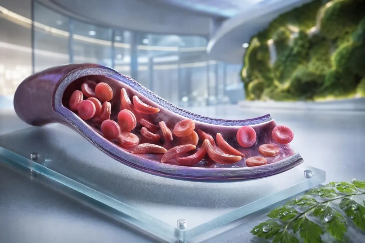
Anemija srpastih stanica nije jedan test, jedan simptom ili jedna razina hitnosti. Ovaj vodič usmjeren na pacijenta razdvaja status nositelja, potvrđenu bolest, rutinsko praćenje i situacije koje zahtijevaju hitnu pomoć.


- Srpasta stanica (sickle cell trait) znači jedan gen HbS; obično ne uzrokuje kroničnu anemiju ili ponavljajuće bolne krize.
- Probir novorođenčadi identificira većinu pogođene dojenčadi prije početka simptoma, omogućujući rani početak primjene penicilina i specijalističkog praćenja.
- Elektroforeza hemoglobina razdvaja tipove hemoglobina i pomaže u potvrđivanju HbSS, HbSC, HbS-beta talasemije ili statusa nositelja.
- Jaka bol u prsima, nedostatak zraka, temperatura od 38,5 °C ili viša, zbunjenost ili nova slabost zahtijevaju hitnu procjenu u hitnoj službi kod osobe s anemijom srpastih stanica.
- Sindrom akutnog prsnog koša može započeti kašljem, vrućicom, bolovima u prsima ili niskim kisikom i može se pogoršati tijekom sati, a ne dana.
- Nagli pad hemoglobina od 2 g/dL ili više od uobičajene razine osobe može signalizirati aplastičnu krizu, sekvestraciju, krvarenje ili ubrzanu hemolizu.
- Broj retikulocita pomaže u razlikovanju aktivnog odgovora koštane srži od aplastične krize; nizak broj tijekom pogoršanja anemije upozoravajući je obrazac.
- Hidroksilureja je lijek koji modificira tijek bolesti, a ne samo lijek protiv bolova; praćenje obično uključuje potpunu krvnu sliku svaka 4 tjedna tijekom prilagodbe doze.
Što anemija srpastih stanica znači – i što ne znači
Anemija srpastih stanica obično se odnosi na bolest HbSS, kod koje osoba nasljeđuje dva gena koji proizvode hemoglobin S; crvene stanice mogu postati krute, prerano se razgrađuju i začepljuju male krvne žile. Srpastih stanica je drugačije: jedan gen HbS obično je dovoljan za genetski prijenos, ali nije dovoljan za izazivanje kronične anemije i ponavljajućih vazo-okluzivnih epizoda viđenih kod HbSS.
Bolest srpastih stanica je krovni pojam koji uključuje HbSS, HbSC, HbS-beta-zero talasemiju i HbS-beta-plus talasemiju. HbSS često uzrokuje najizraženiju anemiju, ali težina uvelike varira čak i unutar jedne obitelji; bazalni hemoglobin od 7,5 g/dL može biti stabilan za jednu odraslu osobu, a opasan za drugu ako je brzo pao.
Mutacija hemoglobina S mijenja jednu aminokiselinu u beta-globinskom lancu: valin zamjenjuje glutaminsku kiselinu na položaju 6. Pod niskim kisikom, dehidracijom, acidozom ili vrućicom, HbS molekule se mogu polimerizirati unutar stanice. Ta fizička promjena objašnjava zašto se kriza može razviti nakon dugog leta, gastrointestinalne bolesti ili noći nedovoljnog unosa tekućine.
Kantesti AI je Analizator krvnih testova s umjetnom inteligencijom koji stavlja CBC, bilirubin, LDH, retikulocite, kreatinin i prethodne rezultate u jedan longitudinalni prikaz; ne dijagnosticira bolest srpastih stanica samo iz CBC-a. Za praktični podsjetnik o izvještavanju o hemoglobinu, pogledajte što HGB znači.
Od 4. rujna 2026. još uvijek viđam pacijente kojima se pogrešno govori da su trajt i bolest zamjenjivi. Nisu. Kliničko pravilo dr. Thomasa Kleina je jednostavno: tražite točan genotip hemoglobina, a ne nejasnu izjavu da je test bio “pozitivan”.”
Zašto genotip mijenja skrb
HbSC može imati viši hemoglobin nego HbSS, a opet uzrokovati retinalne komplikacije, bol ili avaskularnu nekrozu. HbS-beta talasemija može nalikovati traitu ili HbSS, ovisno o tome je li proizvodnja beta-globina djelomično prisutna, zbog čega molekularno testiranje ponekad rješava nejasnu elektroforetsku sliku.
Simptomi anemije srpastih stanica prema dobi i početnom stanju
Simptomi bolesti srpastih stanica obično uključuju povremenu bol u kostima ili prsima, umor, žuticu, nedostatak daha, usporen rast i česte infekcije, ali nitko ne doživljava isti obrazac. Simptomi se obično pojavljuju nakon 4 do 6 mjeseci starosti jer fetalni hemoglobin izvorno štiti dojenčad od polimerizacije HbS.
Daktilitis – bolno oticanje ruku ili nogu – može biti prvi vidljivi znak kod dojenčadi. Kod školske djece, bol u trbuhu zaslužuje posebnu pažnju jer zatvor, bolest žučnog mjehura, povećanje slezene i vazo-okluzija mogu kod kuće zvučati iznenađujuće slično.
Žutica kod bolesti srpastih stanica odražava tekuću razgradnju crvenih krvnih stanica i povišenu razinu neizravnog bilirubina; ne znači automatski hepatitis. Tamni urin, blijeda stolica, svrbež, vrućica ili bol u gornjem desnom dijelu trbuha mijenjaju tu procjenu i zahtijevaju procjenu jetrenih i bilijarnih uzroka; naš vodič za obrasci izravnog bilirubina objašnjava razliku.
Odrasli mogu normalizirati bol koja je postupno postajala sve češća. Prema mojem iskustvu, pomak s dvije kućne epizode godišnje na dvije mjesečne epizode klinički je značajan čak i ako se broj posjeta hitnoj pomoći nije povećao. Poremećaj spavanja, propušteni posao, erektilni simptomi, loše raspoloženje i smanjena tolerancija na vježbanje trebaju biti dokumentirani jer često prethode formalnoj promjeni liječenja.
Koncentracija hemoglobina od 6 do 9 g/dL može biti uobičajeni raspon u stanju mirovanja kod HbSS, dok bi vrijednost ispod 10 g/dL bila neočekivana kod većine ljudi bez bolesti. Broj je važan, ali promjena u odnosu na osobnu osnovnu razinu važnija je.
Tko bi trebao razmotriti probir na nositeljstvo srpastih stanica
Probir na srpaste stanice je razuman prije trudnoće ili rano u trudnoći za svaku osobu čiji status nositelja nije poznat, bez obzira na izgled, prezime ili pretpostavljeno podrijetlo. Status nositelja se nasljeđuje i često ostaje nepoznat do novorođenačkog probira ili neočekivano abnormalnog testa hemoglobina.
Ako oba biološka roditelja nose varijantu povezanu s HbS ili beta-talasemijom, svaka trudnoća može imati 25% šanse za nasljeđivanje klinički značajnog poremećaja hemoglobina, ovisno o uključenim varijantama. Nositelj može imati normalnu energiju, normalne nalaze CBC-a i povijest bez epizoda boli.
Probir je posebno koristan kada jedan partner ima HbAS, HbC traita, beta-talasemijski traita, neobjašnjivu mikrocitozu ili obiteljsku povijest bolesti srpastih stanica. Nizak srednji volumen stanice ne treba pripisivati nedostatku željeza bez provjere feritina i razmatranja talasemije; usporedite MCV i MCH tragove.
Globalno izvješće Svjetske zdravstvene organizacije iz 2022. procjenjuje da poremećaji hemoglobina ostaju glavno opterećenje nasljednim bolestima, posebno tamo gdje je pristup probiru novorođenčadi neujednačen. Najpoštovaniji pristup je univerzalni pristup informiranom probiru umjesto korištenja etničke pripadnosti kao čuvara pristupa.
Kantesti je Platforma za tumačenje krvnih nalaza pomoću AI-a koji mogu objasniti prijavljeni HbS rezultat jednostavnim jezikom, ali potvrda nositelja i genetičko savjetovanje zahtijevaju akreditirani laboratorij i kliničara. Ako par planira trudnoću, pitajte može li testiranje partnera i genetičko savjetovanje obaviti prije začeća, a ne nakon što rezultat postane vremenski osjetljiv.
Kako elektroforeza hemoglobina potvrđuje dijagnozu
Elektroforeza hemoglobina razdvaja frakcije hemoglobina i ključni je test za potvrdu anemije srpastih stanica ili traita, često uz visokoučinsku tekućinsku kromatografiju i, kada je potrebno, genetičko testiranje. Rutinski CBC ne može potvrditi HbSS ili traita jer nedostatak željeza i talasemija mogu proizvesti preklapajuće eritrocitne indekse.
U neliječenom HbSS, elektroforeza obično pokazuje pretežno HbS bez HbA, iako postotak HbF varira. HbAS obično pokazuje i HbA i HbS, često s višim udjelom HbA od HbS; točni postoci mogu se mijenjati s ko-naslijeđenom alfa-talasemijom ili nedavnom transfuzijom.
Nedavna transfuzija može učiniti rezultat elektroforeze pogrešnim otprilike 3 mjeseca jer cirkulira donirani HbA. To je također razlog zašto HbA1c može podcijeniti prosječni glukozu nakon transfuzije ili kod hemolize s visokim prometom; vidi kada HbA1c zavede.
Periferni razmaz može pokazati ciljne stanice, polikromaziju i srpaste oblike, ali mikroskopija podupire, a ne zamjenjuje testiranje frakcija. Dr. Klein vidio je “normalne” hitne razmaze koji su odgodili razjašnjenje kada je pacijentu transfuzija obavljena tjednima ranije—uvijek obavijestite laboratorij i tim za hematologiju o transfuzijama.
Metode probira novorođenčadi identificiraju HbS prije kliničke bolesti, ali rezultat još uvijek zahtijeva pravovremeno potvrđujuće testiranje. Smjernica utemeljena na dokazima iz 2014. autora Yawn et al. preporučuje rano uključivanje specijalista u život, s preventivnim mjerama koje počinju prije prve veće komplikacije (Yawn et al., 2014.).
Rutinsko praćenje krvi: što kliničari zapravo prate
Rutinska kontrola kod bolesti srpastih stanica obično prati hemoglobin, retikulocite, bijele krvne stanice, trombocite, bilirubin, LDH, bubrežnu funkciju i proteine u urinu – ne izolirani rezultat. Najsigurnija usporedba je s vlastitom stabilnom početnom vrijednosti bolesnika, idealno određenom kada je dobro i nije nedavno primio transfuziju.
Hemoliza često dovodi do povišenog LDH, indirektnog bilirubina i retikulocita uz nizak haptoglobin. Haptoglobin može biti nizak iz drugih razloga, uključujući bolesti jetre, stoga je kombinirani obrazac jači od pojedinačnog markera; naš vodič za tumačenje haptoglobina pokazuje zašto.
Samo postotak retikulocita može precjeniti odgovor koštane srži kod značajne anemije. Kliničari često izračunavaju apsolutni broj retikulocita ili korigirani odgovor retikulocita; pad apsolutnog broja tijekom pogoršanja anemije izaziva zabrinutost zbog aplastične krize povezane s parvovirusom B19.
Kantesti je Alat za analizu krvnih nalaza uz pomoć AI-a koji uspoređuje ponovljene hematološke rezultate umjesto da visoki LDH smatra samostalnom dijagnozom. Uzorak označen kao hemoliziran u epruveti može lažno povećati kalij i LDH, stoga iznenađujući rezultat može zahtijevati ponovno uzimanje uzorka; pregled ponovljeno testiranje nakon hemolize.
Hidroksiureja obično povećava MCV i HbF tijekom vremena, dok umjereno smanjuje neutrofile pri učinkovitoj dozi. Ti pomaci mogu biti očekivani, ali apsolutni broj neutrofila ispod individualnog terapijskog praga ili brzo padajući broj trombocita zahtijeva pregled liječnika, a ne samostalno prilagođavanje lijeka.
Kada je bol vazo-okluzivna epizoda – a kada nije
Bol povezana s vazookluzijom često je duboka, jaka i locirana u leđima, prsima, kukovima, udovima ili abdomenu, ali nova bol se ne smije automatski označiti kao srpčasta kriza. Vrućica, žarišna oteklina, ozljeda, trbušna obrana ili jednostrana neurološka simptomatologija ukazuju na probleme koji zahtijevaju drugačiju obradu.
Kućni plan za bol obično uključuje ranu hidrataciju, toplinu, propisanu analgeziju, odmor i osobni prag eskalacije dogovoren sa timom za srpčastu bolest. Pijenje prekomjernih količina nije sigurnije: osobe s bubrežnim oštećenjem ili srčanim bolestima mogu razviti preopterećenje tekućinom, osobito tijekom akutne bolesti.
Bol koja ostaje nekontrolirana nakon uobičajenog plana spašavanja, sprječava zadržavanje tekućine ili lijekova, ili je drugačija po kvaliteti ili lokaciji, treba potaknuti hitan klinički savjet. Otečeni, topli zglob može predstavljati septički artritis ili infekciju kosti, a ne nekompliciranu vazookluziju; naš vodič za testiranje neobjašnjive boli navodi korisne prve pretrage.
U hitnoj skrbi, pravodobna analgezija i ponovna procjena važniji su od dokazivanja težine boli kroz laboratorijski broj. Smjernice NICE-a o akutnim bolnim epizodama srpastih stanica preporučuju brzu procjenu analgezije i česte provjere, jer sam nedovoljan tretman doprinosi produljenoj patnji i odgođenom oporavku (NICE, 2012).
Izbjegavajte hladne obloge izravno na područje vazookluzivne boli, osim ako vam tim to izričito ne savjetuje; hladnoća može suziti periferne krvne žile. Taj praktični detalj zvuči beznačajno, ali mnogi pacijenti mi kažu da im to nitko nije objasnio tek nakon nekoliko teških epizoda.
Sindrom akutnog prsnog koša: hitnoća koju mnogi ljudi propuste
Sindrom akutnog prsnog koša je nova plućna infiltracija na snimci prsnog koša plus respiratorni simptomi, vrućica, bol u prsima ili niska razina kisika kod osobe sa bolešću srpastih stanica, i to je hitno stanje. Može se brzo pogoršati, ponekad nakon prijema zbog epizode boli, a ne u trenutku kada se pojave simptomi u prsima.
Nazovite hitnu službu ili idite na hitnu pomoć zbog novog nedostatka zraka, boli u prsima, plavo-sivih usana, nesvjestice, upornog kašlja s vrućicom ili zasićenja kisikom ispod propisane ciljne vrijednosti osobe. Zasićenje od 92% može biti velik pad za nekoga tko je normalno na 98%, dok neki pacijenti imaju nižu dokumentiranu početnu vrijednost; važna je i apsolutna vrijednost i promjena.
Sindrom akutnog prsnog koša može uključivati infekciju, masnu emboliju iz koštane srži, plućnu vaskularnu opstrukciju, atelektazu ili više procesa odjednom. Kliničari obično procjenjuju oksigenaciju, CBC, retikulocite, kulture kada je prisutna vrućica, snimanje prsnog koša, a ponekad i plinske analize krvi; krvne pretrage za otežano disanje dodaje kontekst ovim rezultatima.
Kod odraslih osoba, pretjerana sedacija opioidima, plitko disanje uzrokovano boli i višak intravenske tekućine mogu pogoršati respiratorni status. Poticanje dubokog disanja pomoću spirometrije tijekom hospitalizacije smanjuje plućne komplikacije u odabranim bolnim epizodama, iako nije zamjena za hitnu procjenu kada su se simptomi već pojavili.
Pregled Katoa i kolega iz 2018. opisuje sindrom akutnog prsnog koša kao glavni uzrok obolijevanja i smrtnosti tijekom cijelog životnog vijeka (Kato et al., 2018). Nemojte voziti ako su nedostatak daha, pospanost ili pritisak u prsima značajni.
Groznica, infekcija i hitne situacije sa slezenom
Temperatura od 38,5°C (101,3°F) ili viša u djeteta ili odrasle osobe sa srpastom anemijom zahtijeva hitnu medicinsku procjenu istog dana, osim ako njihov specijalistički tim nije dao drugačiji plan. Funkcijski gubitak slezenske imunološke funkcije može uzrokovati da neke bakterijske infekcije napreduju mnogo brže nego uobičajena virusna vrućica.
Vrućica uz letargiju, osip, nizak krvni tlak, zbunjenost ili smanjeno izlučivanje mokraće zahtijeva hitnu medicinsku pomoć odmah. Normalan broj bijelih krvnih stanica ne isključuje pouzdano ozbiljnu infekciju kod srpaste anemije, osobito rano u tijeku bolesti ili tijekom liječenja hidroksiurejom.
Sekvestracija slezene najčešće se javlja u male djece i uzrokuje iznenadno povećanje slezene, bljedilo, osjećaj punoće u trbuhu, ubrzano disanje i brzi pad hemoglobina. Roditeljima se ponekad pokazuje kako napipati slezenu djeteta, ali svako novo povećanje uz bolest treba potaknuti hitnu procjenu, a ne ponavljane kućne provjere.
Pad hemoglobina od 2 g/dL ili više ispod uobičajene bazne linije s povećanom slezenom zabrinjavajući je zbog sekvestracije. Nasuprot tome, teška anemija s vrlo niskim brojem retikulocita može ukazivati na aplastičnu krizu, često povezanu s parvovirusom B19; interpretacija broja retikulocita objašnjava signal koštane srži.
Laboratorijske pretrage za sepsu mogu uključivati laktat, kulture, CBC, pretrage bubrega i upalne markere, ali odluke o liječenju nikada ne bi smjele čekati da se svi rezultati vrate. Za kontekst o hitnim obrascima, pročitajte tragovi markera sepse.
Cjepiva i preventivni antibiotici
Cjepiva i dječja profilaksa penicilinom smanjuju rizik, ali ne čine vrućicu niskorizičnom. Potvrdite svoj individualni plan s hematološkim ili pedijatrijskim timom, osobito nakon splenektomije, putovanja, novorođenčeta ili promjene lokalnog rasporeda cijepljenja.
Moždani udar i neurološki znakovi upozorenja zahtijevaju trenutno djelovanje
Svako iznenadno spuštanje lica, slabost u ruci ili nozi, poteškoće s govorom, napadaj, jaka neobična glavobolja, zbunjenost ili gubitak ravnoteže kod srpaste anemije treba tretirati kao moguću moždanu kap. Odmah nazovite hitnu pomoć; nemojte čekati da vidite hoće li simptomi proći ili davati lijekove protiv bolova.
Djeci s HbSS ili HbS-beta-nultom talasemijom obično se nudi ultrazvučni pregled transkranijalnog doplerom od 2. do 16. godine života. Abnormalna prosječna maksimalna brzina od 200 cm/sekundi ili viša ukazuje na značajno povećan rizik od moždanog udara i zahtijeva hitnu specijalističku intervenciju.
Kronična transfuzijska terapija može smanjiti rizik od prvog moždanog udara kod djece s abnormalnim dopler nalazima, ali stvara dodatne potrebe: praćenje preopterećenja željezom, skrining na antitijela i pažljivo podudaranje donorskih stanica. Ovo je specijalizirana njega, a ne rezultat kojim se upravlja putem aplikacije.
Odrasli mogu imati prolazne neurološke simptome, kognitivne promjene ili tiho cerebralno oštećenje bez klasičnog dramatičnog moždanog udara. 10-minutna epizoda poteškoća s pronalaženjem riječi i dalje zahtijeva hitnu procjenu jer prolazni ishemijski simptomi mogu biti upozorenje, a ne olakšanje.
Dr. Thomas Klein savjetuje obiteljima da sačuvaju svoj lokalni broj za hitne slučanke srpaste anemije uz uobičajeni plan za bol. Time se uklanja jedna odluka tijekom nekoliko minuta kada se dijete ili odrasla osoba možda bore komunicirati.
Zaštita bubrega, očiju i pluća između kriza
Srpasta anemija može oštetiti bubrege, mrežnicu, pluća i srce čak i kada je bol rijetka, stoga je rutinsko praćenje organa dio liječenja, a ne opcionalni dodatak. Testiranje urina na proteine, provjere krvnog tlaka, pregledi očiju i procjena pluća vođena simptomima otkrivaju komplikacije prije nego što postanu očite.
Albuminurija može biti rani znak srpčaste nefropatije. Omjer albumina i kreatinina u urinu od 30 mg/g ili više je abnormalan kod većine odraslih i treba ga potvrditi ponovljenim testiranjem jer vrućica, tjelovježba, menstruacija i dehidracija mogu prolazno povisiti te vrijednosti.
Kreatinin može izgledati zavaravajuće normalan kod srpaste anemije jer povećano tubularno izlučivanje i niža mišićna masa mogu maskirati smanjenu filtraciju. Cistatin C ili kombinirane jednadžbe mogu pružiti korisne kontekste, dok uporna pjenasta mokraća zaslužuje procjenu; vidi smjernice za proteine u urinu.
Retinopatija je posebno važna kod HbSC bolesti, koja u drugim aspektima može biti klinički tiša. Nove plutajuće mrlje, bljeskanje, promjena vida poput zavjese ili iznenadna zamućenost vida zahtijevaju hitan oftalmološki pregled, a ne rutinski pregled tjednima kasnije.
Trudnoća dodaje fiziološku hiperfiltraciju i veći majčino-fetalni rizik, stoga bi osnovna procjena bubrega i urina trebala biti obavljena rano. Naše objašnjenje promjena GFR-a u trudnoći pomaže razlikovati očekivanu promjenu od zabrinjavajućeg trenda.
Mogućnosti liječenja, hidroksiureja i sigurnost transfuzije
Hidroksiuja smanjuje bolne epizode i akutni prsni sindrom kod mnogih osoba s HbSS ili HbS-beta-nula talasemijom povećanjem fetalnog hemoglobina, ali zahtijeva strukturirano praćenje. Transfuzija može spasiti život kod odabranih komplikacija, ali ponovljene transfuzije nose rizik od nakupljanja željeza i stvaranja antitijela na crvene krvne stanice.
Hidroksiuja se obično titrira uz CBC i broj retikulocita otprilike svaka 4 tjedna dok se ne postigne stabilna doza, a zatim u dužim intervalima određenim od strane tima koji liječi. Viši MCV je često očekivani učinak liječenja, dok teška citopenija zahtijeva pregled lijekova i ponekad privremeni prekid.
Jednostavna transfuzija povećava sposobnost prijenosa kisika, ali prekomjerna koncentracija hemoglobina može povećati viskoznost kod HbSS. Mnogi akutni protokoli izbjegavaju povećanje hemoglobina nakon transfuzije iznad 10 g/dL, osim ako specijalist ne postavi drugačiji cilj; izmjenjiva transfuzija koristi se za odabrane teške komplikacije.
Preopterećenje željezom postaje vjerojatnije nakon ponovljenih transfuzija i procjenjuje se trendovima feritina te po potrebi MR-om jetre ili srca. Feritin raste s upalom, stoga jedna visoka razina nije dokaz nakupljanja željeza u tkivima; naš vodič za proučavanje željeza objašnjava ograničenja.
Dokazi su najjači za hidroksiuja kod HbSS i HbS-beta-nula bolesti; korist kod HbSC ostaje područje s manje sigurnosti i više individualiziranog donošenja odluka. Plan liječenja lijekovima trebao bi uključivati rasprave o kontracepciji i trudnoći, gdje je to relevantno, prepreke pridržavanju i pisani plan za propuštene doze.
Trudnoća, plodnost i planiranje obitelji s HbS
Trudnoća sa srpastom bolesti stanica zahtijeva ranu zajedničku skrb od opstetricije i hematologije jer su rizici od bolnih epizoda, anemije, tromboze, infekcije, preeklampsije, usporenog rasta fetusa i prijevremenog poroda veći. Probir nositelja je važan prije trudnoće jer genetski rizik ovisi o oba biološka roditelja.
Posjet prije trudnoće trebao bi obuhvatiti genotip, početni hemoglobin, funkciju bubrega, albuminuriju, krvni tlak, povijest transfuzija, antitijela na crvene krvne stanice, cijepljenja i lijekove. Odluke o hidroksiuji u trudnoći su individualizirane i moraju se donijeti sa specijalistima; nemojte je prestati uzimati ili ponovno početi uzimati samo na temelju savjeta s interneta.
Željezo se ne smije automatski propisivati za nizak hemoglobin kod HbSS. Nedostatak željeza može postojati, osobito kod trudnoće ili obilnog menstrualnog krvarenja, ali feritin i zasićenost transferina trebaju voditi liječenje jer kronična hemoliza sama po sebi ne znači nedostatak željeza.
Kada oba roditelja nose relevantne varijante hemoglobina, genetičko savjetovanje može objasniti prirodnu koncepciju, opcije prenatalne dijagnostike, opcije donora i preimplantacijsko genetičko testiranje bez usmjeravanja obitelji prema jednoj odluci. Cilj je informirani izbor, a ne panika.
Za ostale praktične laboratorijske crvene zastavice u trudnoći, pogledajte laboratorijske zabrinutosti u trudnoći iste dan. Poznata početna vrijednost pacijenta treba biti uključena u svako predavanje o trudnoći; to mijenja način na koji se tumači rezultat hemoglobina.
Sigurna upotreba laboratorijskih rezultata između pregleda
Laboratorijski rezultat srpaste bolesti stanica najkorisniji je kada se usporedi s vašim genotipom, stabilnim početnim vrijednostima, simptomima, liječenjima i nedavnim transfuzijama. Visoki bilirubin ili nizak hemoglobin mogu biti očekivani za osobu s HbSS, ali hitni za istu osobu ako se rezultat naglo promijeni.
Zadržite jednodnevni zapis genotipa, uobičajenog hemoglobina, uobičajene zasićenosti kisikom ako je poznata, krvne grupe i antitijela, trenutnih lijekova, datuma transfuzija i hitnih kontakata. Ovo štedi vrijeme u nepoznatim hitnim odjelima i smanjuje mogućnost da se početna vrijednost HbSS pogrešno protumači kao novootkrivena anemija.
Kantesti je usluzi za tumačenje laboratorijskih testova AI koji mogu organizirati učitana laboratorijska izvješća i označiti obrasce za raspravu, uključujući markere hemolize, bubrežne trendove i praćenje lijekova. Ne može vidjeti vaše grudi, mjeriti oksigenaciju niti zamijeniti hitnu skrb kada simptomi ukazuju na akutni prsni sindrom, sepsu ili moždani udar.
Rezultat izvan referentnog laboratorijskog intervala nije automatski opasan, a rezultat unutar njega i dalje može biti opasan ako se naglo promijenio. Za objašnjenje oznaka izvješćivanja i referentnih raspona pročitajte značenje rezultata izvan raspona.
Klinički pristup Kantesti-a pregledava se u odnosu na definirane metode i ograničenja; čitatelji koji žele razumjeti naše zaštitne mjere mogu pregledati standarde kliničke validacije. Ponesite originalni PDF na svoj hematološki pregled – OCR je koristan, ali izvorni izvještaj ostaje zapis.
Jasan plan hitnog djelovanja kod anemije srpastih stanica
Potražite hitnu pomoć odmah zbog bolova u prsima ili otežanog disanja, vrućice od 38,5 °C ili više, novih neuroloških simptoma, nesvjestice, jake bljedoće, nekontrolirane boli, naglo rastućeg trbuha ili nemogućnosti pijenja i uzimanja lijekova. Ovi znakovi upozorenja mogu ukazivati na akutni sindrom prsnog koša, sepsu, moždani udar, tešku anemiju ili sekvestraciju slezene.
Osoblju hitne pomoći recite da imate bolest srpastih stanica, navedite svoj genotip ako je poznat i opišite što se razlikuje od vaše uobičajene epizode. Spomenite nedavnu transfuziju, bazalni hemoglobin, lijekove protiv bolova koji su već uzeti s dozama i vremenima, alergije, trudnoću i bilo kakav prethodni akutni sindrom prsnog koša ili moždani udar.
Nemojte čekati da se kućni pulsni oksimetar nenormalno očita ako se simptomi u prsima pogoršavaju. Potrošački uređaji mogu pogrešno očitati s hladnim prstima, proizvodima za nokte, pokretima, lošom cirkulacijom i nekim tonovima kože; simptomi i klinička procjena nadmašuju jedan umirujući prikaz.
Ako brinete za dijete, pitajte njihov tim za pisani prag temperature, plan analgezije i upute za provjeru slezene. Ako ste odrasla osoba, čuvajte kopiju u telefonu i podijelite je s jednom povjerljivom osobom; planiranje obiteljskog zdravstvenog zapisa može to olakšati.
Medicinski tim Kantesti-a, uključujući naš Medicinski savjetodavni odbor, podržava edukaciju o laboratorijskoj interpretaciji, dok hitne odluke pripadaju kliničkim uslugama koje se obavljaju uživo. O našoj organizaciji i kliničkoj misiji, pogledajte naš tim i pristup.
Često postavljana pitanja
Koji su prvi simptomi anemije srpastih stanica kod beba?
Prvi simptomi anemije srpastih stanica često se pojavljuju nakon 4 do 6 mjeseci života, kada fetalni hemoglobin prirodno opada. Bolno oticanje ruku i nogu, nazvano daklilitsko, vrućica, neobična razdražljivost, bljedilo, žutica, slabo hranjenje i otečeni trbuh uobičajeni su rani znakovi upozorenja. Vrućica od 38,5°C (101,3°F) ili viša u dojenčeta sa srpastom bolešću zahtijeva hitnu liječničku procjenu istog dana jer se teška infekcija može brzo razviti. Probir novorođenčadi identificira zahvaćenu dojenčad prije pojave simptoma, ali potvrđujuće testiranje i praćenje od strane specijalista ostaju neophodni.
Mogu li imati bolest srpastih stanica i još uvijek imati simptome?
Većina ljudi s nasljednom anemijom srpastih stanica nema kroničnu anemiju, nema povratne bolne krize i ima normalnu dnevnu toleranciju na vježbanje. Rijetke komplikacije mogu se pojaviti kod ekstremne dehidracije, teške izloženosti toplini, velikih nadmorskih visina, vrlo intenzivnog napora ili okruženja s niskim udjelom kisika, a krv u mokraći treba procijeniti umjesto pretpostaviti da je bezopasna. Svojstvo znači jedan gen HbS, dok bolest srpastih stanica obično znači bolest HbSS s dva zahvaćena gena za beta-globin. Elektroforeza hemoglobina ili ekvivalentni test frakcioniranja može pojasniti razliku.
Koji test potvrđuje anemiju srpastih stanica?
Elektroforeza hemoglobina, tečna hromatografija visokih performansi ili kapilarna elektroforeza potvrđuju prisutne frakcije hemoglobina i ključni su za dijagnostikovanje srpastocelijske anemije. Neliječeni HbSS obično pokazuje pretežno hemoglobin S bez hemoglobina A, dok osobina obično pokazuje i hemoglobin A i hemoglobin S. CBC može identificirati anemiju ili male crvene krvne stanice, ali ne može samostalno dijagnosticirati HbSS, HbSC ili osobinu. Nedavna transfuzija može iskriviti rezultate frakcija na oko 3 mjeseca, tako da molekularno testiranje može biti potrebno kada je obrazac nejasan.
Kada se osoba sa srpastom bolesti treba uputiti na hitni prijem?
Osoba sa srpastom bolesti treba potražiti hitnu procjenu zbog vrućice od 38,5°C (101,3°F) ili više, bolova u prsima, nedostatka zraka, niske razine kisika, iznenadne slabosti, poteškoća u govoru, napadaja, nesvjestice, naglog bljedila, jakog oticanja trbuha ili boli koja ostaje nekontrolirana prema utvrđenom planu. Ovi simptomi mogu signalizirati akutni torakalni sindrom, infekciju, moždani udar, sekvestraciju slezene ili nagli pad hemoglobina. Nova ili drugačija bol također zahtijeva procjenu jer infekcija kosti, bolest žučnog mjehura, upala slijepog crijeva i ugrušci mogu oponašati bol uzrokovanu vazo-okluzijom. Nemojte sami voziti ako su neurološki ili respiratorni simptomi značajni.
Koja je razina hemoglobina opasna kod srpastocelularne bolesti?
Ne postoji jedinstveni opasan broj hemoglobina kod anemije srpastih stanica jer mnogi ljudi s HbSS imaju stabilnu početnu vrijednost između otprilike 6 i 9 g/dL. Pad od 2 g/dL ili više u odnosu na uobičajenu razinu hemoglobina kod pojedinca često je zabrinjavajući od apsolutne vrijednosti i može ukazivati na aplastičnu krizu, sekvestraciju slezene, krvarenje ili ubrzanu hemolizu. Teški simptomi poput nedostatka daha, nesvjestice, bolova u prsima, ubrzanog rada srca ili zbunjenosti zahtijevaju hitnu procjenu na bilo kojoj razini hemoglobina. Odluke o transfuziji ovise o simptomima, početnoj vrijednosti, uzroku anemije, oksigenaciji i riziku od hiperviskoznosti.
Hidroksiureja liječi srpastocelularnu anemiju?
Hidroksoureja ne liječi anemiju srpastih stanica, ali može smanjiti bolne epizode, akutni prsni sindrom, potrebu za transfuzijama i hospitalizaciju kod mnogih osoba s HbSS ili HbS-beta-zero talasemijom. Djeluje djelomično povećanjem fetalnog hemoglobina i obično zahtijeva praćenje CBC i retikulocita otprilike svaka 4 tjedna dok se doza prilagođava. Povišeni MCV i razina HbF mogu ukazivati na biološki učinak, iako nijedan rezultat ne dokazuje savršenu pridržavanje terapiji. Kurativni pristupi poput transplantacije matičnih stanica i nekih gen-temeljenih terapija zahtijevaju visoko specijaliziranu procjenu podobnosti i dugoročno praćenje.
Isprobajte analizu krvne slike uz AI već danas
Pridružite se više od 2 milijuna korisnika diljem svijeta koji vjeruju Kantesti-u za trenutačnu i točnu analizu laboratorijskih nalaza. Prenesite svoje rezultate krvne slike i dobijte sveobuhvatno tumačenje 15,000+ biomarkera u sekundama.
📚 Referirane znanstvene publikacije
Klein, T., Mitchell, S., & Weber, H. (2026). Vodič za krvni test komplementa C3 C4 i ANA titra. Kantesti AI Medical Research.
Klein, T., Mitchell, S., & Weber, H. (2026). Test krvi na virus Nipah: Vodič za rano otkrivanje i dijagnozu 2026.. Kantesti AI Medical Research.
📖 Vanjske medicinske reference
⚕️ Medicinska izjava o odricanju odgovornosti
Ovaj je članak samo u obrazovne svrhe i ne predstavlja medicinski savjet. Za odluke o dijagnozi i liječenju uvijek se posavjetujte s kvalificiranim pružateljem zdravstvenih usluga.
E-E-A-T signal(i) povjerenja
Iskustvo
Liječnički vođena klinička revizija radnih tokova tumačenja laboratorijskih nalaza.
Stručnost
Fokus laboratorijske medicine na to kako se biomarkeri ponašaju u kliničkom kontekstu.
Autoritativnost
Napisao dr. Thomas Klein, uz recenziju dr. Sarah Mitchell i prof. dr. Hans Weber.
Pouzdanost
Tumačenje utemeljeno na dokazima, s jasnim putovima praćenja kako bi se smanjila uzbuna.