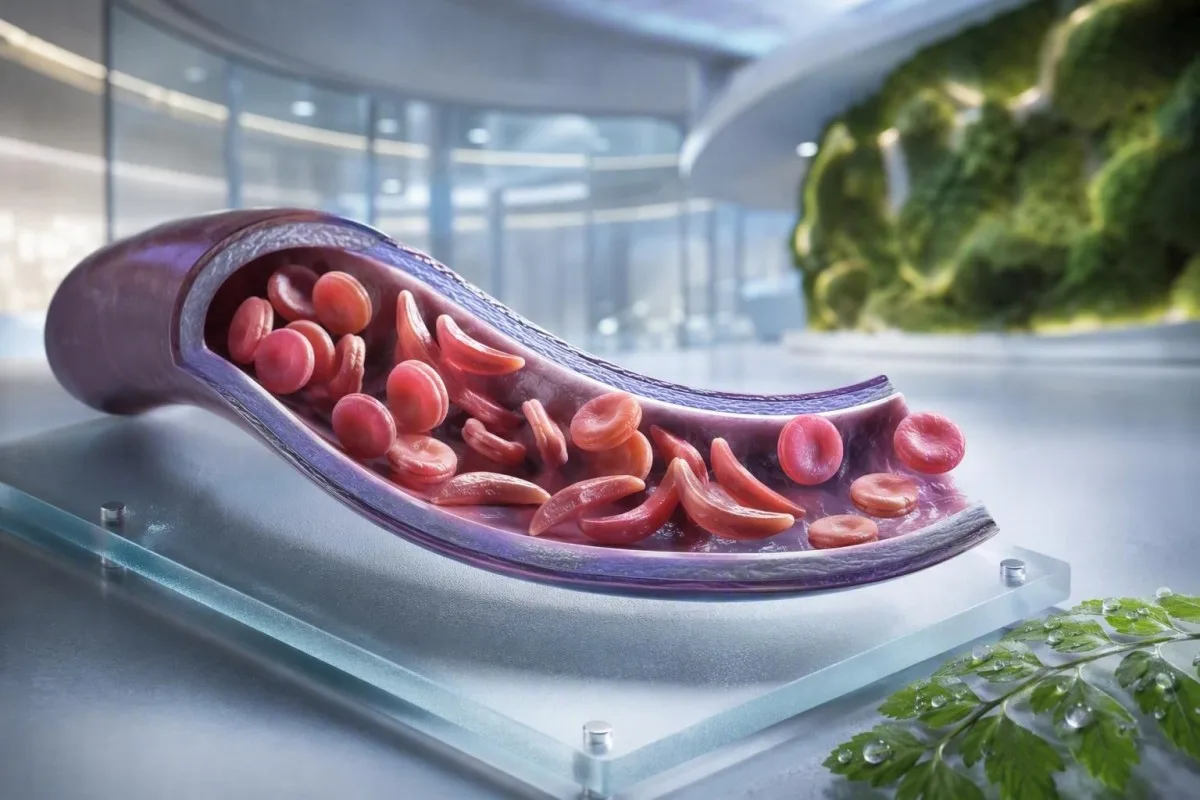
Anemija srpastih celic ni en test, en simptom ali ena stopnja nujnosti. Ta vodnik, ki daje prednost bolniku, loči status nosilca, potrjeno bolezen, rutinsko spremljanje in situacije, ki zahtevajo nujno oskrbo.


- Srpastocelična lastnost pomeni en gen HbS; običajno ne povzroča kronične anemije ali ponavljajočih se bolečinskih kriz.
- Pregled novorojenčkov identificira večino prizadetih dojenčkov pred pojavom simptomov, kar omogoča zgodnji začetek penicilina in specialističnega spremljanja.
- Hemoglobinska elektroforeza loči vrste hemoglobina in pomaga potrditi HbSS, HbSC, HbS-beta talasemijo ali status nosilca.
- Huda bolečina v prsih, težko dihanje, vročina 38,5 °C ali višja, zmedenost ali nova oslabelost zahtevajo nujno oceno v nujnih primerih pri osebi z anemijo srpastih celic.
- Sindrom akutnega prsnega koša se lahko začne s kašljem, vročino, bolečino v prsih ali nizko količino kisika in se lahko poslabša v nekaj urah namesto v dneh.
- Nenaden padec hemoglobina za 2 g/dL ali več od običajne ravni osebe lahko signalizira srpastičnostno krizo, sekvestracijo, krvavitev ali pospešeno hemolizo.
- Število retikulocitov pomaga ločiti aktivni odziv kostnega mozga od srpastičnostne krize; nizko število med poslabšanjem anemije je opozorilni vzorec.
- Hidroksiureja je bolezen spreminjajoče zdravilo, ne zgolj zdravilo proti bolečinam; spremljanje običajno vključuje popolno krvno sliko vsake 4 tedne med prilagajanjem odmerka.
Kaj pomeni anemija srpastih celic – in kaj ne
Srpastiа anemiја običajno pomeni HbSS bolezen, kjer oseba podeduje dva gena, ki proizvajata hemoglobin S; rdeče krvničke lahko postanejo toge, se zgodaj razpadejo in zamašijo majhne žile. Srpastiа celicа je drugačna: en gen HbS je običajno dovolj za genetski prenos, vendar ni dovolj, da bi povzročil kronično anemijo in ponavljajoče se epizode vazo-okluzije, ki jih vidimo pri HbSS.
Srpastiа celična bolezen je krovni izraz, ki vključuje HbSS, HbSC, HbS-beta-ničta talasemijo in HbS-beta-plus talasemijo. HbSS pogosto povzroča najbolj izrazito anemijo, vendar se resnost močno razlikuje tudi znotraj ene družine; osnovni hemoglobin 7,5 g/dL je lahko stabilen za enega odraslega in nevaren za drugega, če je hitro padel.
Mutacija hemoglobina S spremeni eno aminokislino v beta-globinu: valin nadomesti glutaminsko kislino na 6. položaju. Pod nizkim kisikom, dehidracijo, acidozo ali vročino se molekule HbS lahko polimerizirajo znotraj celice. Ta fizikalna sprememba pojasnjuje, zakaj se kriza lahko razvije po dolgem letu, gastrointestinalni bolezni ali noči s pomanjkljivim vnosom tekočine.
Kantesti AI je AI analizator krvi ki postavi CBC, bilirubin, LDH, retikulocite, kreatinin in prejšnje rezultate v en longitudinalni pogled; ne diagnosticira srpastiа celične bolezni samo iz CBC. Za praktično osvežitev o poročanju hemoglobina glejte kaj pomeni HGB.
Od 4. septembra 2026 še vedno opažam, da bolnikom napačno govorijo, da sta trajt in bolezen zamenljiva. Nista. Klinično pravilo dr. Thomasa Kleina je preprosto: prosite za natančen genotip hemoglobina, ne za nejasno trditev, da je bil test “pozitiven”.”
Zakaj genotip spremeni nego
HbSC ima lahko višji hemoglobin kot HbSS, vendar še vedno povzroča mrežnične zaplete, bolečine ali avaskularno nekrozo. HbS-beta talasemija lahko posnema trajt ali HbSS, odvisno od tega, ali je proizvodnja beta-globina delno prisotna, zato molekularno testiranje včasih reši nedoumeven elektroforetski vzorec.
Simptomi anemije srpastih celic po starosti in osnovni vrednosti
Simptomi srpastiа celične bolezni običajno vključujejo občasne bolečine v kosteh ali prsih, utrujenost, zlatenico, zasoplost, zmanjšano rast in pogoste okužbe, vendar nobena dva človeka ne doživljata enakega vzorca. Simptomi se običajno pojavijo po 4 do 6 mesecih starosti, ker fetalni hemoglobin sprva ščiti dojenčke pred polimerizacijo HbS.
Daklititis – boleča oteklina rok ali nog – je lahko prvi vidni znak pri dojenčkih. Pri šoloobveznih otrocih abdominalna bolečina zahteva posebno pozornost, ker so lahko zaprtost, bolezen žolčnika, povečanje vranice in vazo-okluzija doma presenetljivo podobni.
Zlatenica pri srpastiа celični bolezni odraža stalno razgradnjo rdečih krvničk in zvišanje indirektnega bilirubina; ne pomeni samodejno hepatitisa. Temen urin, svetlo blato, srbenje, vročina ali bolečina v zgornjem desnem delu trebuha spremenijo to oceno in zahtevajo oceno jetrnih in žolčnih vzrokov; naš vodnik po vzorcih direktnega bilirubina razlaga razliko.
Odrasli lahko normalizirajo bolečino, ki je postopoma postala pogostejša. Po mojih izkušnjah je premik od dveh epizod na leto, ki jih obvladujejo doma, do dveh epizod na mesec klinično pomemben, tudi če se obiski v nujnih primerih niso povečali. Motnje spanja, izostanek z dela, erektilni simptomi, slabo razpoloženje in zmanjšana toleranca za vadbo je treba dokumentirati, ker pogosto predhodijo formalni spremembi zdravljenja.
Koncentracija hemoglobina od 6 do 9 g/dL je lahko običajno območje v stanju mirovanja pri HbSS, medtem ko bi vrednost pod 10 g/dL bila nepričakovana pri večini ljudi brez bolezni. Številka je pomembna, vendar je sprememba od osebne osnove bolj pomembna.
Kdaj naj razmislijo o testiranju na nosilce anemije srpastih celic
Srpastiа celična presejalna metoda je smiselna pred nosečnostjo ali zgodaj v nosečnosti za vsakogar, čigar status nosilca ni znan, ne glede na videz, priimek ali domnevno poreklo. Status nosilca je podedovan in pogosto ostane neznan do novorojenčka presejalne metode ali nepričakovano nenormalnega testa hemoglobina.
Če oba biološka starša prenašata varianto, povezano s HbS ali beta-talasemijo, ima vsaka nosečnost 25%T možnost podedovanja klinično pomembne krvne bolezni, odvisno od vpletenih variant. Nosilec lahko ima normalno energijo, normalne CBC rezultate in zgodovino brez bolečin.
Testiranje je še posebej koristno, ko ima eden od partnerjev HbAS, HbC, beta-talasemijo, nerazložljivo mikrocitozo ali družinsko anamnezo srpastične bolezni. Nizka povprečna prostornina krvničk (MCV) ne sme biti pripisana pomanjkanju železa brez preverjanja feritina in upoštevanja talasemije; primerjajte MCV in MCH namigi.
Svetovna zdravstvena organizacija je v svojem globalnem poročilu iz leta 2022 ocenila, da krvne bolezni še vedno predstavljajo veliko breme dednih bolezni, zlasti tam, kjer je dostop do presejanja novorojenčkov neenakomeren. Najbolj spoštljiv pristop je univerzalen dostop do informiranega presejanja, namesto da se etničnost uporablja kot izgovor.
Kantesti je platforma za razlaga krvne slike z AI ki lahko pojasnijo poročani rezultat HbS v preprostem jeziku, vendar potrditev nosilcev in reproduktivno svetovanje zahtevata akreditiran laboratorij in zdravnika. Če par načrtuje nosečnost, vprašajte, ali se testiranje partnerja in genetsko svetovanje lahko izvedeta pred spočetjem, namesto po tem, ko rezultat postane časovno občutljiv.
Kako elektroforeza hemoglobina potrdi diagnozo
Elektrofereza hemoglobina loči hemoglobinske frakcije in je ključni test za potrditev srpastične anemije ali ($HbS$), pogosto skupaj z visokofrekvenčno tekočinsko kromatografijo in, ko je potrebno, genetskim testiranjem. Rutinski CBC ne more potrditi ($HbSS$) ali ($HbS$) zaradi ($HbS$) in talasemije, ker lahko pomanjkanje železa in talasemija povzročita prekrivajoče se indekse rdečih krvničk.
Pri nezdravljenem ($HbSS$) je na elektroforezi običajno večinoma ($HbS$) brez ($HbA$), čeprav se odstotek ($HbF$) razlikuje. ($HbAS$) običajno kaže tako ($HbA$) kot ($HbS$), pogosto z višjim odstotkom ($HbA$) kot ($HbS$); natančni odstotki se lahko spremenijo zaradi so-podedovane alfa-talasemije ali nedavne transfuzije.
Nedavna transfuzija lahko naredi rezultat elektroforeze zavajajoč približno 3 mesece, ker ($HbA$) iz donacije še kroži. To je tudi razlog, zakaj ($HbA1c$) lahko podcenjuje povprečno raven glukoze po transfuziji ali pri hemolizi z visoko presnovo; glejte ko ($HbA1c$) zavaja.
Periferna razmazna mikroskopija lahko pokaže tarčne celice, polikromazijo in srpaste oblike, vendar mikroskopija podpira, namesto da bi nadomestila testiranje frakcij. Dr. Klein je videl “normalne” nujne razmaze, ki so zavlekle pojasnitev, ko je bil bolniku pred tedni opravljena transfuzija – vedno obvestite laboratorij in ekipo za hematologijo o transfuzijah.
Metode presejanja novorojenčkov identificirajo ($HbS$) pred klinično boleznijo, vendar rezultat še vedno potrebuje pravočasno potrditveno testiranje. Doktrina iz leta 2014, ki temelji na dokazih, avtorjev Yawn et al. priporoča zgodnje vključevanje specialistov v življenju, z preventivnimi ukrepi, ki se začnejo pred prvo večjo zaporo (Yawn et al., 2014).
Rutinsko spremljanje krvi: kaj kliniki dejansko spremljajo
Rutinsko spremljanje pri srpastocelični bolezni običajno vključuje sledenje hemoglobina, retikulocitov, belih krvnih celic, trombocitov, bilirubina, LDH, ledvične funkcije in beljakovin v urinu – ne enega samega rezultata. Najvarnejša primerjava je s pacientovim lastnim stabilnim izhodiščem, idealno opravljeno, ko je pacient dobro in ni bil nedavno transfuziran.
Hemoliza pogosto povzroči zvišano LDH, indirektni bilirubin in retikulocite z nizkim haptoglobinom. Haptoglobin je lahko nizek tudi iz drugih razlogov, vključno z boleznijo jeter, zato je kombinirani vzorec močnejši od posameznega markerja; naša vodnik za interpretacijo haptoglobina pokaže, zakaj.
Sam odstotek retikulocitov lahko preceni odziv kostnega mozga pri pomembni anemiji. Klinični zdravniki pogosto izračunajo absolutno število retikulocitov ali popravljen odziv retikulocitov; padajoče absolutno število med poslabšujočo se anemijo vzbuja skrb zaradi apastične krize, povezane s parvovirusom B19.
Kantesti je Orodje za analizo krvnih testov z umetno inteligenco ki primerja ponovljene hematološke rezultate, namesto da bi visoko LDH obravnavali kot samostojno diagnozo. Vzorec, označen kot hemoliziran v epruveti, lahko lažno zviša kalij in LDH, zato lahko presenetljiv rezultat zahteva ponovno odvzem; pregled ponovljeno testiranje po hemolizi.
Hidroksiureja običajno sčasoma poveča MCV in HbF, medtem ko pri učinkovitem odmerku skromno zniža nevtrofilce. Ti premiki so lahko pričakovani, vendar absolutno število nevtrofilcev pod posamezno pragom zdravljenja ali hitro padajoče število trombocitov zahteva pregled s strani predpisovalca, ne samostojnega prilagajanja zdravil.
Kdaj je bolečina epizoda vazookluzije – in kdaj ni
Boleče vazooktuzivne epizode so pogosto globoke, hude in locirane v hrbtu, prsih, bokih, okončinah ali trebuhu, vendar novo bolečino ne smemo samodejno označiti kot srpasto krizo. Vročina, žariščna oteklina, poškodba, obrambna napetost trebušne stene ali enostranski nevrološki simptomi kažejo na težave, ki zahtevajo drugačen preiskovalni postopek.
Načrt za obravnavo bolečine na domu običajno vključuje zgodnje hidracijo, toploto, predpisano analgezijo, počitek in osebni prag za poslabšanje, dogovorjen s srpastocelično ekipo. Pitje prekomernih količin ni varnejše: ljudje z ledvično okvaro ali boleznijo srca lahko razvijejo preobremenitev s tekočino, še posebej med akutno boleznijo.
Bolečina, ki ostane nenadzorovana po običajnem načrtu reševanja, preprečuje zadrževanje tekočin ali zdravil v želodcu ali je drugačna po kakovosti ali lokaciji, naj sproži nujni klinični nasvet. Otekla, vroča sklepa lahko predstavljata septični artritis ali kostno okužbo, ne pa nekomplicirano vazooktuzijo; naša smernice za testiranje nepojasnjene bolečine opisujejo uporabne prve preiskave.
V nujni oskrbi je pravočasna analgezija in ponovna ocena pomembnejša od dokazovanja jakosti bolečine s pomočjo laboratorijske številke. Smernice NICE o akutnih bolečih srpastoceličnih epizodah priporočajo hitro oceno analgezije in pogosto ponovno oceno, ker sam podzdravljenje prispeva k podaljšani stiski in počasnemu okrevanju (NICE, 2012).
Izogibajte se hladnim obkladkom neposredno na področje vazooktuzivne bolečine, razen če jih vaša ekipa posebej priporoča; mraz lahko zoži periferne žile. Ta praktični podatek se zdi majhen, vendar mi mnogi bolniki pravijo, da jim tega nihče ni pojasnil do po več težkih epizodah.
Sindrom akutnega prsnega koša: nujno stanje, ki ga mnogi spregledajo
Sindrom akutne prsne bolezni je nova pljučna infiltracija na slikovni diagnostiki prsnega koša, skupaj z respiratornimi simptomi, vročino, bolečinami v prsih ali nizko količino kisika pri osebi s srpastocelično boleznijo in je nujno stanje. Lahko se hitro poslabša, včasih po sprejemu zaradi epizode bolečine, ne pa v trenutku, ko se pojavijo simptomi v prsih.
Pokličite nujno pomoč ali pojdite v nujno oskrbo ob novi kratki sapi, bolečinah v prsih, modro-sivih ustnicah, omedlevici, vztrajnem kašlju z vročino ali nasičenosti kisika pod predpisano ciljno vrednostjo. Nasičenost 92% je lahko velik padec za nekoga, ki je običajno pri 98%, medtem ko imajo nekateri bolniki nižjo dokumentirano izhodiščno vrednost; pomembna sta tako absolutna vrednost kot sprememba.
Sindrom akutne prsne bolezni lahko vključuje okužbo, maščobno embolizacijo iz kostnega mozga, pljučno žilno obstrukcijo, atelectazo ali več procesov hkrati. Klinični zdravniki običajno ocenijo oksigenacijo, CBC, retikulocite, kulture ob vročini, slikovno diagnostiko prsnega koša in včasih krvne pline; krvnih testih za kratko sapo dodaja kontekst tem rezultatom.
Pri odraslih lahko prekomerna sedacija zaradi opioidov, plitko dihanje zaradi bolečine in odvečna intravenska tekočina poslabšajo respiratorni status. Incentive spirometrija med hospitalizacijo zmanjša pljučne zaplete pri izbranih bolečih epizodah, čeprav ni nadomestilo za nujno oceno, ko so se simptomi že začeli.
Pregled Kato in sodelavcev iz leta 2018 opisuje sindrom akutne prsne bolezni kot glavni vzrok obolevnosti in umrljivosti skozi vse življenje (Kato et al., 2018). Ne vozite sami, če je kratka sapa, omotica ali pritisk v prsih močan.
Vročina, okužbe in nujna stanja vranice
Vročina 38,5 °C (101,3 °F) ali višja pri otroku ali odraslem s srpastično celično boleznijo potrebuje nujno zdravniško oceno še isti dan, razen če je njihova specialistična ekipa določila drugačen načrt. Okrnjena imunska funkcija vranice lahko povzroči, da nekatere bakterijske okužbe napredujejo veliko hitreje kot običajna virusna vročina.
Vročina v kombinaciji z letargijo, izpuščajem, nizkim krvnim tlakom, zmedenostjo ali zmanjšanim izločanjem urina zahteva nujno takojšnjo oskrbo. Normalna vrednost belih krvničk zanesljivo ne izključuje resne okužbe pri srpastični celični bolezni, zlasti zgodaj v poteku bolezni ali med zdravljenjem s hidroksikarbamidom.
Sekvestracija vranice se najpogosteje pojavi pri majhnih otrocih in povzroči nenadno povečanje vranice, bledo polt, občutek polnosti v trebuhu, hitro dihanje in hiter upad hemoglobina. Staršem včasih pokažejo, kako otipati otrokovo vranico, vendar naj vsako novo povečanje ob bolezni sproži nujno oceno namesto ponovnih domačih pregledov.
Upad hemoglobina za 2 g/dL ali več pod običajno osnovno vrednostjo ob povečani vranici je zaskrbljujoč glede sekvestracije. V nasprotju s tem lahko huda anemija z zelo nizkim številom retikulocitov kaže na aplastično krizo, pogosto povezano s parvovirusom B19; interpretacija števila retikulocitov pojasnjuje signal kostnega mozga.
Laboratorijske preiskave pri sepci lahko vključujejo laktat, ku-lture, CBC, preiskave ledvic in vnetne označevalce, vendar se odločitve o zdravljenju nikoli ne smejo čakati, dokler se ne vrnejo vse vrednosti. Za kontekst glede nujnih vzorcev preberite namigi za markerje sepse.
Cepiva in preventivni antibiotiki
Cepiva in profilaktična uporaba penicilina v otroštvu zmanjšujeta tveganje, vendar vročine ne uvrščata med nizko tveganje. Svojo individualizirano načrt potrdite pri hematološki ali pediatrični ekipi, zlasti po splenektomiji, potovanju, ob novem dojenčku ali spremembi lokalnega urnika cepljenja.
Možganska kap in nevrološki opozorilni znaki zahtevajo takojšnje ukrepanje
Vsako nenadno povešanje obraza, šibkost v roki ali nogi, težave z govorjenjem, napad, hud nenavaden glavobol, zmedenost ali izguba ravnotežja pri srpastični celični bolezni je treba obravnavati kot možno možgansko kap. Takoj pokličite reševalce; ne čakajte, da simptomi minejo, ali najprej dajte protibolečinska zdravila.
Otrokom z HbSS ali HbS-beta-zero talasemijo pogosto ponudijo presejalno ultrazvočno preiskavo transkranialnega doplerja od 2. do 16. leta starosti. nenormalna srednja maksimalna hitrost (time-averaged mean maximum velocity) 200 cm/sekundo ali višja kaže na znatno povečano tveganje za možgansko kap in zahteva takojšnje specialistično ukrepanje.
Kronična transfuzijska terapija lahko zmanjša tveganje za prvi možgansko kap pri otrocih z nenormalnimi izidi doplerja, vendar ustvarja dodatne potrebe: spremljanje preobremenjenosti z železom, presejalni pregled na protitelesa in skrbno ujemanje donorskih celic. To je specialistična oskrba, ne rezultat, ki ga je mogoče upravljati preko aplikacije.
Odrasli lahko imajo prehodne nevrološke simptome, kognitivne spremembe ali tiho poškodbo možganov brez klasične dramatične možganske kapi. 10-minutni epizoda težav z iskanjem besed še vedno potrebuje nujno oceno, ker so lahko prehodni ishemični simptomi opozorilo, ne zagotovilo.
Dr. Thomas Klein družinam svetuje, naj si shranijo lokalno srpastično kontaktno številko za nujne primere poleg običajnega načrta za obvladovanje bolečine. To odpravi eno odločitev v tistih nekaj minutah, ko se otrok ali odrasel morda težko sporazumeva.
Zaščita ledvic, oči in pljuč med krizami
Srpasto celična bolezen lahko poškoduje ledvice, mrežnico, pljuča in srce tudi, ko bolečina ni pogosta, zato je rutinsko spremljanje organov del zdravljenja in ne dodatna možnost. Testiranje urinskega albumina, meritve krvnega tlaka, očesni pregledi in ocena pljuč na podlagi simptomov odkrijejo zaplete, preden postanejo očitni.
Albuminurija je lahko zgodnji znak srpastične nefropatije. Razmerje med album in kreatininom v urinu 30 mg/g ali več je pri večini odraslih nenormalno in ga je treba potrditi s ponovnim testiranjem, ker ga lahko začasno zvišajo vročina, telesna dejavnost, menstruacija in dehidracija.
Kreatinin je lahko pri srpastični celični bolezni varljivo normalen, ker lahko povečano tubulno izločanje in manjša mišična masa prikrijeta zmanjšano filtracijo. Cistatin C ali kombinirane enačbe lahko dodajo uporaben kontekst, medtem ko vztrajno penast urin zahteva oceno; glejte navodila za beljakovine v urinu.
Retinopatija je še posebej pomembna pri bolezni HbSC, ki je lahko v drugih pogledih klinično manj izrazita. Nove "mušice", bliski, zavesast sprememba vida ali nenadna zameglitev vida zahtevajo nujni pregled pri okulistu, ne rutinskega obiska čez nekaj tednov.
Nosečnost doda fiziološko hiperfiltracijo in poveča tveganje za mater in plod, zato je treba zgodaj opraviti osnovno oceno ledvic in urina. Naša razlaga sprememb GFR med nosečnostjo pomaga razlikovati pričakovane spremembe od zaskrbljujočega trenda.
Možnosti zdravljenja, hidroksiureja in varnost transfuzije
Hidroksikarbamid pri mnogih bolnikih s HbSS ali talasemijo HbS-beta-zero zvišuje fetalni hemoglobin, kar zmanjšuje boleče epizode in akutni pljučni sindrom, vendar zahteva strukturirano spremljanje. Transfuzija je lahko rešilna pri izbranih zapletih, vendar ponavljajoče se transfuzije prinašajo tveganje za nalaganje železa in protitelesa proti rdečim krvničkam.
Hidroksikarbamid se običajno titrira s CBC in štetjem retikulocitov približno vsake 4 tedne, dokler se ne doseže stabilen odmerek, nato pa v daljših intervalih, ki jih določi zdravstvena ekipa. Višji MCV je pogosto pričakovani učinek zdravljenja, medtem ko huda citopenija zahteva pregled zdravil in včasih začasno prekinitev.
Navadna transfuzija poveča sposobnost prenašanja kisika, vendar lahko prekomerna koncentracija hemoglobina poveča viskoznost pri HbSS. Številni akutni protokoli se izogibajo zvišanju hemoglobina po transfuziji nad 10 g/dL, razen če specialist določi drugačen cilj; izmenjalna transfuzija se uporablja pri izbranih hudih zapletih.
Preobremenitev z železom postane bolj verjetna po ponavljajočih se transfuzijah in se ocenjuje s trendi feritina ter po potrebi z MRI jeter ali srca. Feritin se z vnetjem zviša, zato ena sama visoka vrednost ni dokaz za obremenitev tkiv z železom; naš vodnik za študije železa pojasnjuje omejitve.
Dokazi so najmočnejši za hidroksikarbamid pri boleznih HbSS in HbS-beta-zero; korist pri HbSC ostaja področje z manj gotovosti in bolj individualiziranimi odločitvami. Načrt zdravljenja mora vključevati pogovor o kontracepciji in nosečnosti, kadar je to relevantno, ovire pri upoštevanju zdravljenja in pisni načrt za zamujene odmerke.
Nosečnost, plodnost in načrtovanje družine pri HbS
Nosečnost s srpastocelično boleznijo zahteva zgodnje skupno oskrbo porodništva in hematologije, ker so tveganja bolečih epizod, anemije, tromboze, okužb, preeklampsije, zastoja rasti ploda in prezgodnjega poroda višja. Preiskave za nosilce so pomembne pred nosečnostjo, ker je genetsko tveganje odvisno od obeh bioloških staršev.
Obisk pred nosečnostjo mora vključevati pregled genotipa, osnovnega hemoglobina, delovanja ledvic, albumina v urinu, krvnega tlaka, zgodovine transfuzij, protiteles proti rdečim krvničkam, cepljenj in zdravil. Odločitve o hidroksikarbamidu med nosečnostjo so individualne in jih je treba sprejeti s specialisti; ne prekinjajte ali ponovno ne začnite jemati zdravila samo na podlagi internetnih nasvetov.
Železa ne smemo samodejno predpisovati pri nizkem hemoglobinu pri HbSS. Hkrati je lahko prisotna tudi pomanjkanje železa, zlasti pri nosečnosti ali močnih menstrualnih krvavitvah, vendar morata feritin in transferinska nasičenost voditi zdravljenje, ker kronična hemoliza sama po sebi še ne pomeni pomanjkanja železa.
Kadar oba starša nosita ustrezne hemoglobinske različke, lahko genetsko svetovanje pojasni naravno zanositev, možnosti prenatalne diagnostike, možnosti darovalcev in predporodno genetsko testiranje, ne da bi družino usmerjalo k eni sami odločitvi. Cilj je informirana izbira, ne pa panika.
Za druge praktične laboratorijske opozorilne znake pri nosečnosti glejte dnevne pomisleke glede laboratorijskih izvidov med nosečnostjo. Znano osnovno stanje bolnika mora biti vključeno v vsako predajo v porodnišnici; to spremeni način interpretacije izvida hemoglobina.
Varno uporabljanje laboratorijskih rezultatov med pregledi
Laboratorijski izvid srpastocelične bolezni je najbolj uporaben, ko ga primerjate s svojim genotipom, stabilnim osnovnim stanjem, simptomi, zdravljenji in nedavnimi transfuzijami. Visok bilirubin ali nizek hemoglobin je lahko pričakovan pri nekaterih osebah s HbSS, vendar je nujno za isto osebo, če se rezultat nenadoma spremeni.
Shranjujte enostranski zapis genotipa, običajnega hemoglobina, običajne nasičenosti s kisikom (če je znana), krvne skupine in protiteles, trenutnih zdravil, datumov transfuzij in nujnih stikov. To prihrani čas v neznanih oddelkih za nujno obravnavo in zmanjša možnost, da se osnovno stanje pri HbSS zamenja za novo odkrito anemijo.
Kantesti je storitvi za interpretacijo laboratorijskih testov AI ki lahko organizira naložena laboratorijska poročila in označi vzorce za razpravo, vključno z označevalci hemolize, ledvičnimi trendi in spremljanjem zdravil. Ne more videti vaših prsi, meriti nasičenosti s kisikom ali nadomestiti nujne oskrbe, ko simptomi kažejo na akutni pljučni sindrom, sepsi ali možgansko kap.
Rezultat zunaj referenčnega laboratorijskega intervala ni samodejno nevaren, in rezultat znotraj njega je lahko še vedno nevaren, če se je ostro spremenil. Razlago oznak poročanja in referenčnih območij preberite pomen rezultata zunaj območja.
Pristop Kantesti k bolnikom je pregledan glede na opredeljene metode in omejitve; bralci, ki želijo razumeti naše zaščitne ukrepe, lahko pregledajo standarde klinične validacije. Prinesite izvirni PDF na svoj hematološki pregled – OCR je uporaben, vendar ostane poročilo izvirnik.
Jasen načrt ukrepanja v nujnih primerih pri anemiji srpastih celic
Takoj poiščite nujno pomoč pri bolečinah v prsih ali težkem dihanju, vročini 38,5 °C ali višji, novih nevroloških simptomih, omedlevici, hudi bledici, nenadzorovani bolečini, hitro povečujočem se trebuhu ali nezmožnosti pitja in jemanja zdravil. Ti znaki opozorila lahko kažejo na akutni prsni sindrom, septikemijo, možgansko kap, hudo anemijo ali sekvestracijo vranice.
Osebju v nujni pomoči povejte, da imate srpastocelično bolezen, navedite svojo genotip, če je znan, in opišite, kaj se razlikuje od vaše običajne epizode. Omenite nedavno transfuzijo, osnovni hemoglobin, protibolečinska zdravila, ki ste jih že vzeli, z odmerki in časi, alergije, nosečnost ter morebitni predhodni akutni prsni sindrom ali možgansko kap.
Ne čakajte, da domači pulzni oksimeter postane nenormalen, če se simptomi v prsnem košu poslabšajo. Potrošniške naprave se lahko zmotijo s hladnimi prsti, izdelki za nohte, gibanjem, slabo cirkulacijo in nekaterimi toni kože; simptomi in klinična ocena prekašajo en sam pomirjujoč prikaz.
Če skrbite za otroka, prosite njihovo ekipo za pisni prag vročine, načrt analgezije in navodila za preverjanje vranice. Če ste odrasli, imejte kopijo v telefonu in jo delite z eno zaupanja vredno osebo; načrtovanje družinskega zdravstvenega kartona lahko to olajša.
Zdravstvena ekipa Kantesti, vključno z našo Zdravniški svetovalni odbor, podpira izobraževanje o laboratorijski interpretaciji, medtem ko odločitve o nujnih stanjih pripadajo osebnim kliničnim storitvam. O naši organizaciji in klinični misiji si oglejte naša ekipa in pristop.
Pogosto zastavljena vprašanja
Kateri so prvi znaki anemije srpastih celic pri dojenčkih?
Prvi znaki anemije srpastih celic se pogosto pojavijo po 4 do 6 mesecih starosti, ko naravno upade fetalni hemoglobin. Pogosti zgodnji opozorilni znaki so boleče otekanje rok in stopal (daklititis), vročina, nenavadna razdražljivost, bleda polt, zlatenica, slabo hranjenje in oteklo trebuh. Vročina 38,5 °C (101,3 °F) ali višja pri dojenčku z anemijo srpastih celic zahteva nujno zdravniško oceno še isti dan, saj se lahko resna okužba hitro razvije. Novorojenčki presejalni program identificira dojenčke s to boleznijo že pred pojavom simptomov, vendar so potrditveno testiranje in specialistično spremljanje še vedno potrebni.
Lahko imate srpastocelično bolezen in še vedno imate simptome?
Večina ljudi s srpastocelično dednostjo nima kronične anemije, ponavljajočih se bolečinskih kriz in normalne dnevne telesne zmogljivosti. Redke zaplete lahko povzročijo izjemna dehidracija, huda izpostavljenost vročini, velika nadmorska višina, zelo intenzivna obremenitev ali okolja z nizko vsebnostjo kisika, in kri v urinu je treba oceniti, namesto da bi jo domnevali kot neškodljivo. Dednost pomeni en gen HbS, medtem ko srpastocelična anemija običajno pomeni bolezen HbSS z dvema priz.
Kateri test potrdi srpastocelično anemijo?
Elektroforeza hemoglobina, tekočinska kromatografija visoke ločljivosti ali kapilarna elektroforeza potrdi prisotne frakcije hemoglobina in je ključna pri diagnosticiranju srpastocelične anemije. Nepredelana oblika HbSS običajno kaže pretežno hemoglobin S brez hemoglobina A, medtem ko oblika srpastocelične anemije običajno kaže tako hemoglobin A kot hemoglobin S. CBC lahko zazna anemijo ali majhne rdeče krvničke, vendar ne more samostojno diagnosticirati HbSS, HbSC ali oblike srpastocelične anemije. Nedavna transfuzija lahko popači rezultate frakcij za približno 3 mesece, zato je molekularno testiranje morda potrebno, kadar je vzorec nejasen.
Kdaj naj oseba s srpastocelično anemijo obišče urgentni oddelek?
Oseba s srpastocelično boleznijo naj poišče nujno oceno ob vročini 38,5 °C ali višji, bolečini v prsih, težkem dihanju, nizki ravni kisika, nenadni oslabelosti, težavah z govorom, epileptičnem napadu, omedlevici, hitro napredujoči bledici, močnem napenjanju trebuha ali bolečini, ki je ne obvladuje uveljavljeni načrt. Ti simptomi lahko kažejo na sindrom akutnega prsnega koša, okužbo, kap, sekvestracijo vranice ali nenaden upad hemoglobina. Potrebna je ocena tudi nove ali drugačne bolečine, saj lahko kostna okužba, bolezen žolčnika, apendicitis in krvni strdki posnemajo vazookluzivno bolečino. Ne vozite sami, če so nevrološki ali respiratorni simptomi izraziti.
Kakšna je nevarna raven hemoglobina pri srpastocelični anemiji?
Ni ene same nevarne vrednosti hemoglobina pri srpastocelični anemiji, saj imajo mnogi bolniki s HbSS stabilno osnovno vrednost med približno 6 in 9 g/dL. Padec za 2 g/dL ali več glede na običajno raven hemoglobina posameznika je pogosto bolj zaskrbljujoč kot absolutna vrednost in lahko kaže na aplastično krizo, sekvestracijo vranice, krvavitev ali pospešeno hemolizo. Hudi simptomi, kot so težko dihanje, omedlevica, bolečina v prsih, hiter srčni utrip ali zmedenost, zahtevajo nujno oceno pri kateri koli ravni hemoglobina. Odločitve o transfuziji so odvisne od simptomov, osnovne vrednosti, vzroka anemije, oksigenacije in tveganja za hiperviskoznost.
Zhydroxyurea zdravi srpastocelično anemijo?
Hidroksikarbamid ne pozdravi srpastocelične anemije, lahko pa zmanjša boleče epizode, akutni prsni sindrom, potrebo po transfuzijah in bolnišnično oskrbo pri mnogih ljudeh s HbSS ali HbS-beta-ničelno talasemijo. Deluje delno tako, da poveča fetalni hemoglobin in običajno zahteva spremljanje CBC in retikulocitov približno vsake 4 tedne med prilagajanjem odmerka. Naraščajoč MCV in raven HbF lahko kažejo na biološki učinek, čeprav noben rezultat ne dokazuje popolne privrženosti. Kurativni pristopi, kot je presaditev matičnih celic in nekatere genetske terapije, zahtevajo zelo specializirano oceno upravičenosti in dolgoročno spremljanje.
Danes pridobite analizo krvnih preiskav z umetno inteligenco
Pridružite se več kot 2 milijonoma uporabnikov po vsem svetu, ki zaupajo Kantesti za takojšnjo, natančno analizo laboratorijskih preiskav. Naložite svoje rezultate krvnih preiskav in v nekaj sekundah prejmite celovito razlago biomarkerjev 15,000+.
📚 Citirane raziskovalne publikacije
Klein, T., Mitchell, S., & Weber, H. (2026). Vodnik za preiskavo komplementa C3 in C4 ter titra ANA. Kantesti AI Medical Research.
Klein, T., Mitchell, S., & Weber, H. (2026). Krvni test za virus Nipah: Vodnik za zgodnje odkrivanje in diagnozo 2026. Kantesti AI Medical Research.
📖 Zunanje medicinske reference
⚕️ Medicinska izjava o omejitvi odgovornosti
Ta članek je namenjen izobraževalnim namenom in ne predstavlja medicinskega nasveta. Za odločitve o diagnozi in zdravljenju se vedno posvetujte z usposobljenim izvajalcem zdravstvenih storitev.
E-E-A-T zaupanja vredni signali
Izkušnje
Zdravniški klinični pregled delovnih postopkov za interpretacijo laboratorijskih izvidov.
Strokovno znanje
Laboratorijska medicina s poudarkom na tem, kako se biomarkerji obnašajo v kliničnem kontekstu.
Avtoritativnost
Napisal dr. Thomas Klein, pregledala dr. Sarah Mitchell in prof. dr. Hans Weber.
Zanesljivost
Interpretacija na podlagi dokazov z jasnimi nadaljnjimi potmi za zmanjšanje alarmiranja.