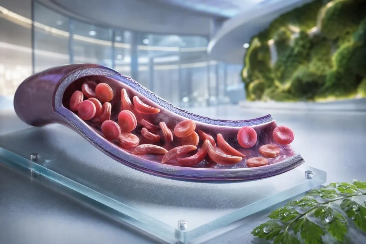
אנמיה חרמשית אינה בדיקה אחת, סימפטום אחד, או רמת דחיפות אחת. מדריך זה, המתמקד בחולה, מפריד בין סטטוס נשא, מחלה מאובחנת, מעקב שגרתי ומצבים הדורשים טיפול חירום.


- תכונת מגל פירושה גן HbS אחד; בדרך כלל אינה גורמת לאנמיה כרונית או למשברי כאב חוזרים.
- בדיקת סקר ביילודים מזהה את רוב התינוקות החולים לפני הופעת תסמינים, ומאפשרת התחלה מוקדמת של טיפול בפניצילין ומעקב מומחים.
- אלקטרופורזה של המוגלובין מפרידה בין סוגי המוגלובין ועוזרת לאשר HbSS, HbSC, HbS-beta תלסמיה, או סטטוס נשא.
- כאב חזה חמור, קוצר נשימה, חום של 38.5°C או יותר, בלבול, או חולשה חדשה דורשים הערכת חירום דחופה אצל אדם עם אנמיה חרמשית.
- תסמונת חזה חריפה יכולה להתחיל בשיעול, חום, כאב חזה, או רמת חמצן נמוכה ועשויה להחמיר במשך שעות ולא ימים.
- ירידה פתאומית של המוגלובין של 2 גרם/ד"ל או יותר מרמה רגילה של אדם יכולה לאותת על משבר אפלסטי, אגירה, דימום או המוליזה מואצת.
- ספירת רטיקולוציטים מסייע להבחין בתגובת מח עצם פעילה ממשבר אפלסטי; ספירה נמוכה במהלך אנמיה מחמירה היא דפוס אזהרה.
- הידרוקסיאוריאה היא תרופה המשנה את מהלך המחלה, ולא רק תרופה לשיכוך כאב; ניטור כולל בדרך כלל ספירת דם מלאה כל 4 שבועות במהלך התאמת המינון.
מהי אנמיה חרמשית – ומה היא לא
אנמיה של תאי חרמש מתייחסת בדרך כלל למחלת HbSS, שבה אדם יורש שני גנים המייצרים המוגלובין S; תאי דם אדומים יכולים להפוך קשיחים, להתפרק מוקדם ולחסום כלי דם קטנים. תכונת תאי חרמש שונה: גן HbS אחד מספיק בדרך כלל להעברה גנטית, אך לא מספיק כדי לגרום לאנמיה הכרונית ולאפיזודות החוזרות של חסימת כלי דם הנראות ב-HbSS.
מחלת תאי חרמש היא מונח מטרייה הכולל HbSS, HbSC, HbS-beta-zero thalassemia, ו-HbS-beta-plus thalassemia. HbSS גורמת לעיתים קרובות לאנמיה הבולטת ביותר, אך החומרה משתנה באופן קיצוני אפילו בתוך משפחה אחת; רמת המוגלובין בסיסית של 7.5 גרם/ד"ל עשויה להיות יציבה עבור מבוגר אחד ומסוכנת עבור אחר אם היא ירדה במהירות.
מוטציית ההמוגלובין S משנה חומצת אמינו אחת בבטא-גלובולין: ולין מחליפה חומצה גלוטמית במיקום 6. תחת חמצן נמוך, התייבשות, חמצת או חום, מולקולות HbS יכולות לפולימריזציה בתוך התא. שינוי פיזי זה מסביר מדוע משבר יכול להתפתח לאחר טיסה ארוכה, מחלת עיכול, או לילה של צריכת נוזלים לקויה.
Kantesti AI הוא מנתח בדיקות דם בינה מלאכותית המציב CBC, בילירובין, LDH, רטיקולוציטים, קריאטינין, ותוצאות קודמות בתצוגה אורכית אחת; הוא אינו מאבחן מחלת תאי חרמש רק מ-CBC. לרענון מעשי בדיווח על המוגלובין, ראה מה HGB אומר.
נכון ל-4 בספטמבר 2026, אני עדיין רואה מטופלים שאומרים להם בטעות שתכונה ומחלה ניתנות להחלפה. הן אינן. הכלל הקליני של ד“ר תומאס קליין פשוט: בקש את הגנוטיפ המדויק של ההמוגלובין, לא אמירה מעורפלת שבדיקה הייתה ”חיובית".”
מדוע גנוטיפ משנה את הטיפול
HbSC עשוי להיות בעל המוגלובין גבוה יותר מ-HbSS ועדיין לגרום לסיבוכים ברשתית, כאב, או נמק אספקטי. HbS-beta תלסמיה יכולה לדמות תכונה או HbSS בהתאם לכך שהאם קיימת חלקית ייצור בטא-גלובולין, זו הסיבה שבדיקות מולקולריות לפעמים מיישבות דפוס אלקטרופורזה דו-משמעי.
תסמיני אנמיה חרמשית לפי גיל ובסיס
תסמיני מחלת תאי חרמש כוללים בדרך כלל כאבי עצמות או חזה אפיזודיים, עייפות, צהבת, קוצר נשימה, עיכוב גדילה וזיהומים תכופים, אך אין שני אנשים החווים את אותו הדפוס. תסמינים מופיעים בדרך כלל לאחר 4 עד 6 חודשי חיים מכיוון שהמוגלובין עוברי מגן בתחילה על תינוקות מפני פולימריזציה של HbS.
דקטיליטיס—נפיחות כואבת של הידיים או הרגליים—עשויה להיות הסימן הנראה הראשון אצל תינוקות. אצל ילדים בגיל בית הספר, כאבי בטן דורשים טיפול מיוחד מכיוון שעצירות, מחלת כיס המרה, הגדלת טחול וחסימת כלי דם יכולים להרגיש דומים להפליא בבית.
צהבת במחלת תאי חרמש משקפת פירוק מתמשך של תאי דם אדומים ועלייה בבילירובין עקיף; היא אינה מעידה בהכרח על דלקת כבד. שתן כהה, צואה חיוורת, גירוד, חום, או כאב בבטן הימנית העליונה משנים הערכה זו ומצדיקים הערכה של סיבות בכבד ובדרכי המרה; המדריך שלנו ל דפוסי בילירובין ישיר מסבירה את ההבדל.
מבוגרים עשויים לנרמל כאב שהפך בהדרגה תכוף יותר. מניסיוני, מעבר משני אירועים המטופלים בבית בשנה לשני אירועים בחודש הוא משמעותי קלינית גם אם לא חל גידול בביקורי חירום. הפרעות שינה, אי עבודה, תסמינים זקפתיים, מצב רוח ירוד וסבילות מופחתת לפעילות גופנית יש לתעד מכיוון שהם לעיתים קרובות מקדימים שינוי רשמי בטיפול.
ריכוז המוגלובין של 6 עד 9 גרם/ד"ל יכול להיות טווח יציב רגיל ב-HbSS, בעוד שערך מתחת ל-10 גרם/ד"ל יהיה בלתי צפוי ברוב האנשים ללא מחלה. המספר חשוב, אך השינוי מהבסיס האישי חשוב יותר.
מי צריך לשקול בדיקת נשאות לאנמיה חרמשית
בדיקת סקר להמוגלובין S סבירה לפני הריון או בתחילת הריון לכל מי שסטטוס הנשאות שלו אינו ידוע, ללא קשר למראה, שם משפחה, או מוצא משוער. סטטוס נשאות הוא תורשתי ולעיתים קרובות נשאר לא ידוע עד בדיקת סקר של יילוד או בדיקת המוגלובין חריגה באופן בלתי צפוי.
אם שני ההורים הביולוגיים נושאים וריאנט הקשור ל-HbS או לתלסמיה בטא, לכל הריון יכולה להיות סיכוי של 25% לרשת מצב המוגלובין בעל משמעות קלינית, בהתאם לווריאנטים המעורבים. נשא יכול להיות בעל אנרגיה תקינה, תוצאות CBC תקינות, וללא היסטוריה של התקפי כאב.
הבדיקה שימושית במיוחד כאשר אחד מבני הזוג נושא HbAS, תכונת HbC, תכונת תלסמיה בטא, מיקרוציטוזיס בלתי מוסברת, או היסטוריה משפחתית של מחלת תאים חרמשיים. נפח כדוריות קטן (MCV) לא צריך להיות מיוחס למחסור בברזל ללא בדיקת פריטין והתחשבות בתלסמיה; השווה רמזי MCV ו-MCH.
דוח עולמי של ארגון הבריאות העולמי משנת 2022 העריך שהפרעות המוגלובין נותרו עומס מחלה תורשתי עיקרי, במיוחד במקומות בהם הגישה לבדיקת יילודים אינה אחידה. הגישה המכבדת ביותר היא גישה אוניברסלית לבדיקה מושכלת במקום להשתמש במוצא אתני כשומר סף.
קנטסטי הוא פלטפורמת פענוח בדיקות דם של AI שיכולים להסביר תוצאת HbS מדווחת בשפה פשוטה, אך אישור נשאות וייעוץ רבייתי דורשים מעבדה מוסמכת ורופא. אם זוג מתכנן הריון, יש לשאול האם ניתן לבצע בדיקת בן זוג וייעוץ גנטי לפני ההתעברות ולא לאחר שהתוצאה הופכת תלויה בזמן.
כיצד אלקטרופורזה של המוגלובין מאשרת את האבחנה
אלקטרופורזה של המוגלובין מפרידה מקטעי המוגלובין והיא בדיקה מרכזית לאישור אנמיה חרמשית או תכונה., לעתים קרובות יחד עם כרומטוגרפיית נוזל בביצועים גבוהים (HPLC), וכאשר יש צורך, בדיקות גנטיות. CBC שגרתי אינו יכול לאשר HbSS או תכונה מכיוון שמחסור בברזל ותלסמיה יכולים לייצר מדדי תאי דם אדומים חופפים.
ב-HbSS בלתי מטופל, אלקטרופורזה בדרך כלל מראה בעיקר HbS ללא HbA, אם כי אחוז HbF משתנה. HbAS בדרך כלל מראה גם HbA וגם HbS, לעתים קרובות עם HbA גבוה יותר מ-HbS; אחוזים מדויקים יכולים להשתנות עם תלסמיה אלפא תורשתית משותפת או עירוי דם אחרון.
עירוי דם אחרון יכול להפוך תוצאת אלקטרופורזה למטעה למשך כ-3 חודשים מכיוון ש-HbA של התורם עדיין במחזור. זו גם הסיבה לכך ש-HbA1c יכול להמעיט בערך הממוצע של גלוקוז לאחר עירוי דם או בהמוליזה עם תחלופה גבוהה; ראה מתי HbA1c מטעה.
משטח היקפי עשוי להראות תאים מטרה, פוליכרומזיה, וצורות חרמש, אך מיקרוסקופיה תומכת במקום להחליף בדיקת מקטעים. ד“ר קליין ראה מריחות חירום ”תקינות" שעיכבו הבהרה כאשר מטופל עבר עירוי דם שבועות קודם לכן — תמיד יש לדווח למעבדה ולצוות ההמטולוגיה על עירויי דם.
שיטות בדיקת יילודים מזהות HbS לפני מחלה קלינית, אך התוצאה עדיין דורשת בדיקה מאשרת בזמן. ההנחיה מבוססת הראיות משנת 2014 מאת Yawn et al. ממליצה על מעורבות מומחים בשלב מוקדם בחיים, כאשר אמצעי מניעה מתחילים לפני הסיבוך העיקרי הראשון (Yawn et al., 2014).
מעקב דם שגרתי: מה קלינאים באמת עוקבים אחריו
ניטור שגרתי במחלת תרשים התאים נוטה בדרך כלל להמוגלובין, רטיקולוציטים, תאי דם לבנים, טסיות, בילירובין, LDH, תפקוד כליות, וחלבון בשתן — לא תוצאה בודדת. ההשוואה הבטוחה ביותר היא עם קו הבסיס היציב של המטופל עצמו, שנלקח באופן אידיאלי כאשר הוא חש בטוב ולא עבר עירוי דם לאחרונה.
המוליזה מייצרת לעיתים קרובות LDH מוגבר, בילירובין עקיף, ורטיקולוציטים עם הפטוגלובין נמוך. הפטוגלובין יכול להיות נמוך מסיבות אחרות, כולל מחלת כבד, כך שהתבנית המשולבת חזקה יותר מכל סמן בודד; שלנו מדריך פענוח ההפטוגלובין מראה למה.
אחוז רטיקולוציטים לבדו יכול להגזים בתגובת מח העצם באנמיה משמעותית. רופאים מחשבים לעיתים קרובות ספירת רטיקולוציטים מוחלטת או תגובת רטיקולוציטים מתוקנת; ספירה מוחלטת יורדת במהלך אנמיה מחמירה מעוררת דאגה למשבר אפלסטי הקשור לפרבווירוס B19.
קנטסטי הוא כלי לניתוח בדיקות דם מבוסס AI המשווה תוצאות המטולוגיות חוזרות במקום לטפל ב-LDH גבוה כאבחנה עצמאית. דגימה המסומנת כהמוֹליטית במבחנה יכולה להגביר באופן כוזב את רמת האשלגן וה-LDH, כך שתוצאה מפתיעה עשויה לדרוש בדיקה חוזרת; סקירה בדיקות חוזרות לאחר המוליזה.
הידרוקסיאוריאה מגדילה בדרך כלל את ה-MCV וה-HbF לאורך זמן, תוך הורדה מתונה של נויטרופילים במינון יעיל. שינויים אלה עשויים להיות צפויים, אך ספירת נויטרופילים מוחלטת מתחת לסף הטיפול האישי או ספירת טסיות יורדת במהירות דורשת סקירה של הרושם ולא התאמה עצמית של התרופה.
מתי כאב הוא אירוע חסימתי-וסקולרי – ומתי לא
כאב וזואוקלוסיבי הוא לעיתים קרובות עמוק, חמור, וממוקם בגב, בחזה, בירכיים, בגפיים, או בבטן, אך כאב חדש אסור לסמן אוטומטית כמשבר תרשים. חום, נפיחות ממוקדת, פציעה, נוקשות בטנית, או תסמינים נוירולוגיים חד-צדדיים מצביעים על בעיות הדורשות בירור שונה.
תוכנית כאב ביתית כוללת בדרך כלל הידרציה מוקדמת, חום, שיכוך כאבים במרשם, מנוחה, וסף הסלמה אישי מוסכם עם צוות התרשים. שתיית כמויות מופרזות אינה בטוחה יותר: אנשים עם פגיעה בכליות או מחלות לב יכולים לפתח עומס נוזלים, במיוחד במהלך מחלה חריפה.
כאב שנשאר בלתי נשלט לאחר תוכנית ההצלה הרגילה, מונע החזקת נוזלים או תרופות בקיבה, או שונה באיכותו או במיקומו, צריך להביא לייעוץ קליני דחוף. מפרק נפוח וחם עשוי לייצג דלקת מפרקים ספטית או זיהום בעצם ולא וזואוקלוזיה לא מסובכת; שלנו מדריך לבדיקת כאב בלתי מוסבר מפרט חקירות ראשוניות שימושיות.
בטיפול חירום, שיכוך כאבים בזמן הנכון והערכה מחדש חשובים יותר מהוכחת חומרת הכאב באמצעות מספר מעבדה. ההנחיה של NICE בנוגע לאפיזודות כאב חריפות בתרשים ממליצה על הערכת שיכוך כאבים מהירה וסקירה תכופה, מכיוון שטיפול תת-יעיל עצמו תורם למצוקה ממושכת ולהחלמה איטית (NICE, 2012).
הימנעו מקומפרסים קרים ישירות על אזור של כאב וזואוקלוסיבי, אלא אם כן הצוות שלכם מייעץ להם במפורש; קור יכול לכווץ כלי דם היקפיים. פרט מעשי זה נשמע זניח, אך מטופלים רבים אומרים לי שאף אחד לא הסביר להם זאת עד לאחר מספר אפיזודות קשות.
תסמונת חזה חריפה: החירום שרבים מפספסים
תסמונת חזה חריפה היא חדירת ריאות חדשה בהדמיית חזה בתוספת תסמינים נשימתיים, חום, כאבים בחזה, או חמצן נמוך באדם עם מחלת תרשים, וזהו מצב חירום. זה יכול להתדרדר במהירות, לפעמים לאחר אשפוז בשל אפיזודה של כאב ולא ברגע שהתסמינים בחזה מתחילים.
התקשרו לשירותי חירום או גשו לטיפול חירום במקרה של קוצר נשימה חדש, כאבים בחזה, שפתיים כחולות-אפורות, עילפון, שיעול מתמשך עם חום, או ריווי חמצן מתחת ליעד המרשם של האדם. רווי של 92% עשוי להיות ירידה משמעותית עבור מישהו שבדרך כלל נמצא ב-98%, בעוד שלחלק מהמטופלים יש קו בסיס מתועד נמוך יותר; גם הערך המוחלט וגם השינוי חשובים.
תסמונת חזה חריפה עשויה לכלול זיהום, תסחיף שומן ממח העצם, חסימה של כלי דם ריאתיים, תסספת, או מספר תהליכים בו זמנית. רופאים מעריכים בדרך כלל חמצון, CBC, רטיקולוציטים, תרביות כאשר יש חום, הדמיית חזה, ולעיתים גזי דם; בדיקות דם לקוצר נשימה מוסיף הקשר לתוצאות אלה.
במבוגרים, יתר-סדציה מאופיואידים, נשימה שטוחה מכאב, ועודף נוזלים תוך-ורידי יכולים להחמיר את המצב הנשימתי. ספירומטריה תמריצית במהלך אשפוז מפחיתה סיבוכים ריאתיים באפיזודות כאב נבחרות, אם כי זה אינו תחליף להערכה דחופה כאשר התסמינים כבר החלו.
הסקירה משנת 2018 מאת קאטו ועמיתיו מתארת תסמונת חזה חריפה כגורם עיקרי לתחלואה ותמותה לאורך החיים (Kato et al., 2018). אין לנהוג בעצמך אם קוצר נשימה, ישנוניות, או לחץ בחזה משמעותיים.
חום, זיהום ומצבי חירום בטחול
חום של 38.5°C (101.3°F) או יותר אצל ילד או מבוגר עם אנמיה חרמשית מחייב הערכה רפואית דחופה באותו היום, אלא אם כן צוות המומחים שלהם נתן הנחיות אחרות. אובדן תפקודי של מערכת החיסון הטחולית יכול לגרום לכך שזיהומים חיידקיים מסוימים יתקדמו מהר יותר מחום ויראלי רגיל.
חום בתוספת לעייפות, פריחה, לחץ דם נמוך, בלבול או ירידה בתפוקת השתן מחייב טיפול חירום מיידי. ספירת תאי דם לבנים שנראית תקינה אינה שוללת באופן אמין זיהום חמור במחלת אנמיה חרמשית, במיוחד בשלבים מוקדמים של המחלה או במהלך טיפול בהידרוקסיאוריאה.
התנקזות טחולית מתרחשת לרוב אצל ילדים צעירים וגורמת להגדלת טחול פתאומית, חיוורון, תחושת מלאות בבטן, נשימה מהירה וירידה מהירה בהמוגלובין. הורים לומדים לפעמים כיצד למשש את הטחול של ילדם, אך כל הגדלה חדשה בעת מחלה צריכה להפעיל הערכה דחופה ולא בדיקות ביתיות חוזרות.
ירידה של 2 גרם/ד"ל או יותר בהמוגלובין מתחת לקו הבסיס הרגיל, יחד עם הגדלת טחול, מדאיגה לגבי התנקזות. לעומת זאת, אנמיה חמורה עם ספירת רטיקולוציטים נמוכה מאוד יכולה להצביע על משבר אפלסטי, הקשור לעיתים קרובות לפרבו-וירוס B19; פרשנות ספירת רטיקולוציטים מסבירה את אות המח עצם.
בדיקות מעבדה לספסיס עשויות לכלול לקטט, תרביות, CBC, בדיקות כליה וסמני דלקת, אך החלטות טיפול לעולם אין להשהות עד שכל הערך יתקבל. להקשר על דפוסים דחופים, קראו רמזי סמני ספסיס.
חיסונים ואנטיביוטיקה מונעת
חיסונים ופרופילקטיקה של פניצילין בילדות מפחיתים סיכון אך אינם הופכים חום לסיכון נמוך. אשר את התוכנית האישית שלך עם צוות המטולוגיה או צוות ילדים, במיוחד לאחר כריתת טחול, נסיעה, תינוק חדש, או שינוי בלוח החיסונים המקומי.
שבץ וסימני אזהרה נוירולוגיים דורשים פעולה מיידית
כל צניחה פתאומית בפני הפנים, חולשה בזרוע או ברגל, קשיי דיבור, התקף, כאב ראש חמור וחריג, בלבול, או אובדן שיווי משקל במחלת אנמיה חרמשית יש לטפל בו כמקרה חירום אפשרי של שבץ. התקשרו לשירותי חירום באופן מיידי; אל תחכו לראות אם התסמינים חולפים או תנו תרופה נגד כאבים תחילה.
ילדים עם HbSS או תלסמיה HbS-beta-zero מוצעים בדרך כלל סקר אולטרסאונד דופלר טרנס-קרניאלי מגיל 2 עד 16 שנים. מהירות ממוצעת בזמן של 200 ס"מ/שנייה או יותר מזהה סיכון מוגבר משמעותית לשבץ ומחייבת פעולה מיידית של מומחה.
טיפול בעירויי דם כרוני יכול להפחית את הסיכון לשבץ ראשון אצל ילדים עם ממצאי דופלר חריגים, אך הוא יוצר צרכים נוספים: מעקב אחר עומס ברזל, סקר נוגדנים, והתאמה קפדנית של תאי תורם. זהו טיפול מיוחד, לא תוצאה שיש לנהל דרך אפליקציה.
מבוגרים יכולים לחוות תסמינים נוירולוגיים חולפים, שינויים קוגניטיביים, או פגיעה מוחי שקטה ללא שבץ דרמטי קלאסי. פרק של 10 דקות של קושי במציאת מילים עדיין מחייב הערכה דחופה מכיוון שתסמינים איסכמיים חולפים יכולים להוות אזהרה, לא הרגעה.
ד"ר תומס קליין מייעץ למשפחות לשמור את מספר החירום המקומי לאנמיה חרמשית ליד תוכנית הכאב הרגילה. זה מסיר החלטה אחת במהלך הדקות המעטות שבהן ילד או מבוגר עשויים להיאבק לתקשר.
הגנה על כליות, עיניים וריאות בין משברים
מחלת אנמיה חרמשית עלולה לפגוע בכליות, ברשתית, בריאות ובלב גם כאשר הכאב אינו תדיר, ולכן מעקב שגרתי אחר איברים הוא חלק מהטיפול ולא תוספת אופציונלית. בדיקת חלבון בשתן (אלבומין), בדיקות לחץ דם, בדיקות עיניים, והערכת ריאות מונחית תסמינים מזהים סיבוכים לפני שהם הופכים ברורים.
אלבומינוריה עשויה להיות סימן מוקדם לנפרופתיה חרמשית. יחס אלבומין-קריאטינין בשתן של 30 מ"ג/גרם או יותר הוא חריג ברוב המבוגרים ויש לאשר אותו בבדיקה חוזרת מכיוון שחום, פעילות גופנית, מחזור חודשי והתייבשות יכולים להעלות אותו באופן חולף.
קריאטינין עשוי להיראות תקין באופן מטעה במחלת אנמיה חרמשית מכיוון שהפרשת צינורית מוגברת ומסת שריר נמוכה עשויים למסך על סינון מופחת. ציסטטין C או משוואות משולבות יכולים להוסיף הקשר שימושי, בעוד שתן קצפי מתמשך מצדיק הערכה; ראה הנחיות לחלבון בשתן.
רטינופתיה חשובה במיוחד במחלת HbSC, שיכולה להיות שקטה מבחינה קלינית בהיבטים אחרים. צפים חדשים, הבזקים, שינוי ראייה דמוי וילון, או טשטוש ראייה פתאומי דורשים בדיקה עיניים דחופה, לא פגישה שגרתית בעוד שבועות.
הריון מוסיף היפר-פילטרציה פיזיולוגית וסיכון גבוה יותר לאם ולעובר, ולכן יש לבצע הערכת כליות ושתן בסיסית מוקדם. ההסבר שלנו לשינויי GFR בהריון עוזר להבחין בין שינוי צפוי למגמה מדאיגה.
אפשרויות טיפול, הידרוקסיאוריאה ובטיחות עירוי
הידרוקסיאוריאה מפחיתה התקפי כאב ותסמונת חזה חריפה אצל אנשים רבים עם HbSS או תלסמיה S-בטא-אפס על ידי הגדלת המוגלובין עוברי, אך היא דורשת מעקב מובנה. עירוי דם יכול להציל חיים עבור סיבוכים נבחרים, אך עירויים חוזרים מביאים עימם סיכוני הצטברות ברזל ונוגדנים לתאי דם אדומים.
הידרוקסיאוריאה נ titrated בדרך כלל עם CBC וספירת רטיקולוציטים בערך כל 4 שבועות עד להשגת מינון יציב, ואז במרווחים ארוכים יותר שנקבעים על ידי הצוות המטפל. MCV גבוה יותר הוא לעיתים קרובות אפקט טיפולי צפוי, בעוד שציטופניה חמורה דורשת בחינה מחדש של התרופות ולעיתים הפסקה זמנית.
עירוי פשוט מגביר את יכולת נשיאת החמצן, אך ריכוז המוגלובין מופרז עלול להגביר את צמיגות הדם ב-HbSS. פרוטוקולים חריפים רבים נמנעים מהעלאת המוגלובין לאחר עירוי מעל 10 גרם/ד"ל אלא אם מומחה קובע יעד אחר; עירוי החלפה משמש לסיבוכים חמורים נבחרים.
הצטברות ברזל הופכת סבירה יותר לאחר עירויים חוזרים מוערכת עם מגמות פריטין ובנוסף MRI של הכבד או הלב בעת הצורך. פריטין עולה עם דלקת, כך שרמה גבוהה בודדת אינה הוכחה להעמסת ברזל ברקמות; מדריך ללימודי ברזל מסביר את המגבלות.
העדויות החזקות ביותר הן להידרוקסיאוריאה ב-HbSS ומחלת S-בטא-אפס; התועלת ב-HbSC נותרת תחום עם פחות ודאות ויותר קבלת החלטות אינדיבידואלית. תוכנית תרופתית צריכה לכלול דיונים על אמצעי מניעה והריון היכן שרלוונטי, מחסומי היענות, ותוכנית כתובה למינונים שהוחמצו.
הריון, פוריות ותכנון משפחה עם HbS
הריון עם מחלת תלסמיה חרמשית דורש טיפול משותף מוקדם מגינקולוגיה והמטולוגיה מכיוון שהסיכונים להתקפי כאב, אנמיה, פקקת, זיהום, רעלת הריון, הגבלת גדילה עוברית ולידה מוקדמת גבוהים יותר. סקירת נשאים חשובה לפני ההריון מכיוון שהסיכון הגנטי תלוי בשני ההורים הביולוגיים.
ביקור לפני הריון צריך לכלול סקירה של גנוטיפ, המוגלובין בסיסי, תפקוד כליות, אלבומין בשתן, לחץ דם, היסטוריית עירויים, נוגדנים לתאי דם אדומים, חיסונים ותרופות. החלטות לגבי הידרוקסיאוריאה בהריון הן אינדיבידואליות ויש לקבלן עם מומחים; אין להפסיק או להתחיל מחדש על סמך ייעוץ אינטרנטי בלבד.
אין לרשום ברזל אוטומטית עבור המוגלובין נמוך ב-HbSS. מחסור בברזל יכול להתקיים במקביל, במיוחד עם הריון או דימום וסתי כבד, אך פריטין ורוויית טרנספרין צריכים להנחות את הטיפול מכיוון שהמוליזה כרונית לבדה אינה שווה למחסור בברזל.
כאשר שני ההורים נושאים וריאנטים של המוגלובין רלוונטיים, ייעוץ גנטי יכול להסביר הריון טבעי, אפשרויות אבחון טרום לידתי, אפשרויות תורם ובדיקה גנטית טרום-השתלה ללא הפניית משפחה להחלטה אחת. המטרה היא בחירה מושכלת, לא בהלה.
לדגלים אדומים מעבדתיים נוספים והריון מעשיים, ראה דאגות מעבדת הריון באותו יום. הבסיס הידוע של המטופל צריך להיכלל בכל העברה של מיילדות; זה משנה כיצד תוצאת המוגלובין מפורשת.
שימוש בטוח בתוצאות מעבדה בין פגישות
תוצאת מעבדה של מחלת תלסמיה חרמשית שימושית ביותר כאשר היא מושווית לגנוטיפ שלך, לבסיס היציב, לתסמינים, לטיפולים ולעירויים אחרונים. בילירובין גבוה או המוגלובין נמוך עשויים להיות צפויים עבור אדם אחד עם HbSS אך דחופים עבור אותו אדם אם התוצאה משתנה בפתאומיות.
שמור רשומה של עמוד אחד של גנוטיפ, המוגלובין רגיל, רוויית חמצן רגילה אם ידועה, קבוצת דם ונוגדנים, תרופות נוכחיות, תאריכי עירוי ומספרי קשר לשעת חירום. זה חוסך זמן במחלקות חירום לא מוכרות ומפחית את הסיכוי שבסיס HbSS יתבלבל עם אנמיה שהתגלתה לאחרונה.
קנטסטי הוא שירות פרשנות בדיקות מעבדת ה-AI שיכול לארגן דוחות מעבדה שהועלו ולסמן דפוסים לדיון, כולל סמני המוליזה, מגמות כליה ומעקב אחר תרופות. זה לא יכול לראות את החזה שלך, למדוד חמצון, או להחליף טיפול חירום כאשר תסמינים מצביעים על תסמונת חזה חריפה, אלח דם או שבץ.
תוצאה מחוץ לטווח הייחוס של המעבדה אינה מסוכנת באופן אוטומטי, ותוצאה בתוכו עדיין יכולה להיות מסוכנת אם חלה בה שינוי חד. להסבר על דגלי דיווח וטווחים של ייחוס, קראו משמעות תוצאה מחוץ לטווח.
הגישה הקלינית של Kantesti נסקרת אל מול שיטות ומגבלות מוגדרות; קוראים המעוניינים להבין את אמצעי הזהירות שלנו יכולים לעיין ב , משום ש-. הביאו את קובץ ה-PDF המקורי לפגישת המטולוגיה שלכם — זיהוי תווים אופטי (OCR) מועיל, אך דוח המקור נותר התיעוד.
תוכנית פעולה ברורה למצבי חירום באנמיה חרמשית
חפשו עזרה דחופה כעת לכאבים בחזה או קוצר נשימה, חום של 38.5 מעלות צלזיוס ומעלה, תסמינים נוירולוגיים חדשים, עילפון, חיוורון קשה, כאב בלתי נשלט, התרחבות מהירה של הבטן, או חוסר יכולת לשתות ולקחת תרופות. סימני אזהרה אלה יכולים להצביע על תסמונת חזה חריפה, אלח דם, שבץ מוחי, אנמיה חמורה, או ספיקת טחול.
הודיעו לצוות החירום שיש לכם מחלת תאי חרמש, ציינו את הגנוטיפ שלכם אם ידוע, ותארו מה שונה מהפרק הרגיל שלכם. ציינו עירוי אחרון, רמת המוגלובין בסיסית, תרופות לכאב שכבר נלקחו עם מינונים וזמנים, אלרגיות, הריון, וכל תסמונת חזה חריפה או שבץ מוחי קודם.
אל תחכו עד שאוקסימטר ביתי לפולס יהפוך לא תקין אם תסמיני החזה מחמירים. מכשירי צרכנים יכולים לקרוא לא נכון עם אצבעות קרות, מוצרי ציפורניים, תנועה, זרימת דם לקויה, וגווני עור מסוימים; תסמינים והערכה קלינית עולים על תצוגה מרגיעה אחת.
אם אתם מטפלים בילד, בקשו מהצוות שלו סף חום כתוב, תוכנית לשיכוך כאבים, והוראות לבדיקת טחול. אם אתם מבוגרים, שמרו עותק בטלפון שלכם ושתפו אותו עם אדם אחד מהימן; תכנון רישום בריאות משפחתית יכול להקל על כך.
הצוות הרפואי של Kantesti, כולל המועצה המייעצת הרפואית, שלנו, תומך בחינוך בנוגע לפרשנות מעבדתית, בעוד שהחלטות חירום שייכות לשירותים קליניים פנים אל פנים. אודות הארגון שלנו והמשימה הקלינית שלנו, ראו הצוות והגישה שלנו.
שאלות נפוצות
מהם התסמינים הראשונים של אנמיה חרמשית אצל תינוקות?
התסמינים הראשונים של אנמיה חרמשית מופיעים לרוב לאחר 4 עד 6 חודשי חיים, כאשר המוגלובין עוברי יורד באופן טבעי. נפיחות כואבת של הידיים והרגליים, הנקראת דקטיליטיס, חום, אי שקט חריג, חיוורון, צהבת, קשיי האכלה ובטן נפוחה הם סימני אזהרה מוקדמים נפוצים. חום של 38.5°C (101.3°F) או יותר בתינוק עם מחלת תאים חרמשיים מחייב הערכה רפואית דחופה באותו יום, מכיוון שזיהום חמור יכול להתקדם במהירות. בדיקות סקר יילודים מזהות תינוקות מושפעים לפני הופעת תסמינים, אך בדיקות אישור ומעקב מומחים נותרים נחוצים.
האם ייתכן שיהיה לך גן של תכונה אנמית חרמשית ועדיין יופיעו תסמינים?
לרוב האנשים עם תכונת תאי חרמש אין אנמיה כרונית, אין התקפי כאב חוזרים, וסיבולת תרגול יומיומית תקינה. סיבוכים נדירים עלולים להתרחש במצבי התייבשות קיצונית, חשיפה לחום עז, גובה רב, מאמץ אינטנסיבי מאוד, או סביבות דלות בחמצן, ויש להעריך דם בשתן ולא להניח שהוא שפיר. תכונה פירושה גן HbS אחד, בעוד שאנמיה של תאי חרמש פירושה בדרך כלל מחלת HbSS עם שני גנים פגועים ל-beta-globin. אלקטרופורזה של המוגלובין או בדיקת חלוקה שוות ערך יכולה להבהיר את ההבחנה.
איזה בדיקה מאשרת אנמיה חרמשית?
אלקטרופורזה של המוגלובין, כרומטוגרפיית נוזל בביצועים גבוהים, או אלקטרופורזה נימית מאשרים את מקטעי ההמוגלובין הקיימים והם מרכזיים לאבחון אנמיה חרמשית. HbSS ללא טיפול מראה בדרך כלל המוגלובין S דומיננטי ללא המוגלובין A, בעוד ש-trait מראה בדרך כלל גם המוגלובין A וגם המוגלובין S. CBC יכול לזהות אנמיה או תאי דם אדומים קטנים, אך אינו יכול לאבחן באופן עצמאי HbSS, HbSC, או trait. עירוי דם אחרון עלול לעוות את תוצאות המקטעים למשך כ-3 חודשים, ולכן ייתכן שיידרש בדיקה מולקולרית כאשר התבנית אינה ברורה.
מתי אדם עם אנמיה חרמשית צריך לפנות לחדר מיון?
אדם עם מחלת תרכובת התאים (sickle cell disease) צריך לפנות להערכה דחופה במקרים של חום של 38.5°C (101.3°F) ומעלה, כאבים בחזה, קוצר נשימה, רמות חמצן נמוכות, חולשה פתאומית, קשיי דיבור, פרכוסים, עילפון, חיוורון המחמיר במהירות, נפיחות חמורה בבטן, או כאב שאינו נשלט על ידי התוכנית הטיפולית הקיימת. תסמינים אלו יכולים להצביע על תסמונת חזה חריפה, זיהום, שבץ מוחי, ספיקת טחול, או ירידה פתאומית בהמוגלובין. כאב חדש או שונה גם מחייב הערכה, מכיוון שדלקת בעצם, מחלות כיס מרה, דלקת התוספתן (appendicitis), וקרישי דם יכולים לחקות כאב וזוק-אוקלוסיבי. אין לנהוג ברכב באופן עצמאי אם התסמינים הנוירולוגיים או הנשימתיים משמעותיים.
מהי רמת המוגלובין מסוכנת במחלת תרשים תאים?
אין מספר המוגלובין מסוכן יחיד במחלת תרדמת התאים מכיוון שאנשים רבים עם HbSS (המוגלובין SS) חווים קו בסיס יציב בין בערך 6 ל-9 גרם/ד"ל. ירידה של 2 גרם/ד"ל או יותר מההמוגלובין הרגיל של האדם היא לעיתים קרובות מדאיגה יותר מהערך המוחלט ויכולה להצביע על משבר אפלסטי, ספיחת טחול, דימום או המוליזה מואצת. תסמינים חמורים כגון קוצר נשימה, עילפון, כאבים בחזה, דופק מהיר או בלבול דורשים הערכה דחופה בכל רמת המוגלובין. החלטות על עירוי דם תלויות בתסמינים, בקו הבסיס, בסיבת האנמיה, בחמצון ובסיכון להיפרויקוזיות.
האם הידרוקסיאוריאה מרפאת אנמיה חרמשית?
הידרוקסיאוריאה אינה מרפאת אנמיה חרמשית, אך היא יכולה להפחית התקפי כאב, תסמונת חזה חריפה, צורך בעירויי דם ושימוש בבית חולים עבור אנשים רבים עם HbSS או תלסמיה ביתא-אפס של תא חרמש. היא פועלת בחלקה על ידי הגברת המוגלובין עוברי ובדרך כלל דורשת ניטור CBC ורטיקולוציטים כל 4 שבועות בערך בזמן התאמת המינון. עלייה ב-MCV וברמת HbF יכולה להעיד על השפעה ביולוגית, אם כי אף אחד מהתוצאות אינו מוכיח היענות מושלמת. גישות מרפאות כגון השתלת תאי גזע וטיפולים מסוימים מבוססי גנים דורשות הערכת כשירות מיוחדת מאוד ומעקב ארוך טווח.
קבל ניתוח בדיקות דם מבוסס בינה מלאכותית כבר היום
הצטרף ליותר מ-2 מיליון משתמשים ברחבי העולם שסומכים על Kantesti לצורך ניתוח מיידי ומדויק של בדיקות מעבדה. העלה את תוצאות בדיקות הדם שלך וקבל פרשנות מקיפה של ביומרקרים של 15,000+ בתוך שניות.
📚 פרסומי מחקר עם הפניות
Klein, T., Mitchell, S., & Weber, H. (2026). מדריך בדיקת דם משלים C3 C4 ו-ANA: טיטר. מחקר רפואי של Kantesti בינה מלאכותית.
Klein, T., Mitchell, S., & Weber, H. (2026). בדיקת דם לנגיף ניפה: מדריך לגילוי ואבחון מוקדם 2026. מחקר רפואי של Kantesti בינה מלאכותית.
📖 הפניות רפואיות חיצוניות
⚕️ הצהרת אחריות רפואית
מאמר זה מיועד למטרות חינוכיות בלבד ואינו מהווה ייעוץ רפואי. תמיד יש להתייעץ עם גורם מוסמך בתחום הבריאות לצורך החלטות אבחון וטיפול.
אותות אמון E-E-A-T
הִתנַסוּת
סקירה קלינית בהובלת רופא של תהליכי עבודה לפענוח בדיקות מעבדה.
מוּמחִיוּת
רפואה מעבדתית מתמקדת באופן שבו סמנים ביולוגיים מתנהגים בהקשר קליני.
סמכותיות
נכתב על ידי ד״ר תומאס קליין, עם סקירה על ידי ד״ר שרה מיטשל ופרופ׳ ד״ר האנס וובר.
אֲמִינוּת
פרשנות מבוססת-ראיות עם מסלולי המשך ברורים כדי להפחית בהלה.