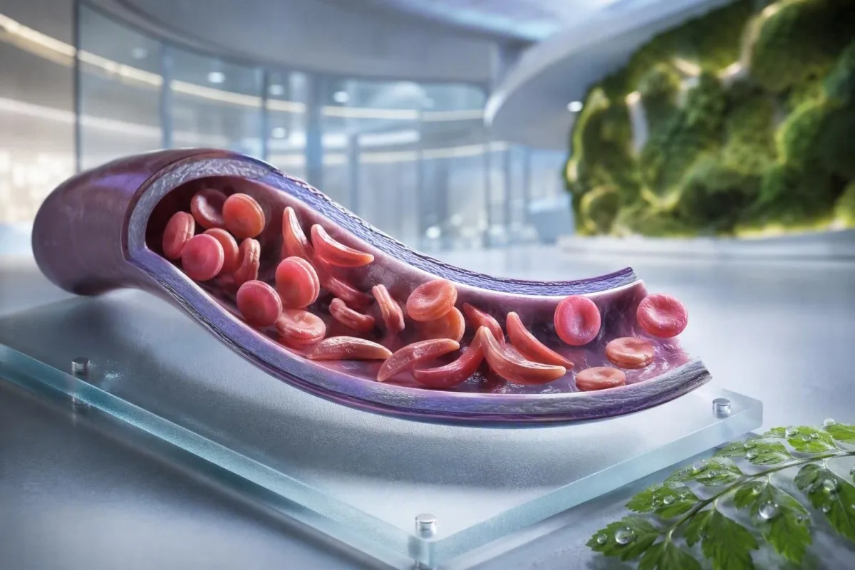
A anemia falciforme non é unha proba, un síntoma nin un nivel de urxencia. Esta guía centrada no paciente separa o estado de portador, a enfermidade confirmada, o seguimento rutinario e as situacións que requiren atención de emerxencia.


- Trazo falciforme significa un xene HbS; normalmente non causa anemia crónica nin crises de dor recurrentes.
- Cribado neonatal identifica a maioría dos lactantes afectados antes de que comecen os síntomas, permitindo que a penicilina e o seguimento por especialistas comecen cedo.
- Electroforese de hemoglobina separa os tipos de hemoglobina e axuda a confirmar HbSS, HbSC, HbS-beta talasemia ou o estado de portador.
- Dor torácica intensa, dificultade para respirar, febre de 38,5 °C ou superior, confusión ou unha nova debilidade requiren avaliación urxente nunha persoa con enfermidade falciforme.
- Síndrome torácica aguda pode comezar con tose, febre, dor torácica ou baixo nivel de osíxeno e pode empeorar en horas en lugar de días.
- Unha caída súbita da hemoglobina de 2 g/dL ou máis dun dun nivel habitual dunha persoa pode sinalar crise aplásica, secuestro, hemorraxia ou hemólise acelerada.
- Conta de reticulocitos axuda a distinguir a resposta medular activa da crise aplásica; un baixo reconto durante a anemia empeorada é un patrón de advertencia.
- Hidroxiurea é un medicamento modificador da enfermidade, non simplemente un analxésico; o seguimento inclúe habitualmente un hemograma completo cada 4 semanas durante o axuste da dose.
Que significa a anemia falciforme, e que non significa
A anemia falciforme adoita referirse á enfermidade HbSS, na que unha persoa herda dous xenes que producen hemoglobina S; os glóbulos vermellos poden volverse ríxidos, descompoñerse cedo e obstruír pequenos vasos. O trazo falciforme é diferente: un xene HbS adoita ser suficiente para transmitilo xeneticamente pero non para causar a anemia crónica e os episodios vasooclusivos recorrentes que se ven na HbSS.
A enfermidade falciforme é un termo xeral que inclúe HbSS, HbSC, HbS-beta-cero talasemia e HbS-beta-máis talasemia. A HbSS adoita causar a anemia máis pronunciada, pero a gravidade varía enormemente incluso dentro dunha familia; unha hemoglobina basal de 7,5 g/dL pode ser estable para un adulto e perigosa para outro se caeu rapidamente.
A mutación da hemoglobina S cambia un aminoácido na beta-globina: a valina substitúe ao ácido glutámico na posición 6. Baixo baixo osíxeno, deshidratación, acidosis ou febre, as moléculas de HbS poden polimerizarse dentro da célula. Ese cambio físico explica por que unha crise pode desenvolverse despois dun voo longo, unha enfermidade gastrointestinal ou unha noite de inxestión de líquidos escasa.
Kantesti AI é un Analizador de análises de sangue con IA que inclúe CBC, bilirrubina, LDH, reticulocitos, creatinina e resultados previos nunha vista lonxitudinal; non diagnostica a enfermidade falciforme só a partir dun CBC. Para un repaso práctico sobre a información da hemoglobina, véxase que significa HGB.
A 4 de setembro de 2026, aínda vexo pacientes aos que se lles di erróneamente que o trazo e a enfermidade son intercambiables. Non o son. A regra clínica do Dr. Thomas Klein é sinxela: pedir o xenotipo exacto da hemoglobina, non unha declaración vaga de que unha proba foi “positiva”.”
Por que o xenotipo cambia a atención
A HbSC pode ter unha hemoglobina máis alta que a HbSS e aínda causar complicacións retinianas, dor ou necrose avascular. A beta talasemia HbS pode parecerse ao trazo ou á HbSS dependendo de se a produción de beta-globina está parcialmente presente, por iso as probas moleculares ás veces resolven un patrón de electroforese ambiguo.
Síntomas da enfermidade falciforme por idade e liña base
Os síntomas da enfermidade falciforme inclúen comúnmente dor ósea ou torácica episódica, fatiga, ictericia, falta de aire, crecemento retardado e infeccións frecuentes, pero non dúas persoas experimentan o mesmo patrón. Os síntomas adoitan aparecer despois dos 4 a 6 meses de idade porque a hemoglobina fetal protexe inicialmente aos lactantes da polimerización da HbS.
A dactilite -inchazón dolorosa das mans ou dos pés- pode ser o primeiro signo visible en lactantes. En nenos en idade escolar, a dor abdominal merece especial atención porque o estreñimento, a colelitiasis, o aumento do bazo e a vaso-oclusión poden sentirse sorprendentemente similares na casa.
A ictericia na enfermidade falciforme reflicte a degradación continua dos glóbulos vermellos e a elevación da bilirrubina indirecta; non significa automaticamente hepatite. A orina escura, as feces pálidas, a coceira, a febre ou a dor na parte superior dereita do abdome cambian esa avaliación e requiren a avaliación de causas hepáticas e biliares; a nosa guía para patróns de bilirrubina directa explica a diferenza.
Os adultos poden normalizar a dor que gradualmente se volveu máis frecuente. Na miña experiencia, un cambio de dous episodios xestionados na casa ao ano a dous episodios ao mes é clinicamente significativo aínda que non aumentasen as visitas de emerxencia. A alteración do sono, o traballo perdido, os síntomas eréctiles, o estado de ánimo baixo e a redución da tolerancia ao exercicio deben documentarse porque a miúdo preceden a un cambio formal no tratamento.
Unha concentración de hemoglobina de 6 a 9 g/dL pode ser un rango habitual de estado de equilibrio na HbSS, mentres que un valor inferior a 10 g/dL sería inesperado na maioría das persoas sen enfermidade. O número importa, pero o cambio respecto á liña base persoal importa máis.
Quen debe considerar a proba de detección de portadores de anemia falciforme
O cribado de sickle cell é razoable antes do embarazo ou no embarazo temperán para calquera persoa cuxo estado de portador sexa descoñecido, independentemente da aparencia, apelido ou ascendencia asumida. O estado de portador é herdado e a miúdo permanece descoñecido ata unha proba de cribado neonatal ou unha proba de hemoglobina inesperadamente anormal.
Se ambos os pais biolóxicos portan unha variante relacionada co HbS ou a beta-talasemia, cada embarazo pode ter unha posibilidade do 25% de herdar unha condición de hemoglobina clinicamente significativa, dependendo das variantes implicadas. Un portador pode ter enerxía normal, resultados normais de CBC e ningunha historia de episodios de dor.
O cribado é particularmente útil cando un membro da parella ten trazo HbAS, trazo HbC, trazo de beta-talasemia, microcitose inexplicable ou antecedentes familiares de anemia falciforme. Un baixo volume corpuscular medio non debe atribuírse a deficiencia de ferro sen comprobar a ferritina e considerar a talasemia; compare pistas de MCV e MCH.
Un informe global da Organización Mundial da Saúde de 2022 estimou que as alteracións da hemoglobina seguen sendo unha carga importante de enfermidades hereditarias, particularmente onde o acceso ao cribado neonatal é desigual. O enfoque máis respectuoso é o acceso universal ao cribado informado en lugar de usar a etnia como garda de acceso.
Kantesti é un plataforma de interpretación de análise de sangue de IA que poden explicar un resultado de HbS reportado en linguaxe sinxela, pero a confirmación do portador e o asesoramento reprodutivo requiren un laboratorio e un clínico acreditados. Se unha parella está a planear un embarazo, pregunte se as probas da parella e o asesoramento xenético poden ocorrer antes da concepción en lugar de despois de que un resultado se volva sensible ao tempo.
Como a electroforese de hemoglobina confirma o diagnóstico
A electroforese de hemoglobina separa as fraccións de hemoglobina e é unha proba fundamental para confirmar a anemia falciforme ou o trazo falciforme, a miúdo xunto coa cromatografía líquida de alta resolución e, cando sexa necesario, as probas xenéticas. Unha CBC de rutina non pode confirmar HbSS ou trazo porque a deficiencia de ferro e a talasemia poden producir índices de glóbulos vermellos superpostos.
En HbSS non tratada, a electroforese mostra tipicamente predominantemente HbS sen HbA, aínda que a porcentaxe de HbF varía. HbAS normalmente mostra tanto HbA como HbS, a miúdo con HbA superior a HbS; as porcentaxes exactas poden cambiar coa alfa-talasemia co-herdada ou transfusión recente.
A transfusión recente pode facer que un resultado de electroforese sexa enganoso durante aproximadamente 3 meses porque a HbA do doador aínda está circulando. Esta é tamén a razón pola que HbA1c pode subestimar a glicosa media despois dunha transfusión ou en hemólise de alta rotación; ver cando HbA1c engana.
Un frote periférico pode mostrar células diana, policromasia e formas falciformes, pero a microscopía apoia en lugar de substituír as probas de fracción. O Dr. Klein viu “esfregados de emerxencia” normais que atrasaron a clarificación cando un paciente fora transfundido semanas antes; sempre informe ao laboratorio e ao equipo de hematoloxía sobre as transfusións.
Os métodos de cribado neonatal identifican a HbS antes da enfermidade clínica, pero o resultado aínda require probas confirmatorias oportunas. A guía baseada en evidencias de 2014 de Yawn et al. recomenda a implicación de especialistas dende cedo na vida, con medidas preventivas que comecen antes da primeira complicación importante (Yawn et al., 2014).
Seguimento sanguíneo rutinario: que monitorizan os clínicos
A monitorización de rutina na doenza falciforme adoita facer un seguimento da hemoglobina, reticulocitos, leucocitos, plaquetas, bilirrubina, LDH, función renal e proteína na urina, non un resultado illado. A comparación máis segura é coa liña de base estable do propio paciente, idealmente recollida cando está ben e non transfundido recentemente.
A hemólise a miúdo produce LDH elevada, bilirrubina indirecta e reticulocitos con baixa haptoglobina. A haptoglobina pode ser baixa por outras razóns, incluída a hepatopatía, polo que o patrón combinado é máis forte que calquera marcador único; a nosa guía de interpretación de haptoglobina mostra por que.
Unha porcentaxe de reticulocitos soa pode sobreestimar a resposta da medula nunha anemia significativa. Os clínicos adoitan calcular unha conta absoluta de reticulocitos ou unha resposta corrixida de reticulocitos; unha conta absoluta decrecente durante unha anemia que empeora suscita preocupación por unha crise aplásica asociada ao parvovirus B19.
Kantesti é un Ferramenta de análise de probas de sangue impulsada por IA que compara resultados hematolóxicos repetidos en lugar de tratar unha LDH alta como un diagnóstico independente. Unha mostra sinalada como hemolizada no tubo pode aumentar falsamente o potasio e a LDH, polo que un resultado sorprendente pode necesitar unha nova extracción; revise probas repetidas despois da hemólise.
A hidroxiurea aumenta comúnmente o MCV e o HbF co tempo, mentres que diminúe modestamente os neutrófilos a unha dose eficaz. Esas variacións poden ser esperadas, pero unha conta absoluta de neutrófilos por debaixo do limiar de tratamento individual ou unha conta de plaquetas que cae rapidamente requiren a revisión do prescriptor en lugar de axustar a medicación por conta propia.
Cando a dor é un episodio vaso-oclusivo, e cando non o é
A dor vaso-oclusiva adoita ser profunda, severa e localizada nas costas, peito, cadeiras, extremidades ou abdome, pero a dor nova non debe ser etiquetada automaticamente como crise falciforme. A febre, a inchazón focal, a lesión, o endurecemento abdominal ou os síntomas neurolóxicos unilaterais apuntan a problemas que requiren un estudo diferente.
Un plan de dor domiciliario inclúe tipicamente hidratación temperá, calor, analgesia prescrita, descanso e un limiar de escalada persoal acordado co equipo de falciformes. Beber volumes excesivos non é máis seguro: as persoas con insuficiencia renal ou enfermidade cardíaca poden desenvolver sobrecarga de líquidos, especialmente durante unha enfermidade aguda.
A dor que permanece sen control despois do plan de rescate habitual, impide a retención de líquidos ou medicamentos, ou é diferente en calidade ou localización, debe levar a un consello clínico urxente. Unha articulación inchada e quente pode representar artrite séptica ou infección ósea en lugar de oclusión vascular non complicada; o noso guía de probas de dor inexplicada describe as primeiras investigacións útiles.
Na atención de urxencia, a analxesia oportuna e a reevaluación importan máis que probar a gravidade da dor a través dun número de laboratorio. A guía do NICE sobre episodios agudos de dor falciforme recomenda unha avaliación rápida da analxesia e unha revisión frecuente, xa que o subtratamento contribúe en si mesmo a unha angustia prolongada e a unha recuperación tardía (NICE, 2012).
Evite as bolsas de xeo directamente sobre unha área de dor vaso-oclusiva a menos que o seu equipo as recomende especificamente; o frío pode constrinxir os vasos periféricos. Ese detalle práctico soa menor, pero moitos pacientes dinme que ninguén o explicou ata despois de varios episodios difíciles.
Síndrome torácica aguda: a emerxencia que moita xente pasa por alto
O síndrome de tórax agudo é un novo infiltrado pulmonar na imaxe torácica máis síntomas respiratorios, febre, dor torácica ou baixa osixenación nunha persoa con doenza falciforme, e é unha emerxencia. Pode deteriorarse rapidamente, ás veces despois do ingreso por un episodio de dor en lugar de no momento en que comezan os síntomas torácicos.
Chame aos servizos de emerxencia ou acuda á atención de urxencia por nova dificultade para respirar, dor torácica, beizos azul-grisáceos, desmaio, tose persistente con febre ou unha saturación de osíxeno por debaixo do obxectivo prescrito. Unha saturación de 92% pode ser unha caída importante para alguén normalmente no 98%, mentres que algúns pacientes teñen unha liña de base documentada máis baixa; tanto o valor absoluto como o cambio importan.
A síndrome de tórax agudo pode implicar infección, embolia graxa da medula ósea, obstrución vascular pulmonar, atelectasia ou varios procesos á vez. Os clínicos avalían comúnmente a osixenación, o CBC, os reticulocitos, os cultivos cando hai febre, a imaxe torácica e ás veces os gases no sangue; análises de sangue para a falta de aire engade contexto a estes resultados.
En adultos, a sedación excesiva por opiodes, a respiración superficial pola dor e o exceso de líquidos intravenosos poden empeorar o estado respiratorio. A espirometría de incentivo durante a hospitalización reduce as complicacións pulmonares en episodios de dor seleccionados, aínda que non é un substituto para a avaliación urxente cando os síntomas xa comezaron.
A revisión de 2018 por Kato e colegas describe a síndrome de tórax agudo como unha das principais causas de morbilidade e mortalidade ao longo da vida (Kato et al., 2018). Non conduza vostede mesmo se a dificultade para respirar, a somnolencia ou a presión no peito é significativa.
Febre, infección e emerxencias esplénicas
Unha temperatura de 38,5 °C (101,3 °F) ou superior nun neno ou adulto con enfermidade falciforme require unha avaliación médica urxente o mesmo día, a menos que o seu equipo especialista proporcionara un plan diferente. A perda funcional da función inmune do bazo pode facer que algunhas infeccións bacterianas progresen moito máis rápido que unha febre viral ordinaria.
Febre máis letarxia, erupción cutánea, presión arterial baixa, confusión ou diminución da produción de ouriños require atención de emerxencia agora. Unha conta de leucocitos de aspecto normal non exclúe de forma fiable unha infección grave na enfermidade falciforme, especialmente ao principio da enfermidade ou durante o tratamento con hidroxiurea.
A secuestración esplénica ocorre con máis frecuencia en nenos pequenos e causa un aumento súbito do bazo, palidez, plenitude abdominal, respiración rápida e unha caída rápida da hemoglobina. Os pais ás veces aprenden a palpar o bazo do seu fillo, pero calquera novo aumento con enfermidade debe desencadear unha avaliación urxente en lugar de controis domésticos repetidos.
Unha diminución da hemoglobina de 2 g/dL ou máis por debaixo da liña base habitual cun bazo agrandado é preocupante por secuestración. Pola contra, a anemia grave cunha conta moi baixa de reticulocitos pode suxerir unha crise aplásica, frecuentemente relacionada co parvovirus B19; interpretación da conta de reticulocitos explica o sinal da medula.
Os laboratorios de sepsis poden incluír lactato, cultivos, CBC, probas renais e marcadores inflamatorios, pero as decisións de tratamento nunca deben esperar a que todos os valores regresen. Para obter contexto sobre patróns urxentes, lea pistas de marcadores de sepsis.
Vacinas e antibióticos preventivos
As vacinas e a profilaxe con penicilina na infancia reducen o risco pero non fan que a febre sexa de baixo risco. Confirme o seu plan individualizado cun equipo de hematoloxía ou pediatría, particularmente despois da esplenectomía, viaxes, un bebé novo ou un cambio no calendario de inmunización local.
A ACV e os signos de alerta neurolóxicos requiren acción inmediata
Calquera debilidade facial súbita, debilidade dun brazo ou perna, dificultade para falar, convulsións, dor de cabeza intensa e inusual, confusión ou perda de equilibrio na enfermidade falciforme debe tratarse como unha posible emerxencia por accidente cerebrovascular. Chame aos servizos de emerxencia inmediatamente; non esperes a ver se os síntomas pasan nin administres analxésicos primeiro.
Os nenos con HbSS ou talasemia HbS-beta-zero adoitan ofrecerlles cribado con ecografía Doppler transcranial desde os 2 ata os 16 anos. Unha velocidade máxima media temporal anormal de 200 cm/segundo ou superior identifica un risco de accidente cerebrovascular substancialmente aumentado e require unha acción especializada e inmediata.
A terapia de transfusión crónica pode reducir o risco de primeiro accidente cerebrovascular en nenos con resultados anormais do Doppler, pero crea necesidades adicionais: monitorización da sobrecarga de ferro, cribado de anticorpos e correspondencia coidadosa das células do doante. Este é un coidado especializado, non un resultado a xestionar a través dunha aplicación.
Os adultos poden ter síntomas neurolóxicos transitorios, cambios cognitivos ou lesión cerebral silenciosa sen un accidente cerebrovascular dramático clásico. Un episodio de 10 minutos de dificultade para atopar palabras aínda require avaliación de emerxencia porque os síntomas isquémicos transitorios poden ser unha advertencia, non un alivio.
O Dr. Thomas Klein aconsella ás familias que garda o seu número de contacto de emerxencia falciforme local xunto ao plan de dor habitual. Elimina unha decisión durante os poucos minutos nos que un neno ou adulto pode estar loitando para comunicarse.
Protexer riles, ollos e pulmóns entre crises
A enfermidade falciforme pode danar os riles, a retina, os pulmóns e o corazón mesmo cando a dor é infrecuente, polo que a vixilancia rutinaria dos órganos forma parte do tratamento e non dun extra opcional. As probas de albúmina na ouriña, os controis da presión arterial, os exames oculares e a avaliación pulmonar baseada en síntomas identifican complicacións antes de que se fagan evidentes.
A albuminuria pode ser un signo temperán de nefroloxía falciforme. Unha relación albúmina-creatinina na ouriña de 30 mg/g ou máis é anormal na maioría dos adultos e debe confirmarse con probas repetidas porque a febre, o exercicio, a menstruación e a deshidratación poden elevala transitoriamente.
A creatinina pode parecer enganosamente normal na enfermidade falciforme porque o aumento da secreción tubular e a menor masa muscular poden enmascarar unha filtración reducida. A cistatina C ou as ecuacións combinadas poden engadir un contexto útil, mentres que a ouriña espumosa persistente merece avaliación; consulte orientación sobre proteínas na ouriña.
A retinopatía é especialmente importante na enfermidade HbSC, que doutros xeitos pode ser clinicamente máis leve. Novos flotadores, flashes, un cambio visual en forma de cortina ou unha perda de visión súbita requiren unha revisión oftalmolóxica urxente, non unha cita rutinaria semanas despois.
O embarazo engade hiperfiltración fisiolóxica e un maior risco materno-fetal, polo que a avaliación renal e urinaria basal debe facerse cedo. A nosa explicación de cambios na GFR durante o embarazo axuda a distinguir o cambio esperado dunha tendencia preocupante.
Opcións de tratamento, hidroxiúrea e seguridade da transfusión
A hidroxiurea reduce os episodios de dor e a síndrome torácica aguda en moitas persoas con talasemia HbSS ou HbS-beta-zero ao aumentar a hemoglobina fetal, pero require unha monitorización estruturada. A transfusión pode salvar vidas para complicacións seleccionadas, pero as transfusións repetidas comportan riscos de carga de ferro e anticorpos contra os glóbulos vermellos.
A hidroxiurea adoita axustarse cunha CBC e un recuento de reticulocitos aproximadamente cada 4 semanas ata acadar unha dose estable, despois a intervalos máis longos determinados polo equipo de tratamento. Un MCV máis alto adoita ser un efecto esperado do tratamento, mentres que a citopenia grave require unha revisión da medicación e, ás veces, unha interrupción temporal.
A transfusión simple aumenta a capacidade de transporte de osíxeno, pero unha concentración excesiva de hemoglobina pode aumentar a viscosidade na HbSS. Moitos protocolos agudos evitan elevar a hemoglobina post-transfusión moito por riba de 10 g/dL a menos que un especialista estableza un obxectivo diferente; a transfusión de intercambio úsase para complicacións graves seleccionadas.
A sobrecarga de ferro faise máis probable despois de transfusións repetidas e avalíase con tendencias de ferritina máis RM hepática ou cardíaca cando sexa apropiado. A ferritina aumenta coa inflamación, polo que un nivel alto único non é proba de carga de ferro nos tecidos; a nosa guía de estudos sobre o ferro explica os límites.
A evidencia é máis forte para a hidroxiurea na enfermidade HbSS e HbS-beta-zero; o beneficio na HbSC segue sendo unha área con menos certeza e toma de decisións máis individualizada. Un plan de medicación debe incluír discusións sobre anticoncepción e embarazo cando sexa relevante, barreiras de adherencia e un plan escrito para doses omitidas.
Embarazo, fertilidade e planificación familiar con HbS
O embarazo con enfermidade falciforme require atención conxunta temperá de obstetricia e hematoloxía porque os riscos de episodios de dor, anemia, trombose, infección, preeclampsia, restrición do crecemento fetal e parto prematuro son maiores. O cribado de portadores é importante antes do embarazo porque o risco xenético depende de ambos os proxenitores biolóxicos.
Unha consulta previa ao embarazo debe revisar o xenotipo, a hemoglobina basal, a función renal, a albuminuria, a presión arterial, o historial de transfusións, os anticorpos contra os glóbulos vermellos, as vacinas e os medicamentos. As decisións sobre a hidroxiurea durante o embarazo son individualizadas e deben tomarse con especialistas; non a suspenda nin a reinicie baseándose unicamente en consellos de internet.
O ferro non debe ser prescrito automaticamente para unha hemoglobina baixa na HbSS. A deficiencia de ferro pode coexistir, especialmente co embarazo ou sangrado menstrual abundante, pero a ferritina e a saturación da transferrina deben guiar o tratamento porque a hemólise crónica soa non equivale a deficiencia de ferro.
Cando ambos os proxenitores portan variantes de hemoglobina relevantes, o asesoramento xenético pode explicar a concepción natural, as opcións de diagnóstico prenatal, as opcións de doadores e as probas xenéticas preimplantacionais sen dirixir a unha familia cara a unha decisión. O obxectivo é unha elección informada, non a alarma.
Para outras bandeiras vermellas de laboratorio prácticas durante o embarazo, consulte preocupacións de laboratorio de embarazo o mesmo día. A liña de base coñecida do paciente debe incluírse en cada traspaso de maternidade; cambia a forma en que se interpreta un resultado de hemoglobina.
Uso seguro dos resultados de laboratorio entre citas
Un resultado de laboratorio de enfermidade falciforme é máis útil cando se compara co seu xenotipo, liña de base estable, síntomas, tratamentos e transfusións recentes. Unha bilirrubina alta ou unha hemoglobina baixa pode ser esperada para unha persoa con HbSS, pero urxente para a mesma persoa se o resultado cambia abruptamente.
Manteña un rexistro dunha páxina do xenotipo, a hemoglobina habitual, a saturación de osíxeno habitual se se coñece, o grupo sanguíneo e os anticorpos, os medicamentos actuais, as datas das transfusións e os contactos de emerxencia. Isto aforra tempo en departamentos de urxencias descoñecidos e reduce a posibilidade de que unha liña de base de HbSS se confunda cunha anemia recentemente descuberta.
Kantesti é un servizo de interpretación de probas de laboratorio de IA que pode organizar informes de laboratorio cargados e sinalar patróns para discusión, incluíndo marcadores de hemólise, tendencias renais e monitorización de medicacións. Non pode ver o seu peito, medir a osixenación nin substituír a atención de emerxencia cando os síntomas apuntan a síndrome torácica aguda, sepsis ou accidente cerebrovascular.
Un resultado fóra dun intervalo de referencia de laboratorio non é automaticamente perigoso, e un resultado dentro del aínda pode ser perigoso se cambiou bruscamente. Para unha explicación das bandeiras de notificación e os rangos de referencia, lea o significado dun resultado fóra de rango.
O enfoque clínico de Kantesti revísase fronte a métodos e limitacións definidos; os lectores que queiran comprender as nosas medidas de seguridade poden revisar estándares de validación clínica. Leve o PDF orixinal á súa cita de hematoloxía: a OCR é útil, pero o informe orixinal segue sendo o rexistro.
Un plan de acción claro ante emerxencias para a enfermidade falciforme
Busque axuda de emerxencia agora en caso de dor no peito ou dificultade para respirar, febre de 38.5 °C ou superior, síntomas neurolóxicos novos, desmaio, palidez severa, dor incontrolada, un abdome que se expande rapidamente ou incapacidade para beber e tomar medicamentos. Estes sinais de advertencia poden indicar síndrome torácica aguda, sepsis, ictus, anemia grave ou secuestro esplénico.
Informe ao persoal de emerxencia que ten sickle cell disease, indique o seu xenotipo se o coñece e describa o que é diferente do seu episodio habitual. Mencione transfusión recente, hemoglobina basal, analxésicos xa tomados coas doses e horas, alerxias, embarazo e calquera síndrome torácica aguda ou ictus previo.
Non agarde a que un oxímetro de pulso doméstico se volva anormal se os síntomas torácicos empeoran. Os dispositivos de consumo poden ler mal cos dedos fríos, produtos para as uñas, movemento, mala circulación e algúns tons de pel; os síntomas e a avaliación clínica superan unha pantalla tranquilizadora.
Se coida dun neno, pida ao seu equipo un limiar de febre escrito, un plan de analxesia e instrucións para a revisión do bazo. Se é un adulto, garde unha copia no seu teléfono e compártaa cunha persoa de confianza; a planificación do rexistro de saúde familiar pode facilitar isto.
O equipo médico de Kantesti, incluído o noso Consello Asesor Médico, apoia a educación sobre a interpretación de laboratorio, mentres que as decisións de emerxencia pertencen aos servizos clínicos presenciais. Sobre a nosa organización e misión clínica, vexa o noso equipo e enfoque.
Preguntas frecuentes
cales son os primeiros síntomas da anemia falciforme en bebés?
Os primeiros síntomas da anemia falciforme adoitan aparecer despois dos 4 a 6 meses de idade, cando a hemoglobina fetal diminúe de forma natural. Hinchazón dolorosa das mans e dos pés, chamada dactilite, febre, irritabilidade inusual, palidez, ictericia, dificultade para alimentarse e un abdome inchado son sinais de alerta temperáns comúns. Unha febre de 38,5 °C (101,3 °F) ou superior nun lactante con enfermidade falciforme require unha avaliación médica urxente o mesmo día, xa que unha infección grave pode progresar rapidamente. A detección neonatal identifica aos lactantes afectados antes de que comecen os síntomas, pero as probas de confirmación e o seguimento especializado seguen sendo necesarios.
Podes ter trazo falciforme e aínda así ter síntomas?
A maioría das persoas con trazo falciforme non teñen anemia crónica, nin crises de dor recorrentes, nin tolerancia normal ao exercicio diario. Poden ocorrer complicacións raras en casos de deshidratación extrema, exposición severa ao calor, altitude elevada, esforzo moi intenso ou ambientes de baixo osíxeno, e a sangue na urina debe ser avaliada en lugar de asumirse como benigna. Trazo significa un xene HbS, mentres que a anemia falciforme normalmente significa enfermidade HbSS con dous xenes beta-globina afectados. A electroforese de hemoglobina ou unha proba de fraccionamento equivalente pode aclarar a distinción.
Que proba confirma a anemia falciforme?
A electroforese de hemoglobina, a cromatografía líquida de alta resolución ou a electroforese capilar confirman as fraccións de hemoglobina presentes e son centrais para o diagnóstico da anemia falciforme. O HbSS non tratado adoita mostrar predominantemente hemoglobina S sen hemoglobina A, mentres que o trazo adoita mostrar tanto hemoglobina A como hemoglobina S. Unha CBC pode identificar anemia ou glóbulos vermellos pequenos, pero non pode diagnosticar de forma independente HbSS, HbSC ou trazo. Transfusións recentes poden distorsionar os resultados das fraccións durante uns 3 meses, polo que poden ser necesarias probas moleculares cando o padrón non está claro.
Cando debe acudir a urxencias unha persoa con anemia falciforme?
Unha persoa con drepanocitose debe buscar avaliación de emerxencia ante febre de 38,5 °C (101,3 °F) ou superior, dor no peito, disnea, hipoxemia, debilidade súbita, dificultade para falar, convulsións, desmaio, palidez que empeora rapidamente, inchazo abdominal severo ou dor que non se controla co plan establecido. Estes síntomas poden indicar síndrome torácica aguda, infección, ictus, secuestro esplénico ou unha diminución súbita da hemoglobina. A dor nova ou diferente tamén precisa avaliación porque a infección ósea, a enfermidade da vesícula biliar, a apendicite e os coágulos poden imitar a dor vaso-oclusiva. Non conduza vostede mesmo se os síntomas neurolóxicos ou respiratorios son significativos.
Que nivel de hemoglobina é perigoso na doenza falciforme?
Non hai un único número perigoso de hemoglobina na doenza falciforme porque moitas persoas con HbSS teñen unha liña de base estable entre aproximadamente 6 e 9 g/dL. Unha caída de 2 g/dL ou máis respecto á hemoglobina habitual do individuo é a miúdo máis preocupante que o valor absoluto e pode suxerir crise aplásica, secuestro esplénico, hemorraxia ou hemólise acelerada. Síntomas graves como falta de aire, desmaio, dor no peito, latexo cardíaco rápido ou confusión requiren unha avaliación urxente a calquera nivel de hemoglobina. As decisións de transfusión dependen dos síntomas, da liña de base, da causa da anemia, da osixenación e do risco de hiperviscosidade.
A hidroxourea cura a anemia falciforme?
A hidroxiurea non cura a anemia falciforme, pero pode reducir os episodios de dor, a síndrome torácica aguda, a necesidade de transfusións e o uso do hospital para moitas persoas con HbSS ou talasemia HbS-beta-cero. Funciona en parte aumentando a hemoglobina fetal e normalmente require seguimento de CBC e reticulocitos aproximadamente cada 4 semanas mentres se axusta a dose. Un aumento do MCV e do nivel de HbF pode indicar un efecto biolóxico, aínda que ningún resultado demostra unha adhesión perfecta. As abordaxes curativas, como o transplante de células nai e algunhas terapias baseadas en xenes, requiren unha avaliación de elegibilidade altamente especializada e un seguimento a longo prazo.
Obtén hoxe unha análise de sangue con IA
Únete a máis de 2 millóns de usuarios en todo o mundo que confían en Kantesti para obter unha análise instantánea e precisa das análises de laboratorio. Carga os teus resultados de análise de sangue e recibe unha interpretación completa de biomarcadores de 15,000+ en segundos.
📚 Publicacións de investigación citadas
Klein, T., Mitchell, S., & Weber, H. (2026). Guía de proba de sangue de complemento C3 C4 e título de ANA. Kantesti Investigación médica con IA.
Klein, T., Mitchell, S., & Weber, H. (2026). Análise de sangue do virus Nipah: guía de detección e diagnóstico precoz 2026. Kantesti Investigación médica con IA.
📖 Referencias médicas externas
⚕️ Aviso médico
Este artigo é só para fins educativos e non constitúe asesoramento médico. Consulta sempre un/ha profesional sanitario/a cualificado/a para decisións de diagnóstico e tratamento.
Sinais de confianza E-E-A-T
Experiencia
Revisión clínica dirixida por un médico dos fluxos de interpretación de análises.
Experiencia
Foco en medicina de laboratorio sobre como se comportan os biomarcadores no contexto clínico.
Autoridade
Escrito polo Dr. Thomas Klein, con revisión da Dra. Sarah Mitchell e do Prof. Dr. Hans Weber.
Fiabilidade
Interpretación baseada en evidencias con vías de seguimento claras para reducir a alarma.