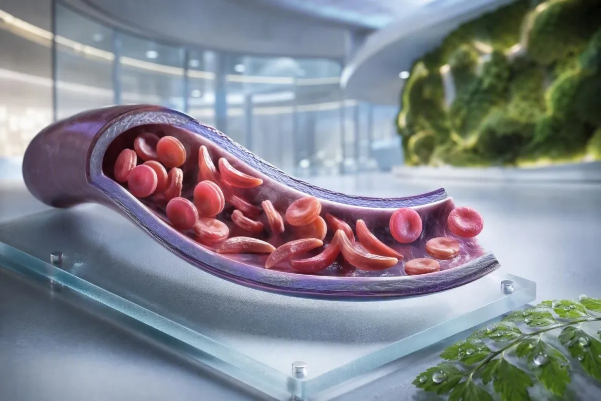
Anemija srpastih ćelija nije jedan test, jedan simptom ili jedan nivo hitnosti. Ovaj vodič fokusiran na pacijenta razdvaja status nosioca, potvrđenu bolest, rutinsko praćenje i situacije koje zahtevaju hitnu negu.


- Osobina srpastih ćelija znači jedan HbS gen; obično ne uzrokuje hroničnu anemiju ili ponavljajuće bolne krize.
- skrining novorođenčadi identifikuje većinu pogođenih beba pre nego što simptomi počnu, omogućavajući rano započinjanje penicilina i specijalističkog praćenja.
- Електрофореза хемоглобина razdvaja tipove hemoglobina i pomaže u potvrđivanju HbSS, HbSC, HbS-beta talasemije ili statusa nosioca.
- jak bol u grudima, nedostatak vazduha, temperatura od 38.5°C ili viša, konfuzija ili nova slabost zahtevaju hitnu urgentnu procenu kod osobe sa bolešću srpastih ćelija.
- Sindrom akutnog grudnog koša može početi kašljem, groznicom, bolom u grudima ili niskim nivoom kiseonika i može se pogoršati tokom sati, a ne dana.
- Nagli pad hemoglobina od 2 g/dL ili više od uobičajenog nivoa osobe može ukazivati na aplastičnu krizu, sekvestraciju, krvarenje ili ubrzanu hemolizu.
- Број ретикулоцита pomaže u razlikovanju aktivnog odgovora koštane srži od aplastične krize; nizak broj tokom pogoršanja anemije je obrazac upozorenja.
- Hidroksiurea je lek koji menja tok bolesti, a ne samo lijek protiv bolova; praćenje obično uključuje kompletnu krvnu sliku svake 4 nedelje tokom prilagođavanja doze.
Šta anemija srpastih ćelija znači – a šta ne znači
Anemija srpastih ćelija se obično odnosi na HbSS bolest, gde osoba nasleđuje dva gena koja proizvode hemoglobin S; crvena krvna zrnca mogu postati kruta, prerano se raspasti i blokirati male krvne sudove. Osobina srpastih ćelija je drugačija: jedan HbS gen je obično dovoljan za genetski prenos, ali nije dovoljan da izazove hroničnu anemiju i ponavljajuće vazo-okluzivne epizode viđene kod HbSS.
Bolest srpastih ćelija je krovni termin koji uključuje HbSS, HbSC, HbS-beta-nultu talasemiju i HbS-beta-plus talasemiju. HbSS često uzrokuje najizraženiju anemiju, ali težina se enormno razlikuje čak i unutar jedne porodice; bazalni hemoglobin od 7,5 g/dL može biti stabilan za jednu odraslu osobu, a opasan za drugu ako je brzo opao.
Mutacija hemoglobina S menja jednu aminokiselinu u beta-globu: valin zamenjuje glutaminsku kiselinu na poziciji 6. Pod niskim nivoom kiseonika, dehidratacijom, acidozom ili groznicom, HbS molekuli se mogu polimerizovati unutar ćelije. Ta fizička promena objašnjava zašto se kriza može razviti nakon dugog leta, gastrointestinalne bolesti ili noći sa slabim unosom tečnosti.
Kantesti AI je АИ анализатор крви koji stavlja CBC, bilirubin, LDH, retikulocite, kreatinin i prethodne rezultate u jedan longitudinalni prikaz; ne postavlja dijagnozu bolesti srpastih ćelija samo na osnovu CBC-a. Za praktičan podsetnik o izveštavanju hemoglobina, pogledajte šta HGB znači.
Od 4. septembra 2026. godine, još uvek viđam pacijente kojima se pogrešno kaže da su osobina i bolest zamenljivi. Nisu. Kliničko pravilo dr. Tomasa Klajna je jednostavno: tražite tačan genotip hemoglobina, a ne nejasnu izjavu da je test bio “pozitivan.”
Zašto genotip menja negu
HbSC može imati viši hemoglobin nego HbSS, a opet uzrokovati retinalne komplikacije, bol ili avaskularnu nekrozu. HbS-beta talasemija može ličiti na osobinu ili HbSS u zavisnosti od toga da li je proizvodnja beta-globina delimično prisutna, što je razlog zašto molekularno testiranje ponekad rešava nejasnu elektroforetsku sliku.
Simptomi bolesti srpastih ćelija prema uzrastu i osnovnim vrednostima
Simptomi bolesti srpastih ćelija obično uključuju povremeni bol u kostima ili grudima, umor, žuticu, kratak dah, usporen rast i česte infekcije, ali niko ne doživljava istu sliku. Simptomi obično izbijaju nakon 4 do 6 meseci starosti jer fetalni hemoglobin inicijalno štiti dojenčad od HbS polimerizacije.
Daktilitis—oteklina ruku ili stopala—može biti prvi vidljivi znak kod dojenčadi. Kod dece školskog uzrasta, bol u stomaku zaslužuje posebnu pažnju jer zatvor, bolesti žučne kese, uvećanje slezine i vazo-okluzija mogu kod kuće delovati iznenađujuće slično.
Žutica kod bolesti srpastih ćelija odražava kontinuirano razaranje crvenih krvnih zrnaca i povišen indirektni bilirubin; ne znači automatski hepatitis. Tamni urin, bledo stolica, svrab, groznica ili bol u gornjem desnom delu stomaka menjaju tu procenu i zahtevaju procenu jetre i žučnih uzroka; naš vodič za obrasci direktnog bilirubina објашњава разлику.
Odrasli mogu normalizovati bol koji je postao postepeno češći. Po mom iskustvu, prelazak sa dve epizode lečene kod kuće godišnje na dve epizode mesečno je klinički značajan, čak i ako se broj hitnih poseta nije povećao. Neredovan san, propušten posao, erektilni problemi, loše raspoloženje i smanjena tolerancija na vežbanje treba dokumentovati jer često prethode formalnoj promeni lečenja.
Koncentracija hemoglobina od 6 do 9 g/dL može biti uobičajen opseg u mirovanju kod HbSS, dok bi vrednost ispod 10 g/dL bila neočekivana kod većine osoba bez bolesti. Broj je bitan, ali promena od lične bazne linije je bitnija.
Ko bi trebalo da razmotri skrining na nosioce srpastih ćelija
Skrining na srpastu bolest je razuman pre trudnoće ili rano u trudnoći za svakoga čiji status nosioca nije poznat, bez obzira na izgled, prezime ili pretpostavljeno poreklo. Status nosioca se nasleđuje i često ostaje nepoznat do skrininga novorođenčeta ili neočekivano abnormalnog testa hemoglobina.
Ако оба биолошка родитеља носе варијанту у вези са HbS или бета-таласемијом, свака трудноћа може имати 25% шансе да наследи клинички значајну хемоглобинску болест, у зависности од укључених варијанти. Носилац може имати нормалну енергију, нормалне CBC резултате и историју без епизода бола.
Скрининг је посебно користан када један партнер има HbAS, HbC траит, бета-таласемија траит, необјашњиву микроцитозу или породичну историју српасте анемије. Низак средњи корпус LULAR (MCV) не треба приписивати недостатку гвожђа без провере феритина и разматрања таласемије; упоредите MCV и MCH назнаке.
Глобални извештај Светске здравствене организације из 2022. године проценио је да хемоглобински поремећаји остају велико оптерећење наследног обољевања, посебно тамо где је приступ скринингу новорођенчади неравномeран. Најпоштованији приступ је универзални приступ информисаном скринингу уместо коришћења етничке припадности као капије.
Кантести је AI платформа за тумачење крвне слике што може објаснити пријављени HbS резултат једноставним језиком, али потврда носиоца и репродуктивно саветовање захтевају акредитовану лабораторију и клиничара. Ако пар планира трудноћу, питајте да ли тестирање партнера и генетско саветовање могу да се обаве пре зачећа, а не након што резултат постане временски осетљив.
Kako elektroforeza hemoglobina potvrđuje dijagnozu
Електрофореза хемоглобина раздваја фракције хемоглобина и представља кључни тест за потврду српасте анемије или траита, често заједно са течном хроматографијом високих перформанси и, када је потребно, генетским тестирањем. Рутински CBC не може потврдити HbSS или траит јер недостатак гвожђа и таласемија могу произвести сличне индексе црвених крвних зрнаца.
У нетретираној HbSS, електрофореза обично показује претежно HbS без HbA, иако се проценат HbF разликује. HbAS обично показује и HbA и HbS, често са вишим уделом HbA него HbS; тачни проценти се могу мењати са ко-наслеђеном алфа-таласемијом или недавном трансфузијом.
Недавна трансфузија може учинити резултат електрофорезе погрешним око 3 месеца јер се донорски HbA још увек циркулише. То је такође разлог зашто A1C може потценити просечан ниво глукозе након трансфузије или код хемолизе високог обрта; видети када A1C доводи у заблуду.
Периферни размаз може показати циљне ћелије, полихромазију и српасте облике, али микроскопија подржава, а не замењује тестирање фракција. Др. Клајн је видео “нормалне” хитне размазе који су одложили разјашњење када је пацијенту трансфузија дата недељама раније — увек обавестите лабораторију и тим за хематологију о трансфузијама.
Методе скрининга новорођенчади идентификују HbS пре клиничке болести, али резултат и даље захтева благовремено потврдну тестирање. Смерница заснована на доказима из 2014. године од стране Yawn et al. препоручује укључивање специјалиста рано у животу, са превентивним мерама које почињу пре прве велике компликације (Yawn et al., 2014).
Rutinsko praćenje krvi: šta kliničari zapravo prate
Uobičajeno praćenje kod bolesti srpastih ćelija obično prati hemoglobin, retikulocite, bele krvne ćelije, trombocite, bilirubin, LDH, bubrežnu funkciju i proteine u urinu — ne jedan izolovani rezultat. Najsigurnije poređenje je sa sopstvenom stabilnom osnovnom linijom pacijenta, idealno uzetom kada je pacijent dobro i nije nedavno transfuzionisan.
Hemoliza često dovodi do povišenog LDH, indirektnog bilirubina i retikulocita sa niskim haptoglobinom. Haptoglobin može biti nizak iz drugih razloga, uključujući bolest jetre, tako da je kombinovani obrazac jači od bilo kog pojedinačnog markera; naša водич за тумачење хаптоглобина показује зашто.
Samo procenat retikulocita može preceniti odgovor koštane srži u značajnoj anemiji. Kliničari često izračunavaju apsolutni broj retikulocita ili korigovani odgovor retikulocita; opadajući apsolutni broj tokom pogoršanja anemije izaziva zabrinutost zbog aplastične krize povezane sa parvovirusom B19.
Кантести је Алат за анализу крвних тестова уз помоћ AI koji poredi ponovljene hematološke rezultate umesto da visoki LDH tretira kao samostalnu dijagnozu. Uzorak označen kao hemolizovan u epruveti može lažno povećati kalijum i LDH, tako da iznenađujući rezultat može zahtevati ponovno uzimanje; pregled ponovljeno testiranje nakon hemolize.
Hidroksiurea obično povećava MCV i HbF tokom vremena, dok umereno snižava neutrofile pri efikasnoj dozi. Ti pomaci mogu biti očekivani, ali apsolutni broj neutrofila ispod individualnog terapijskog praga ili brzo opadajući broj trombocita zahteva pregled od strane lekara, a ne samostalno prilagođavanje leka.
Kada je bol vaso-okluzivna epizoda – a kada nije
Bol prouzrokovan vazookluzijom je često dubok, jak i lokalizovan u leđima, grudima, kukovima, udovima ili stomaku, ali novi bol se ne sme automatski označiti kao srpasta kriza. Temperatura, lokalno oticanje, povreda, stezanje trbušnih mišića ili jednostrani neurološki simptomi ukazuju na probleme koji zahtevaju drugačiju obradu.
Kućni plan za bol obično uključuje ranu hidrataciju, toplotu, propisanu analgeziju, odmor i lični prag eskalacije dogovoren sa timom za srpastu bolest. Pijenje prekomernih količina tečnosti nije sigurnije: osobe sa bubrežnim oštećenjem ili srčanom bolešću mogu razviti preopterećenje tečnošću, posebno tokom akutne bolesti.
Bol koji ostaje nekontrolisan nakon uobičajenog plana lečenja, sprečava zadržavanje tečnosti ili lekova, ili je drugačijeg kvaliteta ili lokacije, treba da podstakne hitan klinički savet. Otečeni, topli zglob može predstavljati septični artritis ili koštanu infekciju, a ne nekomplikovanu vazookluziju; naša vodič za testiranje neobjašnjivog bola opisuje korisne prve dijagnostičke postupke.
U hitnoj nezi, blagovremena analgezija i ponovna procena su važniji od dokazivanja jačine bola kroz laboratorijski broj. Smernica NICE-a o akutnim bolnim epizodama srpastih ćelija preporučuje brzu procenu analgezije i česte provere, jer sam undertretman doprinosi produženoj patnji i odloženom oporavku (NICE, 2012).
Izbegavajte hladne obloge direktno na područje vazookluzivne boli, osim ako vam tim to izričito ne savetuje; hladnoća može suziti periferne krvne sudove. Taj praktični detalj zvuči beznačajno, ali mnogi pacijenti mi kažu da im niko to nije objasnio do nakon nekoliko teških epizoda.
Sindrom akutnog grudnog koša: hitno stanje koje mnogi propuste
Sindrom akutnog grudnog koša je nova plućna infiltracija na snimku grudnog koša uz respiratorne simptome, groznicu, bol u grudima ili nizak nivo kiseonika kod osobe sa bolešću srpastih ćelija, i predstavlja hitno stanje. Može se brzo pogoršati, ponekad nakon prijema zbog epizode bola, a ne u trenutku kada počnu simptomi u grudima.
Pozovite hitnu pomoć ili idite na hitnu pomoć zbog nove kratkoće daha, bola u grudima, plavo-sivih usana, nesvestice, upornog kašlja sa groznicom ili zasićenosti kiseonikom ispod cilja propisanog za osobu. Zasićenost od 92% može biti značajan pad za nekoga ko je normalno na 98%, dok neki pacijenti imaju nižu dokumentovanu osnovnu vrednost; i apsolutna vrednost i promena su bitni.
Sindrom akutnog grudnog koša može uključivati infekciju, emboliju masti iz koštane srži, plućnu vaskularnu opstrukciju, atelektazu ili više procesa istovremeno. Kliničari obično procenjuju oksigenaciju, CBC, retikulocite, kulture kada postoji groznica, snimanje grudnog koša, a ponekad i gasove u krvi; testove krvi zbog otežanog disanja dodaje kontekst ovim rezultatima.
Kod odraslih, prekomerna sedacija opioidima, plitko disanje zbog bola i višak intravenske tečnosti mogu pogoršati respiratorni status. Incentivna spirometrija tokom hospitalizacije smanjuje plućne komplikacije kod odabranih bolnih epizoda, iako nije zamena za hitnu procenu kada su se simptomi već pojavili.
Revizija Katoa i kolega iz 2018. opisuje sindrom akutnog grudnog koša kao glavni uzrok oboljevanja i smrtnosti tokom životnog veka (Kato et al., 2018). Nemojte sami upravljati vozilom ako su nedostatak daha, pospanost ili pritisak u grudima značajni.
Groznica, infekcija i hitna stanja slezine
Temperatura od 38,5°C (101,3°F) ili viša kod deteta ili odrasle osobe sa srpastom anemijom zahteva hitnu lekarsku procenu istog dana, osim ako njihov specijalistički tim nije dao drugačiji plan. Funkcionalni gubitak imunološke funkcije slezine može učiniti da neke bakterijske infekcije napreduju mnogo brže nego obična virusna groznica.
Povišena telesna temperatura uz letargiju, osip, nizak krvni pritisak, konfuziju ili smanjeno izlučivanje urina zahteva hitnu medicinsku pomoć odmah. Normalan broj belih krvnih zrnaca ne isključuje pouzdano ozbiljnu infekciju kod srpaste anemije, posebno u ranoj fazi bolesti ili tokom lečenja hidroksiureom.
Sekvestracija slezine se najčešće javlja kod male dece i uzrokuje iznenadno uvećanje slezine, bljedilo, punoću abdomena, ubrzano disanje i nagli pad hemoglobina. Roditeljima se ponekad uči kako da opipaju slezinu svog deteta, ali svako novo uvećanje povezano sa bolešću treba da pokrene hitnu procenu, a ne ponovljene kućne provere.
Pad hemoglobina od 2 g/dL ili više ispod uobičajene bazne linije sa uvećanom slezinom je zabrinjavajući zbog sekvestracije. Nasuprot tome, teška anemija sa veoma niskim brojem retikulocita može ukazivati na aplastičnu krizu, često povezanu sa parvovirusom B19; tumačenje broja retikulocita objašnjava signal koštane srži.
Laboratorijske analize za sepsu mogu uključivati laktat, kulture, CBC, testove bubrega i inflamatorne markere, ali odluke o lečenju nikada ne treba odlagati dok se ne dobiju svi rezultati. Za kontekst o hitnim obrascima, pročitajte tragovi markera sepse.
Vakcine i preventivni antibiotici
Vakcine i profilaksa penicilinom kod dece smanjuju rizik, ali ne čine povišenu telesnu temperaturu niskorizičnom. Potvrdite svoj individualni plan sa hematološkim ili pedijatrijskim timom, posebno nakon splenektomije, putovanja, novorođenčeta ili promene lokalnog rasporeda imunizacije.
Moždani udar i neurološki znaci upozorenja zahtevaju hitnu akciju
Svako iznenadno spuštanje lica, slabost ruke ili noge, poteškoće u govoru, konvulzije, jak neobičan glavobolja, konfuzija ili gubitak ravnoteže kod srpaste anemije treba tretirati kao moguću moždanu kap hitne pomoći. Pozovite hitnu pomoć odmah; nemojte čekati da vidite da li će simptomi proći ili prvo dajte lek protiv bolova.
Deci sa HbSS ili HbS-beta-zero talasemijom se obično nudi skrining transkranijalnim dopler ultrazvukom od 2 do 16 godina starosti. Abnormalna prosečna maksimalna brzina od 200 cm/sekundi ili viša identifikuje značajno povećan rizik od moždanog udara i zahteva hitnu specijalističku intervenciju.
Hronična transfuziona terapija može smanjiti rizik od prvog moždanog udara kod dece sa abnormalnim Doppler nalazima, ali stvara dodatne potrebe: praćenje preopterećenja gvožđem, skrining antitela i pažljivo podudaranje donorskih ćelija. Ovo je specijalizovana nega, a ne rezultat kojim se upravlja putem aplikacije.
Odrasli mogu imati prolazne neurološke simptome, kognitivne promene ili tiho cerebralno oštećenje bez klasičnog dramatičnog moždanog udara. Epizoda poteškoća u pronalaženju reči od 10 minuta i dalje zahteva hitnu procenu jer prolazni ishemijski simptomi mogu biti upozorenje, a ne uteha.
Dr. Thomas Klein savetuje porodicama da sačuvaju svoj lokalni hitni broj za srpastu bolest pored uobičajenog plana za bol. Time se uklanja jedna odluka tokom nekoliko minuta kada se dete ili odrasla osoba može boriti da komunicira.
Zaštita bubrega, očiju i pluća između kriza
Srpasta anemija može oštetiti bubrege, retinu, pluća i srce čak i kada bol nije česta, tako da je rutinsko praćenje organa deo lečenja, a ne opcioni dodatak. Testiranje urina na albumin, provera krvnog pritiska, očni pregledi i procena pluća vođena simptomima identifikuju komplikacije pre nego što postanu očigledne.
Albuminurija može biti rani znak srpne nefropatije. Odnos urina za albumin prema kreatininu od 30 mg/g ili više je abnormalan kod većine odraslih i treba ga potvrditi ponovljenim testiranjem jer groznica, vežbanje, menstruacija i dehidracija mogu ga prolazno povisiti.
Kreatinin može izgledati lažno normalan kod srpaste anemije jer povećano tubularno lučenje i niža mišićna masa mogu maskirati smanjenu filtraciju. Cistatin C ili kombinovane jednačine mogu dodati korisni kontekst, dok uporna penušava mokraća zaslužuje procenu; pogledajte smernice za proteine u urinu.
Retinopatija je posebno važna kod HbSC bolesti, koja u drugim aspektima može biti klinički tiša. Nova plutajuća tela (floaters), bljeskovi, promena vida nalik zavesi ili naglo zamućen vid zahtevaju hitan oftalmološki pregled, a ne rutinski pregled nedeljama kasnije.
Trudnoća dodaje fiziološku hiperfiltraciju i veći majčino-fetalni rizik, tako da bi osnovna procena bubrega i urina trebalo da se uradi rano. Naše objašnjenje promena GFR-a u trudnoći pomaže da se razlikuje očekivana promena od zabrinjavajućeg trenda.
Izbor lečenja, hidroksiureja i bezbednost transfuzije
Hidroksiurea smanjuje bolne epizode i akutni grudni sindrom kod mnogih osoba sa HbSS ili HbS-beta-zero talasemijom povećavanjem fetalnog hemoglobina, ali zahteva strukturirano praćenje. Transfuzija može spasiti život kod odabranih komplikacija, ali ponovljene transfuzije donose rizik od preopterećenja gvožđem i antitela na crvena krvna zrnca.
Hidroksiurea se obično titrira pomoću CBC i broja retikulocita otprilike svake 4 nedelje dok se ne postigne stabilna doza, zatim u dužim intervalima određenim od strane tima koji leči. Viši MCV je često očekivani efekat lečenja, dok teška citopenija zahteva reviziju lekova i ponekad privremeni prekid.
Prosta transfuzija povećava kapacitet nošenja kiseonika, ali prekomerna koncentracija hemoglobina može povećati viskoznost kod HbSS. Mnogi akutni protokoli izbegavaju povećanje hemoglobina nakon transfuzije mnogo iznad 10 g/dL, osim ako specijalista ne postavi drugačiji cilj; razmenjiva transfuzija se koristi za odabrane teške komplikacije.
Preopterećenje gvožđem postaje verovatnije nakon ponovljenih transfuzija i procenjuje se trendovima feritina plus MRI jetre ili srca kada je to prikladno. Feritin raste sa upalom, tako da jedan visoki nivo nije dokaz opterećenja tkiva gvožđem; naš Водич за проучавање гвожђа objašnjava ograničenja.
Dokazi su najjači za hidroksiureu kod HbSS i HbS-beta-zero bolesti; korist kod HbSC ostaje oblast sa manje izvesnosti i više individualizovanog donošenja odluka. Plan medikacije treba da uključi kontracepciju i razgovore o trudnoći gde je relevantno, barijere pridržavanja i pisani plan za propuštene doze.
Trudnoća, plodnost i planiranje porodice sa HbS
Trudnoća sa srpastom anemijom zahteva ranu zajedničku negu od akušerstva i hematologije jer su rizici od bolnih epizoda, anemije, tromboze, infekcije, preeklampsije, ograničenja fetalnog rasta i prevremenog porođaja veći. Skrining na nosioce je važan pre trudnoće jer genetski rizik zavisi od oba biološka roditelja.
Poseta pre trudnoće treba da pregleda genotip, osnovni hemoglobin, funkciju bubrega, urinarni albumin, krvni pritisak, istoriju transfuzija, antitela na crvena krvna zrnca, vakcinacije i lekove. Odluke o hidroksiurei u trudnoći su individualizovane i moraju se doneti sa specijalistima; nemojte je prekidati ili ponovo započinjati samo na osnovu saveta sa interneta.
Gvožđe ne treba automatski propisivati za nizak hemoglobin kod HbSS. Nedostatak gvožđa može koegzistirati, posebno kod trudnoće ili teškog menstrualnog krvarenja, ali feritin i saturacija transferina treba da vode lečenje jer hronična hemoliza sama po sebi ne znači nedostatak gvožđa.
Kada oba roditelja nose relevantne varijante hemoglobina, genetičko savetovanje može objasniti prirodnu koncepciju, opcije prenatalne dijagnostike, opcije donora i preimplantacijsko genetičko testiranje bez usmeravanja porodice ka jednoj odluci. Cilj je informisani izbor, a ne panika.
Za ostale praktične laboratorijske crvene zastavice u trudnoći, pogledajte laboratorijske zabrinutosti za trudnoću istog dana. Poznata osnovna vrednost pacijenta treba da bude uključena u svaku primopredaju u materinstvu; to menja način na koji se interpretira rezultat hemoglobina.
Bezbedno korišćenje laboratorijskih rezultata između pregleda
Rezultat laboratorije za srpastu anemiju je najkorisniji kada se uporedi sa vašim genotipom, stabilnom osnovnom vrednošću, simptomima, tretmanima i nedavnim transfuzijama. Visoki bilirubin ili nizak hemoglobin može biti očekivan za osobu sa HbSS, ali hitan za istu osobu ako se rezultat naglo promeni.
Sačuvajte jedinstveni zapis genotipa, uobičajenog hemoglobina, uobičajene zasićenosti kiseonikom ako je poznata, krvne grupe i antitela, trenutnih lekova, datuma transfuzija i hitnih kontakata. Ovo štedi vreme u nepoznatim hitnim odeljenjima i smanjuje mogućnost da se HbSS osnovna vrednost pogrešno protumači kao novootkrivena anemija.
Кантести је услузи за тумачење лабораторијских тестова заснованој на AI-у koja može organizovati učitane laboratorijske izveštaje i označiti obrasce za diskusiju, uključujući markere hemolize, bubrežne trendove i praćenje lekova. Ona ne može videti vaša prsa, meriti zasićenost kiseonikom, niti zameniti hitnu negu kada simptomi ukazuju na akutni grudni sindrom, sepsu ili moždani udar.
Rezultat van laboratorijskog referentnog intervala nije automatski opasan, a rezultat unutar njega i dalje može biti opasan ako se naglo promenio. Za objašnjenje oznaka izveštavanja i referentnih opsega, pročitajte značenje rezultata van opsega.
Kantesti-ov klinički pristup se pregleda u odnosu na definisane metode i ograničenja; čitaoci koji žele da razumeju naše zaštitne mere mogu pregledati стандарде клиничке валидације. Ponesite originalni PDF na svoj hematološki pregled – OCR je koristan, ali izvorni izveštaj ostaje zapis.
Jasan plan hitnog delovanja za bolest srpastih ćelija
Potražite hitnu pomoć odmah zbog bola u grudima ili nedostatka daha, temperature od 38,5°C ili više, novih neuroloških simptoma, nesvestice, teške blanširanosti, nekontrolisanog bola, brzo rastućeg abdomena ili nemogućnosti da pijete i uzimate lekove. Ovi upozoravajući znaci mogu ukazivati na akutni sindrom grudnog koša, sepsu, moždani udar, tešku anemiju ili sekvestraciju slezine.
Obavestite osoblje hitne pomoći da imate bolest srpastih ćelija, navedite svoj genotip ako je poznat, i opišite šta se razlikuje od vaše uobičajene epizode. Navedite nedavnu transfuziju, bazni hemoglobin, lekove protiv bolova već uzete sa dozama i vremenima, alergije, trudnoću i bilo koji prethodni akutni sindrom grudnog koša ili moždani udar.
Nemojte čekati da kućni pulsni oksimetar postane abnormalan ako se simptomi u grudima pogoršavaju. Potrošački uređaji mogu pogrešno očitavati zbog hladnih prstiju, proizvoda za nokte, pokreta, slabe cirkulacije i nekih tonova kože; simptomi i klinička procena nadmašuju jedan umirujući prikaz.
Ako brinete o detetu, pitajte njihov tim za pisani prag temperature, plan analgezije i uputstva za proveru slezine. Ako ste odrasla osoba, čuvajte kopiju u svom telefonu i podelite je sa jednom pouzdanom osobom; planiranje porodičnog zdravstvenog kartona može to olakšati.
Kantesti-ov medicinski tim, uključujući naš Медицински саветодавни одбор, podržava edukaciju u vezi sa laboratorijskom interpretacijom, dok hitne odluke pripadaju kliničkim službama uživo. O našoj organizaciji i kliničkoj misiji, pogledajte naš tim i pristup.
Често постављана питања
Koji su prvi simptomi srpastocelularne anemije kod beba?
Prvi simptomi srpastocelijske anemije često se javljaju nakon 4 do 6 meseci starosti, kada nivo fetalnog hemoglobina prirodno opada. Bolni otoci šaka i stopala, nazvani daktilitis, povišena telesna temperatura, neobična razdražljivost, bledilo, žutica, slabo hranjenje i oticanje stomaka uobičajeni su rani znakovi upozorenja. Temperatura od 38,5°C (101,3°F) ili viša kod odojčeta sa srpastocelijskom bolešću zahteva hitnu lekarsku procenu istog dana jer se ozbiljna infekcija može brzo razviti. Ispitivanje novorođenčadi identifikuje pogođenu odojčad pre nego što se simptomi pojave, ali i dalje su neophodna potvrđujuća testiranja i specijalističko praćenje.
Могу ли носиоци особине српастих ћелија и даље имати симптоме?
Većina ljudi sa osobinom srpastih ćelija nema hroničnu anemiju, nema ponavljajuće bolne krize i ima normalnu toleranciju na svakodnevno vežbanje. Retke komplikacije se mogu javiti u uslovima ekstremne dehidratacije, jakog izlaganja toploti, velikih nadmorskih visina, veoma intenzivnog napora ili okruženja sa niskim sadržajem kiseonika, a krv u urinu treba proceniti, a ne smatrati je bezopasnom. Osobina znači jedan gen HbS, dok anemija srpastih ćelija obično znači bolest HbSS sa dva zahvaćena beta-globinska gena. Elektroforeza hemoglobina ili ekvivalentni test frakcionisanja može razjasniti razliku.
Који тест потврђује српасту анемију?
Електрофореза хемоглобина, течна хроматографија високих перформанси или капиларна електрофореза потврђује присуство фракција хемоглобина и од суштинског је значаја за дијагностиковање српасте анемије. Нелечена ХбСС обично показује претежно хемоглобин С без хемоглобина А, док особина обично показује и хемоглобин А и хемоглобин С. CBC може идентификовати анемију или мале црвене крвне ћелије, али не може самостално дијагностиковати ХбСС, ХбСЦ или особину. Недавна трансфузија може искривити резултате фракција током око 3 месеца, тако да може бити потребно молекуларно тестирање када је образац нејасан.
Kada bi osoba sa srpastim ćelijama trebalo da ide na odeljenje hitne pomoći?
Osoba sa srpastom ćelijskom bolešću treba da potraži hitnu procenu zbog temperature od 38,5°C (101,3°F) ili više, bola u grudima, otežanog disanja, niske vrednosti kiseonika, iznenadne slabosti, poteškoća u govoru, napada, nesvestice, naglog pogoršanja blančiranja, jakog oticanja stomaka ili bola koji ostaje nekontrolisan uprkos utvrđenom planu lečenja. Ovi simptomi mogu ukazivati na akutni grudni sindrom, infekciju, moždani udar, sekvestraciju slezine ili nagli pad hemoglobina. Novi ili drugačiji bol takođe zahteva procenu jer infekcija kostiju, bolest žučne kese, upala slepog creva i krvni ugrušci mogu oponašati bol uzrokovan vazookluzijom. Nemojte sami upravljati vozilom ako su neurološki ili respiratorni simptomi značajni.
Какав је ниво хемоглобина опасан код болести српастих ћелија?
Ne postoji jedinstven opasan broj hemoglobina kod srpastocelijske bolesti jer mnogi ljudi sa HbSS imaju stabilnu osnovnu vrednost između otprilike 6 i 9 g/dL. Pad od 2 g/dL ili više u odnosu na uobičajeni hemoglobin pojedinca često je zabrinjavajući od apsolutne vrednosti i može ukazivati na aplastičnu krizu, sekvestraciju slezine, krvarenje ili ubrzanu hemolizu. Teški simptomi kao što su nedostatak daha, nesvestica, bol u grudima, ubrzan rad srca ili konfuzija zahtevaju hitnu procenu na bilo kom nivou hemoglobina. Odluke o transfuziji zavise od simptoma, osnovne vrednosti, uzroka anemije, kiseonične saturacije i rizika od hiperviskoznosti.
Да ли хидроксиуреја лечи српасту анемију?
Hidroksiurea ne leči srpastoceliju, ali može smanjiti bolne epizode, akutni grudni sindrom, potrebu za transfuzijom i hospitalizaciju kod mnogih ljudi sa HbSS ili HbS-beta-zero talasemijom. Delimično deluje povećanjem fetalnog hemoglobina i obično zahteva praćenje CBC i retikulocita otprilike svake 4 nedelje dok se doza prilagođava. Povišen MCV i nivo HbF mogu ukazati na biološki efekat, iako nijedan rezultat ne dokazuje savršenu pridržavanje. Kurativni pristupi kao što su transplantacija matičnih ćelija i neke gen-bazirane terapije zahtevaju visoko specijalizovanu procenu podobnosti i dugoročno praćenje.
Укључите AI анализу крвне слике данас
Придружите се више од 2 милиона корисника широм света који верују Kantesti-у за тренутну и прецизну анализу лабораторијских тестова. Отпремите своје резултате крвне слике и добијте свеобухватно тумачење 15,000+ биомаркера за неколико секунди.
📚 Референциране научне публикације
Klajn, T., Mičel, S., i Veber, H. (2026). Водич за тест комплемента C3 и C4 и ANA титра. Kantesti AI Medical Research.
Klajn, T., Mičel, S., i Veber, H. (2026). Тест крви за вирус Нипах: Водич за рано откривање и дијагнозу 2026. Kantesti AI Medical Research.
📖 Спољне медицинске референце
⚕️ Медицинска одрицање одговорности
Овај чланак је само у образовне сврхе и не представља медицински савет. За одлуке о дијагнози и лечењу увек се консултујте са квалификованим здравственим радником.
Е-Е-А-Т сигнали поверења
Искуство
Клиничка ревизија процеса тумачења лабораторијских налаза коју води лекар.
Експертиза
Фокус на лабораторијску медицину: како се биомаркери понашају у клиничком контексту.
Ауторитативност
Написао др Томас Клајн, уз рецензију др Саре Мичел и проф. др Ханса Вебера.
Поузданост
Тумачење засновано на доказима, са јасним путевима праћења како би се смањила узнемиреност.