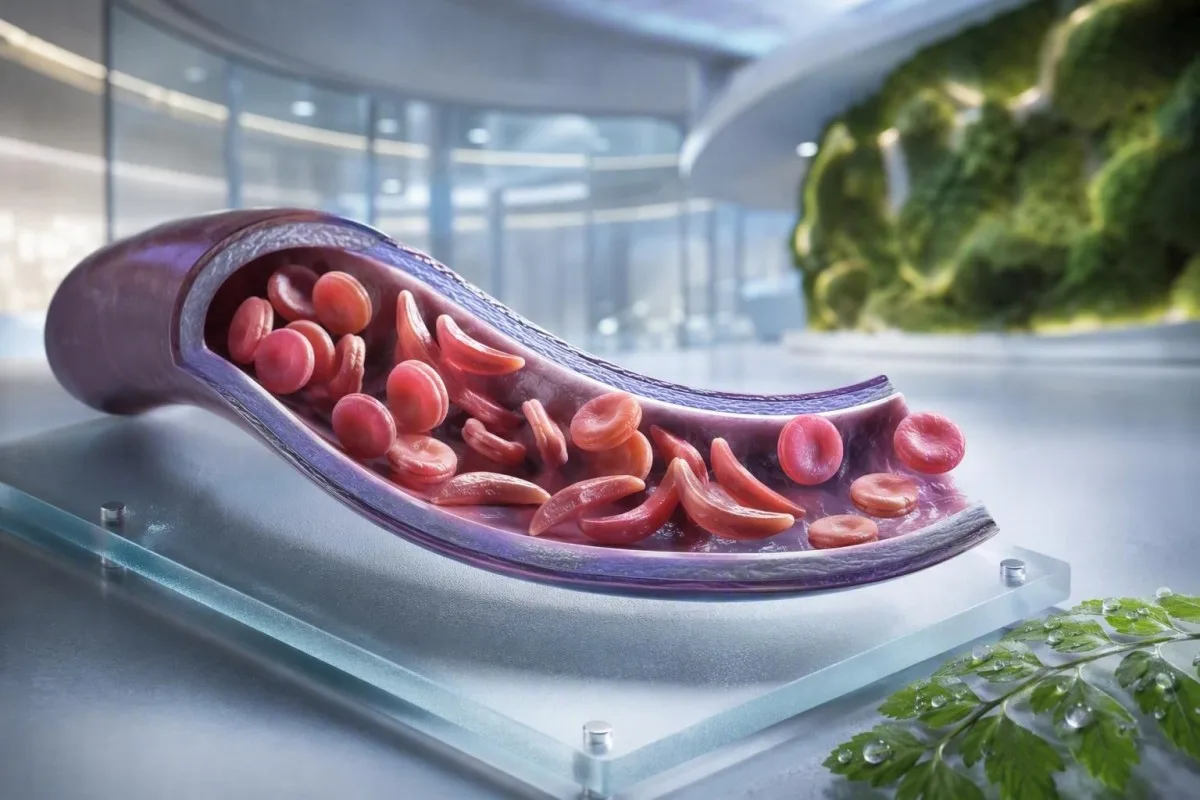
Anemia sel sabit bukanlah satu ujian, satu gejala, atau satu tahap kecemasan. Panduan yang mengutamakan pesakit ini memisahkan status pembawa, penyakit yang disahkan, pemantauan rutin dan situasi yang memerlukan penjagaan kecemasan.


- Trait sel sabit bermaksud satu gen HbS; ia biasanya tidak menyebabkan anemia kronik atau krisis sakit berulang.
- Saringan bayi baru lahir mengenal pasti kebanyakan bayi yang terjejas sebelum gejala bermula, membolehkan penisilin dan susulan pakar bermula lebih awal.
- Hemoglobin electrophoresis memisahkan jenis hemoglobin dan membantu mengesahkan status HbSS, HbSC, HbS-beta thalassemia, atau pembawa.
- Sakit dada yang teruk, sesak nafas, demam 38.5°C atau lebih tinggi, kekeliruan, atau kelemahan baharu memerlukan penilaian kecemasan segera pada seseorang dengan penyakit sel sabit.
- Sindrom dada akut boleh bermula dengan batuk, demam, sakit dada, atau oksigen rendah dan boleh menjadi lebih teruk dalam tempoh beberapa jam berbanding beberapa hari.
- Penurunan hemoglobin mendadak sebanyak 2 g/dL atau lebih daripada tahap biasa seseorang boleh menandakan krisis aplastik, sekuestrasi, pendarahan, atau hemolisis yang dipercepatkan.
- Kiraan retikulosit membantu membezakan tindak balas sumsum aktif daripada krisis aplastik; kiraan rendah semasa anemia yang semakin teruk adalah corak amaran.
- Hidroksiurea ialah ubat pengubahsuai penyakit, bukan sekadar ubat sakit; pemantauan biasanya termasuk kiraan darah penuh setiap 4 minggu semasa pelarasan dos.
Apa yang dimaksudkan dengan anemia sel sabit—dan apa yang tidak dimaksudkan
Anemia sel sabit biasanya merujuk kepada penyakit HbSS, di mana seseorang mewarisi dua gen yang menghasilkan hemoglobin S; sel merah boleh menjadi kaku, pecah awal, dan menghalang saluran darah kecil. Sifat sel sabit adalah berbeza: satu gen HbS biasanya cukup untuk diwariskan secara genetik tetapi tidak cukup untuk menyebabkan anemia kronik dan episod penutupan saluran darah berulang yang dilihat pada HbSS.
Penyakit sel sabit ialah istilah menyeluruh yang merangkumi HbSS, HbSC, HbS-beta-zero talasemia, dan HbS-beta-plus talasemia. HbSS selalunya menyebabkan anemia yang paling ketara, tetapi keterukan berbeza-beza secara meluas walaupun dalam satu keluarga; hemoglobin asas 7.5 g/dL mungkin stabil untuk seorang dewasa dan berbahaya bagi orang lain jika ia telah jatuh dengan cepat.
Mutasi hemoglobin S menukar satu asid amino dalam beta-globin: valin menggantikan asid glutamat pada kedudukan 6. Di bawah oksigen rendah, dehidrasi, asidosis, atau demam, molekul HbS boleh mengumpul di dalam sel. Perubahan fizikal itu menjelaskan mengapa krisis boleh berlaku selepas penerbangan yang panjang, penyakit gastrointestinal, atau malam pengambilan cecair yang kurang baik.
Kantesti AI ialah an Penganalisis ujian darah AI yang meletakkan CBC, bilirubin, LDH, retikulosit, kreatinin, dan keputusan terdahulu ke dalam satu pandangan longitudinal; ia tidak mendiagnosis penyakit sel sabit hanya daripada CBC. Untuk penyegaran praktikal tentang pelaporan hemoglobin, lihat maksud HGB.
Sehingga 4 September 2026, saya masih melihat pesakit yang secara salah diberitahu bahawa sifat dan penyakit boleh ditukar ganti. Mereka tidak. Peraturan klinikal Dr. Thomas Klein adalah mudah: minta genotip hemoglobin yang tepat, bukan kenyataan samar bahawa ujian itu “positif.”
Mengapa genotip mengubah penjagaan
HbSC mungkin mempunyai hemoglobin yang lebih tinggi daripada HbSS namun masih menyebabkan komplikasi retina, sakit, atau nekrosis avaskular. HbS-beta talasemia boleh menyerupai sifat atau HbSS bergantung pada sama ada pengeluaran beta-globin hadir sebahagiannya, sebab itulah ujian molekul kadang-kadang menyelesaikan corak elektroforesis yang samar.
Gejala penyakit sel sabit mengikut usia dan garis dasar
Gejala penyakit sel sabit biasanya termasuk sakit tulang atau dada episodik, keletihan, jaundis, sesak nafas, pertumbuhan terbantut, dan jangkitan kerap, tetapi tiada dua orang mengalami corak yang sama. Gejala biasanya muncul selepas 4 hingga 6 bulan usia kerana hemoglobin janin pada mulanya melindungi bayi daripada pengumpulan HbS.
Daktilitis—bengkak tangan atau kaki yang sakit—mungkin merupakan tanda pertama yang kelihatan pada bayi. Pada kanak-kanak usia sekolah, sakit perut memerlukan perhatian khusus kerana sembelit, penyakit pundi hempedu, pembesaran limpa, dan penutupan saluran darah boleh terasa serupa secara mengejutkan di rumah.
Jaundis dalam penyakit sel sabit mencerminkan pecahan sel darah merah yang berterusan dan peningkatan bilirubin tidak langsung; ia tidak secara automatik bermakna hepatitis. Air kencing gelap, najis pucat, gatal-gatal, demam, atau sakit perut kanan atas mengubah penilaian itu dan memerlukan penilaian punca hati dan hempedu; panduan kami untuk corak bilirubin langsung menerangkan perbezaannya.
Orang dewasa mungkin menormalkan kesakitan yang secara beransur-ansur menjadi lebih kerap. Dalam pengalaman saya, peralihan daripada dua episod yang diuruskan di rumah setiap tahun kepada dua episod sebulan adalah ketara secara klinikal walaupun lawatan kecemasan tidak meningkat. Gangguan tidur, kehilangan kerja, gejala erektil, mood rendah, dan penurunan toleransi senaman harus didokumentasikan kerana ia sering mendahului perubahan rasmi dalam rawatan.
Kepekatan hemoglobin 6 hingga 9 g/dL boleh menjadi julat keadaan stabil biasa pada HbSS, manakala nilai di bawah 10 g/dL akan tidak dijangka pada kebanyakan orang tanpa penyakit. Angka itu penting, tetapi perubahan daripada tahap asas peribadi lebih penting.
Siapa yang perlu mempertimbangkan saringan pembawa sel sabit
Saringan sel sabit adalah munasabah sebelum kehamilan atau awal kehamilan bagi sesiapa sahaja yang status pembawa tidak diketahui, tanpa mengira penampilan, nama keluarga, atau keturunan yang diandaikan. Status pembawa diwarisi dan sering tidak diketahui sehingga saringan bayi baru lahir atau ujian hemoglobin yang secara tidak disangka-sangka tidak normal.
Jika kedua orang tua biologis membawa varian terkait HbS atau beta-talasemia, setiap kehamilan berpeluang 25% untuk mewarisi kondisi hemoglobin yang signifikan secara klinis, tergantung pada varian yang terlibat. Seorang pembawa dapat memiliki energi normal, hasil CBC normal, dan tidak memiliki riwayat episode nyeri.
Saringan sangat berguna ketika salah satu pasangan memiliki HbAS, sifat HbC, sifat beta-talasemia, mikrositosis yang tidak dapat dijelaskan, atau riwayat keluarga penyakit sel sabit. Volume korpuskular rata-rata yang rendah tidak boleh disalahkan karena kekurangan zat besi tanpa memeriksa feritin dan mempertimbangkan talasemia; bandingkan Petunjuk MCV dan MCH.
Laporan global Organisasi Kesehatan Dunia tahun 2022 memperkirakan bahwa kelainan hemoglobin tetap menjadi beban penyakit bawaan yang besar, terutama di mana akses skrining bayi baru lahir tidak merata. Pendekatan yang paling hormat adalah akses universal ke skrining yang terinformasi daripada menggunakan etnisitas sebagai penjaga gerbang.
Kantesti ialah seorang platform tafsiran ujian darah AI yang dapat menjelaskan hasil HbS yang dilaporkan dalam bahasa awam, tetapi konfirmasi pembawa dan konseling reproduksi memerlukan laboratorium dan dokter terakreditasi. Jika pasangan merencanakan kehamilan, tanyakan apakah pengujian pasangan dan konseling genetik dapat dilakukan sebelum konsepsi daripada setelah hasil menjadi sensitif terhadap waktu.
Bagaimana elektroforesis hemoglobin mengesahkan diagnosis
Elektroforesis hemoglobin memisahkan fraksi hemoglobin dan merupakan tes inti untuk mengkonfirmasi anemia sel sabit atau sifatnya, seringkali bersamaan dengan kromatografi cair kinerja tinggi dan, bila diperlukan, pengujian genetik. CBC rutin tidak dapat mengkonfirmasi HbSS atau sifatnya karena kekurangan zat besi dan talasemia dapat menghasilkan indeks sel darah merah yang tumpang tindih.
Pada HbSS yang tidak diobati, elektroforesis biasanya menunjukkan HbS yang dominan tanpa HbA, meskipun persentase HbF bervariasi. HbAS biasanya menunjukkan HbA dan HbS, seringkali dengan HbA lebih tinggi dari HbS; persentase pasti dapat bergeser dengan alfa-talasemia yang diwariskan bersama atau transfusi baru-baru ini.
Transfusi baru-baru ini dapat membuat hasil elektroforesis menyesatkan selama kurang lebih 3 bulan karena HbA donor masih beredar. Inilah sebabnya mengapa HbA1c dapat meremehkan glukosa rata-rata setelah transfusi atau dalam hemolisis dengan pergantian tinggi; lihat ketika HbA1c menyesatkan.
Apusan perifer dapat menunjukkan sel target, polisitemia, dan bentuk sabit, tetapi mikroskop mendukung daripada menggantikan pengujian fraksi. Dr. Klein telah melihat apusan darurat yang “normal” menunda klarifikasi ketika seorang pasien telah ditransfusikan beberapa minggu sebelumnya—selalu beri tahu laboratorium dan tim hematologi tentang transfusi.
Metode skrining bayi baru lahir mengidentifikasi HbS sebelum penyakit klinis, tetapi hasilnya masih memerlukan pengujian konfirmasi yang tepat waktu. Pedoman berbasis bukti tahun 2014 oleh Yawn et al. merekomendasikan keterlibatan spesialis sejak dini, dengan tindakan pencegahan dimulai sebelum komplikasi besar pertama (Yawn et al., 2014).
Pemantauan darah rutin: apa yang sebenarnya dinilai oleh doktor
Pemantauan rutin dalam penyakit sel sabit biasanya menjejaki hemoglobin, retikulosit, sel darah putih, platelet, bilirubin, LDH, fungsi buah pinggang, dan protein air kencing—bukan satu keputusan terpencil. Perbandingan paling selamat adalah dengan garis dasar stabil pesakit itu sendiri, idealnya diambil apabila mereka sihat dan tidak baru-baru ini ditransfus.
Hemolisis selalunya menghasilkan LDH, bilirubin tidak langsung, dan retikulosit yang tinggi dengan haptoglobin yang rendah. Haptoglobin boleh rendah atas sebab lain, termasuk penyakit hati, jadi corak gabungan lebih kuat daripada mana-mana penanda tunggal; kami panduan tafsiran haptoglobin kami menunjukkan sebabnya.
Peratusan retikulosit sahaja boleh melebih-lebihkan tindak balas sumsum dalam anemia ketara. Doktor selalunya mengira kiraan mutlak retikulosit atau tindak balas mutlak retikulosit yang dikoreksi; kiraan mutlak yang jatuh semasa anemia yang semakin teruk menimbulkan kebimbangan tentang krisis aplastik yang berkaitan dengan parvovirus B19.
Kantesti ialah seorang Alat analisis ujian darah berkuasa AI yang membandingkan keputusan hematologi berulang berbanding merawat LDH yang tinggi sebagai diagnosis tersendiri. Sampel yang ditandakan sebagai hemolisis dalam tiub boleh meningkatkan kalium dan LDH secara palsu, jadi keputusan yang mengejutkan mungkin memerlukan ujian semula; semak ujian semula selepas hemolisis.
Hydroxyurea lazimnya meningkatkan MCV dan HbF dari semasa ke semasa, sambil menurunkan neutrofil secara sederhana pada dos yang berkesan. Pergeseran tersebut mungkin dijangka, tetapi kiraan mutlak neutrofil di bawah ambang rawatan individu atau kiraan platelet yang jatuh dengan cepat memerlukan semakan preskripsi berbanding menyesuaikan ubat sendiri.
Apabila sakit adalah episod penutupan saluran darah—dan apabila ia bukan
Sakit vaskular-oklusif selalunya mendalam, teruk, dan terletak di belakang, dada, pinggul, anggota badan, atau abdomen, tetapi sakit baharu tidak boleh secara automatik dilabelkan sebagai krisis sabit. Demam, bengkak setempat, kecederaan, kekejangan abdomen, atau gejala neurologi sebelah pihak menunjukkan masalah yang memerlukan siasatan berbeza.
Pelan sakit di rumah biasanya termasuk hidrasi awal, kehangatan, analgesia yang ditetapkan, rehat, dan ambang eskalasi peribadi yang dipersetujui dengan pasukan sabit. Minum jumlah yang berlebihan tidak lebih selamat: orang dengan gangguan buah pinggang atau penyakit jantung boleh mengalami beban cecair berlebihan, terutamanya semasa penyakit akut.
Sakit yang tidak terkawal selepas pelan penyelamat biasa, menghalang cecair atau ubat-ubatan daripada ditelan, atau berbeza dari segi kualiti atau lokasi harus mendorong nasihat klinikal segera. Sendi yang bengkak dan panas mungkin mewakili artritis septik atau jangkitan tulang berbanding oklusi vaskular yang tidak rumit; kami ujian sakit yang tidak dapat dijelaskan panduan menggariskan siasatan pertama yang berguna.
Dalam penjagaan kecemasan, analgesia yang tepat pada masanya dan penilaian semula lebih penting daripada membuktikan keterukan sakit melalui nombor makmal. Garis panduan NICE mengenai episod sakit akut sabit mengesyorkan penilaian analgesia pantas dan semakan kerap, kerana rawatan yang kurang memberi sumbangan kepada tekanan yang berpanjangan dan pemulihan yang tertangguh (NICE, 2012).
Elakkan pek sejuk terus pada kawasan sakit vaskular-oklusif melainkan pasukan anda secara khusus menasihatinya; sejuk boleh mengecutkan saluran darah periferal. Butiran praktikal itu nampak remeh, tetapi ramai pesakit memberitahu saya tiada siapa yang menjelaskannya sehingga selepas beberapa episod sukar.
Sindrom dada akut: kecemasan yang ramai orang terlepas pandang
Sindrom dada akut ialah rangsangan paru-paru baharu pada imbasan dada serta gejala pernafasan, demam, sakit dada, atau oksigen rendah pada orang yang mempunyai penyakit sel sabit, dan ia adalah kecemasan. Ia boleh merosot dengan cepat, kadang-kadang selepas kemasukan untuk episod sakit berbanding pada saat gejala dada bermula.
Hubungi perkhidmatan kecemasan atau pergi ke penjagaan kecemasan untuk sesak nafas baharu, sakit dada, bibir kebiruan-kelabu, pitam, batuk berterusan dengan demam, atau ketepuan oksigen di bawah sasaran yang ditetapkan untuk seseorang. Ketepuan 92% mungkin penurunan besar bagi seseorang yang biasanya pada 98%, manakala sesetengah pesakit mempunyai garis dasar yang didokumentasikan lebih rendah; kedua-dua nilai mutlak dan perubahan adalah penting.
Sindrom dada akut mungkin melibatkan jangkitan, embolisme lemak dari sumsum tulang, oklusi vaskular paru-paru, atelectasis, atau beberapa proses serentak. Doktor lazimnya menilai pengoksigenan, CBC, retikulosit, kultur apabila demam, imbasan dada, dan kadang-kadang gas darah; ujian darah untuk kesesakan nafas menambah konteks kepada keputusan ini.
Pada orang dewasa, sedasi berlebihan daripada opioid, pernafasan cetek akibat sakit, dan cecair intravena berlebihan boleh memburukkan status pernafasan. Spirometri insentif semasa dimasukkan ke hospital mengurangkan komplikasi paru-paru dalam episod sakit terpilih, walaupun ia bukan pengganti untuk penilaian segera apabila gejala telah bermula.
Semakan 2018 oleh Kato dan rakan-rakannya menerangkan sindrom dada akut sebagai punca utama morbiditi dan mortaliti sepanjang hayat (Kato et al., 2018). Jangan memandu sendiri jika kesesakan nafas, mengantuk, atau tekanan dada adalah ketara.
Demam, jangkitan dan kecemasan limpa
Suhu 38.5°C (101.3°F) atau lebih tinggi pada kanak-kanak atau orang dewasa dengan penyakit sel sabit memerlukan penilaian perubatan segera pada hari yang sama melainkan pasukan pakar mereka telah memberikan rancangan yang berbeza. Kehilangan fungsi imun limpa boleh menyebabkan jangkitan bakteria tertentu berkembang lebih pantas daripada demam virus biasa.
Demam ditambah lesu, ruam, tekanan darah rendah, kekeliruan, atau penurunan pengeluaran air kencing memerlukan penjagaan kecemasan sekarang. Kiraan sel darah putih yang kelihatan normal tidak dapat mengecualikan jangkitan serius secara pasti dalam penyakit sel sabit, terutamanya pada peringkat awal penyakit atau semasa rawatan hydroxyurea.
Pengasingan limpa berlaku paling kerap pada kanak-kanak kecil dan menyebabkan pembesaran limpa secara tiba-tiba, pucat, rasa penuh pada perut, pernafasan pantas, dan penurunan hemoglobin yang pantas. Ibu bapa kadang-kadang diajar cara merasai limpa anak mereka, tetapi sebarang pembesaran baharu semasa sakit harus mendorong penilaian segera dan bukannya pemeriksaan rumah berulang kali.
Penurunan hemoglobin sebanyak 2 g/dL atau lebih di bawah paras asas biasa dengan limpa yang membesar membimbangkan untuk pengasingan. Sebaliknya, anemia teruk dengan kiraan retikulosit yang sangat rendah boleh menunjukkan krisis aplastik, yang sering dikaitkan dengan parvovirus B19; tafsiran kiraan retikulosit menerangkan isyarat sumsum tulang.
Makmal sepsis mungkin termasuk laktat, kultur, CBC, ujian buah pinggang, dan penanda keradangan, tetapi keputusan rawatan tidak sepatutnya menunggu sehingga setiap nilai kembali. Untuk konteks corak segera, baca petunjuk penanda sepsis.
Vaksin dan antibiotik pencegahan
Vaksin dan profilaksis penisilin kanak-kanak mengurangkan risiko tetapi tidak menjadikan demam berisiko rendah. Sahkan rancangan individu anda dengan pasukan hematologi atau pediatrik, terutamanya selepas splenektomi, perjalanan, bayi baharu, atau perubahan dalam jadual imunisasi tempatan.
Strok dan tanda amaran neurologi memerlukan tindakan segera
Sebarang kelemahan muka, lengan atau kaki yang tiba-tiba, kesukaran bercakap, sawan, sakit kepala teruk yang tidak biasa, kekeliruan, atau kehilangan keseimbangan dalam penyakit sel sabit harus dianggap sebagai kemungkinan kecemasan strok. Hubungi perkhidmatan kecemasan dengan segera; jangan tunggu untuk melihat sama ada simptom berlalu atau berikan ubat sakit dahulu.
Kanak-kanak dengan HbSS atau talasemia HbS-beta-zero biasanya ditawarkan saringan imbasan ultrasound Doppler transkranial dari umur 2 hingga 16 tahun. Halaju maksimum purata masa yang tidak normal sebanyak 200 cm/saat atau lebih tinggi mengenal pasti peningkatan risiko strok yang ketara dan memerlukan tindakan pakar segera.
Terapi pemindahan darah kronik boleh mengurangkan risiko strok pertama pada kanak-kanak dengan penemuan Doppler yang tidak normal, tetapi ia mewujudkan keperluan tambahan: pemantauan bebanan besi, saringan antibodi, dan padanan sel penderma yang teliti. Ini adalah penjagaan khusus, bukan hasil yang perlu diuruskan melalui aplikasi.
Orang dewasa boleh mengalami simptom neurologi sementara, perubahan kognitif, atau kecederaan otak senyap tanpa strok dramatik klasik. Episod kesukaran mencari perkataan selama 10 minit masih memerlukan penilaian kecemasan kerana simptom iskemia sementara boleh menjadi amaran, bukan jaminan.
Dr. Thomas Klein menasihatkan keluarga untuk menyimpan nombor kenalan kecemasan sel sabit tempatan mereka di sebelah rancangan sakit biasa. Ia menghapuskan satu keputusan semasa beberapa minit apabila kanak-kanak atau orang dewasa mungkin bergelut untuk berkomunikasi.
Melindungi buah pinggang, mata dan paru-paru antara krisis
Penyakit sel sabit boleh merosakkan buah pinggang, retina, paru-paru dan jantung walaupun apabila sakit jarang berlaku, jadi pemantauan organ rutin adalah sebahagian daripada rawatan dan bukannya tambahan pilihan. Ujian albumin air kencing, pemeriksaan tekanan darah, pemeriksaan mata, dan penilaian paru-paru berdasarkan simptom mengenal pasti komplikasi sebelum ia menjadi ketara.
Albuminuria mungkin merupakan tanda awal nefropati sel sabit. Nisbah albumin air kencing kepada kreatinin sebanyak 30 mg/g atau lebih adalah tidak normal pada kebanyakan orang dewasa dan harus disahkan dengan ujian berulang kerana demam, senaman, haid, dan dehidrasi boleh meningkatkannya secara sementara.
Kreatinin boleh kelihatan normal secara menipu dalam penyakit sel sabit kerana peningkatan rembesan tubul dan jisim otot yang lebih rendah mungkin menyembunyikan penapisan yang berkurangan. Cystatin C atau persamaan gabungan boleh menambah konteks yang berguna, manakala air kencing berbuih yang berterusan layak mendapat penilaian; lihat panduan protein dalam air kencing.
Retinopati adalah sangat penting dalam penyakit HbSC, yang boleh menjadi lebih senyap secara klinikal dalam aspek lain. Timbulnya objek terapung baharu, kilatan cahaya, perubahan visual seperti langsir, atau penglihatan kabur secara tiba-tiba memerlukan semakan oftalmik segera, bukan janji temu rutin beberapa minggu kemudian.
Kehamilan menambah hiperfiltrasi fisiologi dan risiko ibu-janin yang lebih tinggi, jadi penilaian buah pinggang dan air kencing asas perlu dilakukan lebih awal. Penjelasan kami tentang perubahan GFR kehamilan membantu membezakan perubahan yang dijangka daripada trend yang membimbangkan.
Pilihan rawatan, hydroxyurea dan keselamatan transfusi
Hidroksikarbamida mengurangkan episod sakit dan sindrom dada akut bagi ramai penghidap talasemia HbSS atau HbS-beta-sifar dengan meningkatkan hemoglobin janin, tetapi ia memerlukan pemantauan berstruktur. Transfusi boleh menyelamatkan nyawa bagi komplikasi terpilih, namun transfusi berulang membawa risiko pemuatan besi dan antibodi sel darah merah.
Hidroksikarbamida biasanya dilaraskan dengan CBC dan kiraan retikulosit kira-kira setiap 4 minggu sehingga dos stabil dicapai, kemudian pada selang masa yang lebih lama ditentukan oleh pasukan rawatan. MCV yang lebih tinggi selalunya merupakan kesan rawatan yang dijangka, manakala sitopenia teruk memerlukan semakan ubat dan kadang-kadang penghentian sementara.
Transfusi ringkas meningkatkan kapasiti pembawa oksigen, tetapi kepekatan hemoglobin yang berlebihan boleh meningkatkan kelikatan dalam HbSS. Banyak protokol akut mengelakkan peningkatan hemoglobin selepas transfusi melebihi 10 g/dL melainkan pakar menetapkan matlamat yang berbeza; transfusi pertukaran digunakan untuk komplikasi teruk yang terpilih.
Pemuatan besi menjadi lebih berkemungkinan selepas transfusi berulang dan dinilai dengan trend feritin serta MRI hati atau jantung apabila sesuai. Feritin meningkat dengan keradangan, jadi tahap tinggi tunggal bukanlah bukti bebanan besi tisu; panduan kajian besi menerangkan hadnya.
Bukti paling kukuh adalah untuk hidroksikarbamida dalam penyakit HbSS dan HbS-beta-sifar; faedah dalam HbSC kekal sebagai bidang yang kurang pasti dan memerlukan pembuatan keputusan yang lebih individu. Pelan ubat harus merangkumi perbincangan tentang kontrasepsi dan kehamilan jika relevan, halangan kepatuhan, dan pelan bertulis untuk dos yang terlepas.
Kehamilan, kesuburan dan perancangan keluarga dengan HbS
Kehamilan dengan penyakit sel sabit memerlukan penjagaan bersama awal daripada obstetrik dan hematologi kerana risiko episod sakit, anemia, trombosis, jangkitan, preeklampsia, sekatan pertumbuhan janin dan kelahiran pramatang adalah lebih tinggi. Saringan pembawa adalah penting sebelum kehamilan kerana risiko genetik bergantung pada kedua-dua ibu bapa biologikal.
Lawatan pra-kehamilan harus menyemak genotaip, hemoglobin asas, fungsi buah pinggang, albumin air kencing, tekanan darah, sejarah transfusi, antibodi sel darah merah, vaksinasi, dan ubat-ubatan. Keputusan hidroksikarbamida semasa kehamilan adalah individu dan mesti dibuat dengan pakar; jangan hentikan atau mulakan semula hanya berdasarkan nasihat internet.
Besi tidak sepatutnya dibekalkan secara automatik untuk hemoglobin rendah dalam HbSS. Kekurangan zat besi boleh wujud bersama, terutamanya dengan kehamilan atau pendarahan haid yang banyak, tetapi feritin dan ketepuan transferin harus membimbing rawatan kerana hemolisis kronik sahaja tidak sama dengan kekurangan zat besi.
Apabila kedua-dua ibu bapa membawa varian hemoglobin yang berkaitan, kaunseling genetik boleh menjelaskan konsepsi semula jadi, pilihan diagnostik pranatal, pilihan penderma, dan ujian genetik praimplantasi tanpa mengarahkan keluarga kepada satu keputusan. Tujuannya adalah pilihan yang termaklum, bukan kebimbangan.
Untuk tanda-tanda amaran makmal kehamilan praktikal lain, lihat kebimbangan makmal kehamilan hari yang sama. Asas yang diketahui pesakit harus dimasukkan dalam setiap serahan bersalin; ia mengubah cara keputusan hemoglobin ditafsirkan.
Menggunakan keputusan makmal dengan selamat antara janji temu
Keputusan makmal sel sabit paling berguna apabila dibandingkan dengan genotaip anda, asas yang stabil, gejala, rawatan, dan transfusi baru-baru ini. Bilirubin tinggi atau hemoglobin rendah mungkin dijangka bagi seseorang dengan HbSS tetapi mendesak bagi orang yang sama jika keputusan berubah secara mendadak.
Simpan rekod satu muka surat tentang genotaip, hemoglobin biasa, ketepuan oksigen biasa jika diketahui, kumpulan darah dan antibodi, ubat semasa, tarikh transfusi, dan kenalan kecemasan. Ini menjimatkan masa di jabatan kecemasan yang tidak dikenali dan mengurangkan kemungkinan asas HbSS disalah anggap sebagai anemia yang baru ditemui.
Kantesti ialah seorang perkhidmatan tafsiran ujian makmal AI yang boleh menyusun laporan makmal yang dimuat naik dan menandakan corak untuk perbincangan, termasuk penanda hemolisis, trend buah pinggang, dan pemantauan ubat. Ia tidak dapat melihat dada anda, mengukur oksigenasi, atau menggantikan penjagaan kecemasan apabila gejala menunjukkan sindrom dada akut, sepsis, atau strok.
Keputusan di luar julat rujukan makmal tidak semestinya berbahaya, dan keputusan di dalamnya masih boleh berbahaya jika ia berubah secara mendadak. Untuk penjelasan mengenai bendera pelaporan dan julat rujukan, baca maksud keputusan luar julat.
Pendekatan klinikal Kantesti dikaji terhadap kaedah dan batasan yang ditentukan; pembaca yang ingin memahami langkah keselamatan kami boleh menyemak pengesahan klinikal kami. Bawa PDF asal ke janji temu hematologi anda—OCR berguna, tetapi laporan sumber kekal sebagai rekod.
Pelan tindakan kecemasan yang jelas untuk penyakit sel sabit
Dapatkan bantuan kecemasan sekarang sekiranya mengalami sakit dada atau sesak nafas, demam 38.5°C atau lebih tinggi, gejala neurologi baharu, pitam, pucat teruk, kesakitan yang tidak terkawal, abdomen yang membesar dengan cepat, atau ketidakupayaan untuk minum dan mengambil ubat. Tanda amaran ini boleh menunjukkan sindrom dada akut, sepsis, strok, anemia teruk, atau pengasingan limpa.
Beritahu kakitangan kecemasan bahawa anda mempunyai penyakit sel sabit, nyatakan genotip anda jika diketahui, dan terangkan apa yang berbeza daripada episod biasa anda. Sebutkan pemindahan darah baru-baru ini, hemoglobin asas, ubat sakit yang telah diambil dengan dos dan masa, alahan, kehamilan, dan sebarang sindrom dada akut atau strok sebelum ini.
Jangan tunggu sehingga oksimeter nadi di rumah menjadi tidak normal jika gejala dada semakin teruk. Peranti pengguna boleh salah baca dengan jari sejuk, produk kuku, pergerakan, peredaran darah yang lemah, dan beberapa tona kulit; gejala dan penilaian klinikal mengatasi satu paparan yang meyakinkan.
Jika anda menjaga kanak-kanak, minta pasukan mereka untuk ambang demam bertulis, pelan analgesik, dan arahan pemeriksaan limpa. Jika anda seorang dewasa, simpan salinan pada telefon anda dan kongsi dengan seorang yang dipercayai; perancangan rekod kesihatan keluarga boleh memudahkan perkara ini.
Pasukan perubatan Kantesti, termasuk Lembaga Penasihat Perubatan, kami, menyokong pendidikan mengenai tafsiran makmal, manakala keputusan kecemasan adalah dengan perkhidmatan klinikal secara bersemuka. Mengenai organisasi dan misi klinikal kami, lihat pasukan dan pendekatan kami.
Soalan Lazim
Apakah gejala awal anemia sel sabit pada bayi?
Gejala pertama anemia sel sabit selalunya muncul selepas 4 hingga 6 bulan usia, apabila hemoglobin janin secara semula jadi menurun. Bengkak yang menyakitkan pada tangan dan kaki, dipanggil daklititis, demam, kerenahan yang luar biasa, pucat, jaundis, pemakanan yang lemah, dan perut yang membengkak adalah tanda-tanda amaran awal yang biasa. Demam 38.5°C (101.3°F) atau lebih tinggi pada bayi dengan penyakit sel sabit memerlukan penilaian perubatan segera pada hari yang sama kerana jangkitan serius boleh berlaku dengan cepat. Saringan bayi baru lahir mengenal pasti bayi yang terjejas sebelum gejala bermula, tetapi ujian pengesahan dan susulan pakar tetap perlu.
Bolehkah anda mempunyai trait sel sabit dan masih mengalami simptom?
Kebanyakan individu dengan trait sel sabit tidak mengalami anemia kronik, krisis kesakitan berulang, dan toleransi senaman harian yang normal. Komplikasi jarang berlaku boleh berlaku dalam keadaan dehidrasi ekstrem, pendedahan haba yang teruk, altitud tinggi, aktiviti lasak yang sangat intens, atau persekitaran oksigen rendah, dan darah dalam air kencing perlu dinilai dan bukannya dianggap tidak berbahaya. Trait bermaksud satu gen HbS, manakala anemia sel sabit biasanya bermaksud penyakit HbSS dengan dua gen beta-globin yang terjejas. Elektroforesis hemoglobin atau ujian pecahan yang setara boleh menjelaskan perbezaan.
Ujian apa yang mengesahkan anemia sel sabit?
Elektroforesis hemoglobin, kromatografi cecair berprestasi tinggi, atau elektroforesis kapilari mengesahkan pecahan hemoglobin yang ada dan adalah pusat kepada diagnosis anemia sel sabit. HbSS yang tidak dirawat lazimnya menunjukkan hemoglobin S yang dominan tanpa hemoglobin A, manakala trait biasanya menunjukkan kedua-dua hemoglobin A dan hemoglobin S. CBC boleh mengenal pasti anemia atau sel merah kecil tetapi tidak boleh mendiagnosis HbSS, HbSC, atau trait secara bebas. Transfusi terbaru boleh mengganggu keputusan pecahan selama kira-kira 3 bulan, jadi ujian molekul mungkin diperlukan apabila corak tidak jelas.
Bilakah seseorang dengan penyakit sel sabit perlu pergi ke jabatan kecemasan?
Seseorang pesakit sel sabit perlu mendapatkan penilaian kecemasan sekiranya demam 38.5°C (101.3°F) atau lebih tinggi, sakit dada, sesak nafas, kekurangan oksigen, kelemahan mendadak, kesukaran bertutur, sawan, pitam, pucat yang semakin teruk dengan pantas, bengkak perut yang teruk, atau kesakitan yang tidak terkawal dengan pelan rawatan sedia ada. Simptom-simptom ini boleh menandakan sindrom dada akut, jangkitan, strok, sekuestrasi limpa, atau penurunan hemoglobin yang mendadak. Kesakitan baharu atau berbeza juga memerlukan penilaian kerana jangkitan tulang, penyakit pundi hempedu, apendisitis, dan darah beku boleh menyerupai kesakitan vaso-oklusif. Jangan memandu sendiri jika simptom neurologi atau pernafasan adalah ketara.
Apakah paras hemoglobin yang berbahaya dalam penyakit sel sabit?
Tiada satu nombor hemoglobin berbahaya dalam penyakit sel sabit kerana ramai orang dengan HbSS mempunyai paras asas yang stabil antara kira-kira 6 dan 9 g/dL. Penurunan sebanyak 2 g/dL atau lebih daripada hemoglobin biasa individu sering lebih membimbangkan daripada nilai mutlak dan boleh menunjukkan krisis aplastik, sekuestrasi limpa, pendarahan, atau hemolisis yang dipercepatkan. Gejala teruk seperti sesak nafas, pitam, sakit dada, degupan jantung pantas, atau kekeliruan memerlukan penilaian segera pada mana-mana paras hemoglobin. Keputusan pemindahan bergantung pada gejala, paras asas, punca anemia, pengoksigenan, dan risiko hiperviskositi.
Adakah hidroksiurea menyembuhkan anemia sel sabit?
Hidroksiurea tidak menyembuhkan anemia sel sabit, tetapi ia boleh mengurangkan episod kesakitan, sindrom dada akut, keperluan pemindahan darah, dan penggunaan hospital bagi ramai pesakit HbSS atau talasemia HbS-beta-sifar. Ia berfungsi sebahagiannya dengan meningkatkan hemoglobin janin dan biasanya memerlukan pemantauan CBC dan retikulosit kira-kira setiap 4 minggu semasa dos sedang dilaraskan. Peningkatan tahap MCV dan HbF boleh menunjukkan kesan biologi, walaupun kedua-dua keputusan ini tidak membuktikan kepatuhan yang sempurna. Pendekatan kuratif seperti pemindahan sel stem dan beberapa terapi berasaskan gen memerlukan penilaian kelayakan yang sangat khusus dan pemantauan jangka panjang.
Dapatkan Analisis Ujian Darah Berkuasa AI Hari Ini
Sertai lebih 2 juta pengguna di seluruh dunia yang mempercayai Kantesti untuk analisis ujian makmal segera dan tepat. Muat naik keputusan ujian darah anda dan terima tafsiran menyeluruh biomarker 15,000+ dalam beberapa saat.
📚 Penerbitan Penyelidikan Dirujuk
Klein, T., Mitchell, S., & Weber, H. (2026). Panduan Ujian Darah Komplemen C3 C4 & Titer ANA. Kantesti Penyelidikan Perubatan AI.
Klein, T., Mitchell, S., & Weber, H. (2026). Ujian Darah Virus Nipah: Panduan Pengesanan & Diagnosis Awal 2026. Kantesti Penyelidikan Perubatan AI.
📖 Rujukan Perubatan Luaran
⚕️ Penafian Perubatan
Artikel ini adalah untuk tujuan pendidikan sahaja dan tidak merupakan nasihat perubatan. Sentiasa rujuk pembekal penjagaan kesihatan yang berkelayakan untuk keputusan diagnosis dan rawatan.
Isyarat Kepercayaan E-E-A-T
Pengalaman
Semakan klinikal yang diketuai oleh doktor terhadap aliran kerja tafsiran makmal.
Kepakaran
Fokus perubatan makmal tentang bagaimana biomarker berkelakuan dalam konteks klinikal.
Kewibawaan
Ditulis oleh Dr. Thomas Klein dengan semakan oleh Dr. Sarah Mitchell dan Prof. Dr. Hans Weber.
Kebolehpercayaan
Tafsiran berasaskan bukti dengan laluan susulan yang jelas untuk mengurangkan kebimbangan.