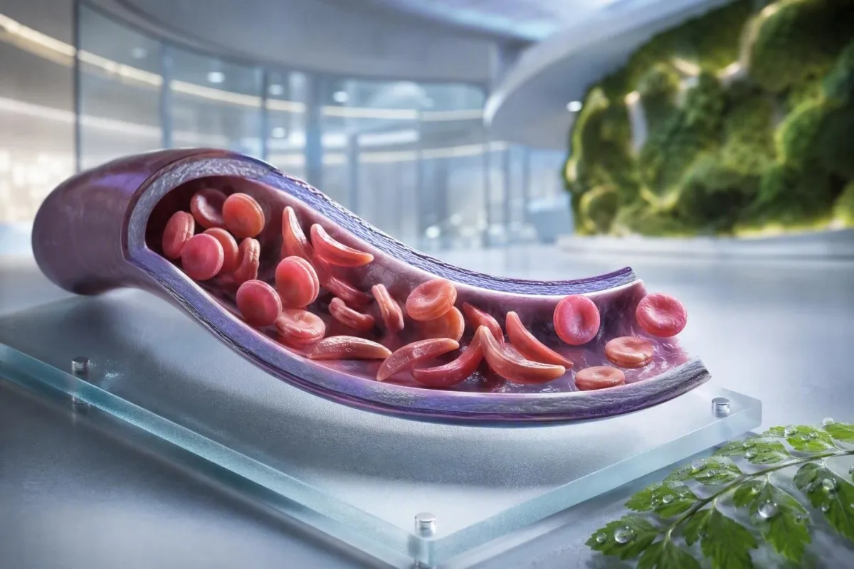
Sickle cell anemia is not one test, one symptom, or one level of urgency. This patient-first guide separates carrier status, confirmed disease, routine monitoring and situations that need emergency care.


- Sickle cell trait means one HbS gene; it usually does not cause chronic anemia or recurrent pain crises.
- Newborn screening identifies most affected infants before symptoms begin, allowing penicillin and specialist follow-up to start early.
- Hemoglobine-elektroforese separates hemoglobin types and helps confirm HbSS, HbSC, HbS-beta thalassemia, or carrier status.
- Severe chest pain, breathlessness, fever of 38.5°C or higher, confusion, or a new weakness need urgent emergency assessment in a person with sickle cell disease.
- Acute chest syndrome can begin with cough, fever, chest pain, or low oxygen and may worsen over hours rather than days.
- A sudden hemoglobin fall of 2 g/dL or more from a person’s usual level can signal aplastic crisis, sequestration, bleeding, or accelerated hemolysis.
- Retikulocyte-telling helps distinguish active marrow response from aplastic crisis; a low count during worsening anemia is a warning pattern.
- Hydroxyurea is a disease-modifying medicine, not simply a pain medicine; monitoring commonly includes a full blood count every 4 weeks during dose adjustment.
What sickle cell anemia means—and what it does not
Sickle cell anemia usually refers to HbSS disease, where a person inherits two genes that produce hemoglobin S; red cells can become rigid, break down early, and obstruct small vessels. Sickle cell trait is different: one HbS gene is usually enough to pass on genetically but not enough to cause the chronic anemia and recurring vaso-occlusive episodes seen in HbSS.
Sickle cell disease is an umbrella term that includes HbSS, HbSC, HbS-beta-zero thalassemia, and HbS-beta-plus thalassemia. HbSS often causes the most pronounced anemia, but severity varies enormously even within one family; a baseline hemoglobin of 7.5 g/dL may be stable for one adult and dangerous for another if it has fallen rapidly.
The hemoglobin S mutation changes one amino acid in beta-globin: valine replaces glutamic acid at position 6. Under low oxygen, dehydration, acidosis, or fever, HbS molecules can polymerize inside the cell. That physical change explains why a crisis can develop after a long flight, a gastrointestinal illness, or a night of poor fluid intake.
Kantesti AI is in AI bloedtestanalysator that places CBC, bilirubin, LDH, reticulocyte, creatinine, and prior results into one longitudinal view; it does not diagnose sickle cell disease from a CBC alone. For a practical refresher on hemoglobin reporting, see wat HGB betsjut.
As of September 4, 2026, I still see patients wrongly told that trait and disease are interchangeable. They are not. Dr. Thomas Klein’s clinical rule is simple: ask for the exact hemoglobin genotype, not a vague statement that a test was “positive.”
Why genotype changes care
HbSC may have a higher hemoglobin than HbSS yet still cause retinal complications, pain, or avascular necrosis. HbS-beta thalassemia can resemble trait or HbSS depending on whether beta-globin production is partly present, which is why molecular testing sometimes settles an ambiguous electrophoresis pattern.
Sickle cell disease symptoms by age and baseline
Sickle cell disease symptoms commonly include episodic bone or chest pain, fatigue, jaundice, breathlessness, delayed growth, and frequent infections, but no two people experience the same pattern. Symptoms usually emerge after 4 to 6 months of age because fetal hemoglobin initially protects infants from HbS polymerization.
Dactylitis—tender swelling of hands or feet—may be the first visible sign in infants. In school-age children, abdominal pain deserves particular care because constipation, gallbladder disease, splenic enlargement, and vaso-occlusion can feel surprisingly similar at home.
Jaundice in sickle cell disease reflects ongoing red-cell breakdown and indirect bilirubin elevation; it does not automatically mean hepatitis. Dark urine, pale stools, itching, fever, or right-upper abdominal pain change that assessment and warrant evaluation of liver and biliary causes; our guide to direct bilirubin patterns ferklearret it ferskil.
Adults may normalize pain that has gradually become more frequent. In my experience, a shift from two home-managed episodes yearly to two monthly episodes is clinically meaningful even if emergency visits have not increased. Sleep disruption, missed work, erectile symptoms, low mood, and reduced exercise tolerance should be documented because they often precede a formal change in treatment.
A hemoglobin concentration of 6 to 9 g/dL can be a usual steady-state range in HbSS, whereas a value under 10 g/dL would be unexpected in most people without disease. The number matters, but the change from personal baseline matters more.
Who should consider sickle cell carrier screening
Sickle cell screening is reasonable before pregnancy or early in pregnancy for anyone whose carrier status is unknown, regardless of appearance, surname, or assumed ancestry. Carrier status is inherited and often remains unknown until a newborn screen or an unexpectedly abnormal hemoglobin test.
If both biological parents carry an HbS-related or beta-thalassemia variant, each pregnancy can have a 25% chance of inheriting a clinically significant hemoglobin condition, depending on the variants involved. A carrier can have normal energy, normal CBC results, and no history of pain episodes.
Screening is particularly useful when one partner has HbAS, HbC trait, beta-thalassemia trait, unexplained microcytosis, or a family history of sickle cell disease. A low mean corpuscular volume should not be blamed on iron deficiency without checking ferritin and considering thalassemia; compare MCV and MCH clues.
A 2022 World Health Organization global report estimated that hemoglobin disorders remain a major inherited disease burden, particularly where newborn screening access is uneven. The most respectful approach is universal access to informed screening rather than using ethnicity as a gatekeeper.
Kantesti is in AI bloedtest-útslachplatfoarm that can explain a reported HbS result in plain language, but carrier confirmation and reproductive counseling require an accredited laboratory and clinician. If a couple is planning a pregnancy, ask whether partner testing and genetic counseling can happen before conception rather than after a result becomes time-sensitive.
How hemoglobin electrophoresis confirms the diagnosis
Hemoglobin electrophoresis separates hemoglobin fractions and is a core test for confirming sickle cell anemia or trait, often alongside high-performance liquid chromatography and, when needed, genetic testing. A routine CBC cannot confirm HbSS or trait because iron deficiency and thalassemia can produce overlapping red-cell indices.
In untreated HbSS, electrophoresis typically shows predominantly HbS with no HbA, although HbF percentage varies. HbAS usually shows both HbA and HbS, often with HbA higher than HbS; exact percentages can shift with co-inherited alpha-thalassemia or recent transfusion.
Recent transfusion can make an electrophoresis result misleading for roughly 3 months because donor HbA is still circulating. This is also why HbA1c can understate average glucose after transfusion or in high-turnover hemolysis; see when HbA1c misleads.
A peripheral smear may show target cells, polychromasia, and sickled forms, but microscopy supports rather than replaces fraction testing. Dr. Klein has seen “normal” emergency smears delay clarification when a patient had been transfused weeks earlier—always tell the laboratory and hematology team about transfusions.
Newborn screening methods identify HbS before clinical illness, but the result still needs timely confirmatory testing. The 2014 evidence-based guideline by Yawn et al. recommends specialist involvement early in life, with preventive measures beginning before the first major complication (Yawn et al., 2014).
Routine blood monitoring: what clinicians actually trend
Routine monitoring in sickle cell disease usually trends hemoglobin, reticulocytes, white cells, platelets, bilirubin, LDH, kidney function, and urine protein—not one isolated result. The safest comparison is with the patient’s own stable baseline, ideally drawn when they are well and not recently transfused.
Hemolysis often produces elevated LDH, indirect bilirubin, and reticulocytes with low haptoglobin. Haptoglobin can be low for other reasons, including liver disease, so the combined pattern is stronger than any single marker; our haptoglobine útslachgids lit sjen wêrom.
A reticulocyte percentage alone can overstate marrow response in significant anemia. Clinicians often calculate an absolute reticulocyte count or corrected reticulocyte response; a falling absolute count during worsening anemia raises concern for parvovirus B19-associated aplastic crisis.
Kantesti is in AI-oandreaune ark foar analyse fan bloedtests that compares repeated hematology results rather than treating a high LDH as a standalone diagnosis. A sample flagged as hemolyzed in the tube can falsely increase potassium and LDH, so a surprising result may need a redraw; review repeat testing after hemolysis.
Hydroxyurea commonly increases MCV and HbF over time, while lowering neutrophils modestly at an effective dose. Those shifts may be expected, but an absolute neutrophil count below the individual treatment threshold or a rapidly falling platelet count requires prescriber review rather than self-adjusting medication.
When pain is a vaso-occlusive episode—and when it is not
Vaso-occlusive pain is often deep, severe, and located in the back, chest, hips, limbs, or abdomen, but new pain must not automatically be labelled a sickle crisis. Fever, focal swelling, injury, abdominal guarding, or one-sided neurologic symptoms point toward problems needing a different work-up.
A home pain plan typically includes early hydration, warmth, prescribed analgesia, rest, and a personal escalation threshold agreed with the sickle team. Drinking excessive volumes is not safer: people with kidney impairment or heart disease can develop fluid overload, especially during an acute illness.
Pain that remains uncontrolled after the usual rescue plan, prevents fluids or medicines being kept down, or is different in quality or location should prompt urgent clinical advice. A swollen, hot joint may represent septic arthritis or bone infection rather than uncomplicated vaso-occlusion; our unexplained pain testing guide outlines useful first investigations.
In emergency care, timely analgesia and reassessment matter more than proving pain severity through a laboratory number. NICE’s guideline on acute painful sickle episodes recommends rapid analgesia assessment and frequent review, because undertreatment itself contributes to prolonged distress and delayed recovery (NICE, 2012).
Avoid cold packs directly on an area of vaso-occlusive pain unless your team specifically advises them; cold can constrict peripheral vessels. That practical detail sounds minor, but many patients tell me nobody explained it until after several difficult episodes.
Acute chest syndrome: the emergency many people miss
Acute chest syndrome is a new lung infiltrate on chest imaging plus respiratory symptoms, fever, chest pain, or low oxygen in a person with sickle cell disease, and it is an emergency. It can deteriorate quickly, sometimes after admission for a pain episode rather than at the moment chest symptoms begin.
Call emergency services or go to emergency care for new shortness of breath, chest pain, blue-gray lips, fainting, persistent cough with fever, or oxygen saturation below the person’s prescribed target. A saturation of 92% may be a major drop for someone normally at 98%, while some patients have a lower documented baseline; both the absolute value and change matter.
Acute chest syndrome may involve infection, fat embolization from bone marrow, pulmonary vascular obstruction, atelectasis, or several processes at once. Clinicians commonly assess oxygenation, CBC, reticulocytes, cultures when febrile, chest imaging, and sometimes blood gases; bloedtests foar sykheljen adds context to these results.
In adults, oversedation from opioids, shallow breathing from pain, and excess intravenous fluid can worsen respiratory status. Incentive spirometry during hospitalization reduces pulmonary complications in selected painful episodes, though it is not a substitute for urgent assessment when symptoms have already started.
The 2018 review by Kato and colleagues describes acute chest syndrome as a major cause of morbidity and mortality across the lifespan (Kato et al., 2018). Do not drive yourself if breathlessness, drowsiness, or chest pressure is significant.
Fever, infection and spleen emergencies
A temperature of 38.5°C (101.3°F) or higher in a child or adult with sickle cell disease needs urgent same-day medical assessment unless their specialist team has given a different plan. Functional loss of splenic immune function can make some bacterial infections progress far faster than an ordinary viral fever.
Fever plus lethargy, rash, low blood pressure, confusion, or reduced urine output requires emergency care now. A normal-looking white cell count does not reliably exclude serious infection in sickle cell disease, especially early in illness or during hydroxyurea treatment.
Splenic sequestration occurs most often in young children and causes sudden spleen enlargement, pallor, abdominal fullness, fast breathing, and a rapid hemoglobin fall. Parents are sometimes taught how to feel for their child’s spleen, but any new enlargement with illness should trigger urgent assessment rather than repeated home checks.
A hemoglobin decline of 2 g/dL or more below usual baseline with an enlarged spleen is concerning for sequestration. By contrast, severe anemia with a very low reticulocyte count can suggest aplastic crisis, frequently related to parvovirus B19; reticulocyte count interpretation explains the marrow signal.
Sepsis laboratories may include lactate, cultures, CBC, kidney tests, and inflammatory markers, but treatment decisions should never wait for every value to return. For context on urgent patterns, read sepsis marker clues.
Vaccines and preventive antibiotics
Vaccines and childhood penicillin prophylaxis reduce risk but do not make fever low-risk. Confirm your individualized plan with a hematology or pediatric team, particularly after splenectomy, travel, a new baby, or a change in local immunization schedule.
Stroke and neurologic warning signs need immediate action
Any sudden face droop, arm or leg weakness, speech difficulty, seizure, severe unusual headache, confusion, or loss of balance in sickle cell disease should be treated as a possible stroke emergency. Call emergency services immediately; do not wait to see whether symptoms pass or give pain medicine first.
Children with HbSS or HbS-beta-zero thalassemia are commonly offered transcranial Doppler ultrasound screening from age 2 through 16 years. An abnormal time-averaged mean maximum velocity of 200 cm/second or higher identifies a substantially increased stroke risk and needs prompt specialist action.
Chronic transfusion therapy can reduce first-stroke risk in children with abnormal Doppler findings, but it creates additional needs: iron-overload monitoring, antibody screening, and careful matching of donor cells. This is specialized care, not a result to manage through an app.
Adults can have transient neurologic symptoms, cognitive changes, or silent cerebral injury without a classic dramatic stroke. A 10-minute episode of word-finding difficulty still needs emergency assessment because transient ischemic symptoms can be a warning, not reassurance.
Dr. Thomas Klein advises families to save their local sickle emergency contact number beside the usual pain plan. It removes one decision during the few minutes when a child or adult may be struggling to communicate.
Protecting kidneys, eyes and lungs between crises
Sickle cell disease can damage kidneys, retina, lungs and heart even when pain is infrequent, so routine organ surveillance is part of treatment rather than an optional extra. Urine albumin testing, blood pressure checks, eye examinations, and symptom-led lung assessment identify complications before they become obvious.
Albuminuria may be an early sign of sickle nephropathy. A urine albumin-to-creatinine ratio of 30 mg/g or more is abnormal in most adults and should be confirmed with repeat testing because fever, exercise, menstruation, and dehydration can transiently raise it.
Creatinine can look deceptively normal in sickle cell disease because increased tubular secretion and lower muscle mass may mask reduced filtration. Cystatin C or combined equations can add useful context, while persistent foamy urine deserves assessment; see protein in urine guidance.
Retinopathy is particularly important in HbSC disease, which can be clinically quieter in other respects. New floaters, flashes, a curtain-like visual change, or sudden blurred vision need urgent ophthalmic review, not a routine appointment weeks later.
Pregnancy adds physiologic hyperfiltration and higher maternal-fetal risk, so baseline kidney and urine assessment should happen early. Our explanation of pregnancy GFR changes helps distinguish expected change from a concerning trend.
Treatment choices, hydroxyurea and transfusion safety
Hydroxyurea reduces painful episodes and acute chest syndrome for many people with HbSS or HbS-beta-zero thalassemia by increasing fetal hemoglobin, but it needs structured monitoring. Transfusion can be lifesaving for selected complications, yet repeated transfusions bring iron loading and red-cell antibody risks.
Hydroxyurea is usually titrated with a CBC and reticulocyte count about every 4 weeks until a stable dose is reached, then at longer intervals determined by the treating team. A higher MCV is often an expected treatment effect, whereas severe cytopenia needs medication review and sometimes a temporary hold.
Simple transfusion raises oxygen-carrying capacity, but excessive hemoglobin concentration can increase viscosity in HbSS. Many acute protocols avoid raising post-transfusion hemoglobin much above 10 g/dL unless a specialist sets a different goal; exchange transfusion is used for selected severe complications.
Iron overload becomes more likely after repeated transfusions and is assessed with ferritin trends plus liver or cardiac MRI when appropriate. Ferritin rises with inflammation, so a single high level is not proof of tissue iron burden; our hantlieding foar izerstúdzjes ferklearret de grinzen.
The evidence is strongest for hydroxyurea in HbSS and HbS-beta-zero disease; benefit in HbSC remains an area with less certainty and more individualized decision-making. A medication plan should include contraception and pregnancy discussions where relevant, adherence barriers, and a written plan for missed doses.
Pregnancy, fertility and family planning with HbS
Pregnancy with sickle cell disease needs early joint care from obstetrics and hematology because risks of pain episodes, anemia, thrombosis, infection, pre-eclampsia, fetal growth restriction and preterm birth are higher. Carrier screening matters before pregnancy because genetic risk depends on both biological parents.
A pre-pregnancy visit should review genotype, baseline hemoglobin, kidney function, urine albumin, blood pressure, transfusion history, red-cell antibodies, vaccinations, and medicines. Hydroxyurea decisions in pregnancy are individualized and must be made with specialists; do not stop or restart it based only on internet advice.
Iron should not be prescribed automatically for a low hemoglobin in HbSS. Iron deficiency can coexist, especially with pregnancy or heavy menstrual bleeding, but ferritin and transferrin saturation should guide treatment because chronic hemolysis alone does not equal iron deficiency.
When both parents carry relevant hemoglobin variants, genetic counseling can explain natural conception, prenatal diagnostic choices, donor options, and preimplantation genetic testing without steering a family toward one decision. The aim is informed choice, not alarm.
For other practical pregnancy laboratory red flags, see same-day pregnancy lab concerns. The patient’s known baseline should be included in every maternity handover; it changes how a hemoglobin result is interpreted.
Using laboratory results safely between appointments
A sickle cell laboratory result is most useful when compared with your genotype, stable baseline, symptoms, treatments, and recent transfusions. A high bilirubin or low hemoglobin may be expected for one person with HbSS but urgent for the same person if the result shifts abruptly.
Keep a one-page record of genotype, usual hemoglobin, usual oxygen saturation if known, blood group and antibodies, current medicines, transfusion dates, and emergency contacts. This saves time in unfamiliar emergency departments and reduces the chance that an HbSS baseline is mistaken for newly discovered anemia.
Kantesti is in AI lab test-ynterpretaasjetsjinst that can organize uploaded laboratory reports and flag patterns for discussion, including hemolysis markers, kidney trends, and medication monitoring. It cannot see your chest, measure oxygenation, or replace emergency care when symptoms point to acute chest syndrome, sepsis, or stroke.
A result outside a laboratory reference interval is not automatically dangerous, and a result inside it can still be dangerous if it has changed sharply. For an explanation of reporting flags and reference ranges, read out-of-range result meaning.
Kantesti’s clinical approach is reviewed against defined methods and limitations; readers who want to understand our safeguards can review klinyske validaasjenormen. Bring the original PDF to your hematology appointment—OCR is useful, but the source report remains the record.
A clear emergency action plan for sickle cell disease
Seek emergency help now for chest pain or breathlessness, fever of 38.5°C or higher, new neurologic symptoms, fainting, severe pallor, uncontrolled pain, a rapidly enlarging abdomen, or inability to drink and take medicines. These warning signs can indicate acute chest syndrome, sepsis, stroke, severe anemia, or splenic sequestration.
Tell emergency staff that you have sickle cell disease, state your genotype if known, and describe what is different from your usual episode. Mention recent transfusion, baseline hemoglobin, pain medicines already taken with doses and times, allergies, pregnancy, and any prior acute chest syndrome or stroke.
Do not wait for a home pulse oximeter to become abnormal if chest symptoms are worsening. Consumer devices can misread with cold fingers, nail products, movement, poor circulation, and some skin tones; symptoms and clinical assessment outweigh one reassuring display.
If you care for a child, ask their team for a written fever threshold, analgesia plan, and spleen-check instructions. If you are an adult, keep a copy in your phone and share it with one trusted person; family health record planning can make this easier.
Kantesti’s medical team, including our Medyske Advysried, supports education around laboratory interpretation, while emergency decisions belong with in-person clinical services. About our organization and clinical mission, see our team and approach.
Faak stelde fragen
Wat binne de earste symptomen fan drúkselbloedarmoede by poppen?
De earste symptomen fan sellesykte komme faak nei 4 oant 6 moanne âldens, as de fetale hemoglobine natuerlik sakket. Pynlike swelling fan 'e hannen en fuotten, dactylitis neamd, koarts, ûngewoane eangst, bleekheid, geelzucht, min iten, en in opsette búk binne gewoane iere warskôgingstekens. In koarts fan 38,5°C (101,3°F) of heger by in poppe mei sellesykte hat driuwend medysk ûndersyk op deselde dei nedich, om't slimme ynfeksje rap foarútgongje kin. Nijberne screening identifisearret troffen poppen foardat de symptomen begjinne, mar befêstigjende tests en spesjalistyske opfolging bliuwe nedich.
Kinne jo sickle cell trait hawwe en dochs symptomen hawwe?
De measte minsken mei sekelcel-eigenskip hawwe gjin chronyske bloedearmoed, gjin weromkommende pinekrisen en in normale deistige oefentolerânsje. Selden kinne komplikaasjes foarkomme by slimme útdroeging, slimme waarmte-eksposysje, hege hichte, tige yntinsive ynspanning of omjouwings mei lege soerstof, en bloed yn urine moat wurde beoardiele ynstee fan oannommen dat it benign is. Eigenskip betsjut ien HbS-gen, wylst sekelcel-bloedearmoed meastal HbSS-sykte betsjut mei twa troffen beta-globine-genen. Hemoglobine-elektroforese of in lykweardige fraksjonaasjetest kin it ûnderskie ferdúdlikje.
Hok test befêstiget sikkelcelanemy?
Hemoglobinelektroforeze, hege-prestaasjelikwide kromografy, of kapillêre elektroforeze befêstiget de oanwêzige hemoglobinefraksjes en is sintraal by it diagnostisearjen fan Sûkersel-anemy. Net behannele HbSS lit meastentiids foaral hemoglobine S sjen sûnder hemoglobine A, wylst in drager meastentiids sawol hemoglobine A as hemoglobine S sjen lit. In CBC kin anemy of lytse reade bloedlegers identifisearje, mar kin HbSS, HbSC, of in drager net selsstannich diagnostisearje. Resinte transfúzje kin fraksjeresultaten sa'n 3 moannen fersteure, sadat molekulêre testen nedich wêze kinne as it patroan ûndúdlik is.
Wannear moat immen mei seldeftige sel sykte nei de ôfdieling needgefallen?
In persoan mei sikkelcelsykte moat need help sykje foar koarts fan 38,5 °C (101,3 °F) of heger, boarstpine, sykheljen, lege soerstof, ynienen swakte, spraakproblemen, oanfal, flauwvallen, rap slimmer wurdende bleekheid, slimme abdominale swelling, of pine dy't net ûnder kontrôle te krijen is mei harren fêste plan. Dizze symptomen kinne wize op akute boarstsyndroom, ynfeksje, beroerte, miltsekwesteraasje, of in ynienen hemoglobine-daling. Nije of oare pine moat ek beoardiele wurde, om't bonkesykte, galblaas sykte, appendisitis, en klots kinne vaso-okklusieve pine neimeitsje. Riden josels net as neurologyske of sykheljen symptomen signifikant binne.
What hemoglobin level is dangerous in sickle cell disease?
There is no single dangerous hemoglobin number in sickle cell disease because many people with HbSS have a stable baseline between roughly 6 and 9 g/dL. A fall of 2 g/dL or more from the individual’s usual hemoglobin is often more concerning than the absolute value and can suggest aplastic crisis, splenic sequestration, bleeding, or accelerated hemolysis. Severe symptoms such as breathlessness, fainting, chest pain, fast heartbeat, or confusion require urgent assessment at any hemoglobin level. Transfusion decisions depend on symptoms, baseline, cause of anemia, oxygenation, and risk of hyperviscosity.
Does hydroxyurea cure sickle cell anemia?
Hydroxyurea does not cure sickle cell anemia, but it can reduce pain episodes, acute chest syndrome, transfusion needs, and hospital use for many people with HbSS or HbS-beta-zero thalassemia. It works partly by increasing fetal hemoglobin and usually requires CBC and reticulocyte monitoring about every 4 weeks while the dose is being adjusted. A rising MCV and HbF level can indicate biologic effect, although neither result proves perfect adherence. Curative approaches such as stem-cell transplantation and some gene-based therapies require highly specialized eligibility assessment and long-term follow-up.
Krij hjoed noch AI-oandreaune bloedtest-analyse
Doch mei oan mear as 2 miljoen brûkers wrâldwiid dy’t Kantesti fertrouwe foar direkte, krekte analyse fan laboratoariumtests. Upload jo bloedtest resultaten en ûntfange wiidweidige ynterpretaasje fan 15,000+-biomarkers yn sekonden.
📚 Ferwiisde ûndersykspublikaasjes
Klein, T., Mitchell, S., & Weber, H. (2026). Gids foar C3- en C4-komplementbloedtest & ANA-titer. Kantesti AI Medical Research.
Klein, T., Mitchell, S., & Weber, H. (2026). Nipah-firusbloedtest: hantlieding foar iere deteksje en diagnoaze 2026. Kantesti AI Medical Research.
📖 Eksterne medyske referinsjes
⚕️ Medyske disclaimer
Dit artikel is allinnich foar ûnderwiisdoelen en foarmet gjin medysk advys. Rieplachtsje altyd in kwalifisearre soarchferliener foar besluten oer diagnoaze en behanneling.
E-E-A-T fertrouwensignalen
Ûnderfining
Dokter-oandreaune klinyske resinsje fan lab-ynterpretaasje-wurkprosessen.
Ekspertize
Fokus fan laboratoariummedisyne op hoe’t biomarkers har gedrage yn in klinyske kontekst.
Autoriteit
Skreaun troch dr. Thomas Klein mei resinsje troch dr. Sarah Mitchell en prof. dr. Hans Weber.
Betrouberens
Bewiis-basearre ynterpretaasje mei dúdlike ferfolchpaadkes om alarm te ferminderjen.