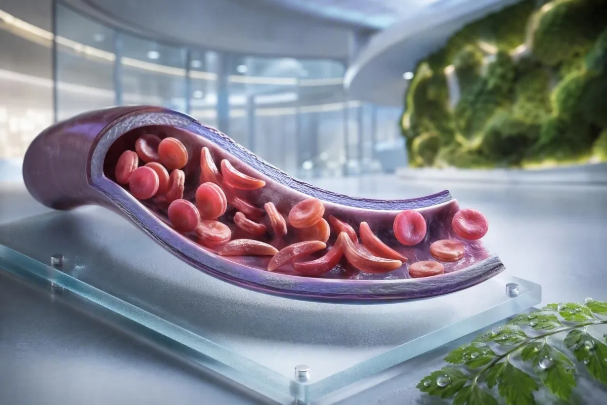
Anemia falciformă nu este un singur test, un singur simptom sau un singur nivel de urgență. Acest ghid centrat pe pacient separă statutul de purtător, boala confirmată, monitorizarea de rutină și situațiile care necesită îngrijiri de urgență.


- Trăsătura falciformă înseamnă o genă HbS; de obicei nu provoacă anemie cronică sau crize de durere recurente.
- Screening neonatal identifică majoritatea sugarilor afectați înainte de debutul simptomelor, permițând începerea precoce a penicilinei și a urmăririi de către un specialist.
- Hemoglobina electroforeză separă tipurile de hemoglobină și ajută la confirmarea HbSS, HbSC, HbS-beta talasemie sau a statutului de purtător.
- Durere toracică severă, dificultăți de respirație, febră de 38,5°C sau mai mare, confuzie sau o nouă slăbiciune necesită evaluare urgentă la urgență la o persoană cu boală falciformă.
- Sindrom toracic acut poate începe cu tuse, febră, durere toracică sau nivel scăzut de oxigen și se poate agrava în decurs de ore, mai degrabă decât zile.
- O scădere bruscă a hemoglobinei de 2 g/dL sau mai mult oare de la nivelul obișnuit al unei persoane pot semnala criză aplastică, sechestrare, sângerare sau hemoliză accelerată.
- Numărul de reticulocite ajută la diferențierea răspunsului activ al măduvei de criza aplastică; un număr scăzut în timpul anemiei în agravare este un model de avertizare.
- Hidroxiuree este un medicament care modifică boala, nu doar un medicament pentru durere; monitorizarea include în mod obișnuit un hemoleucogramă completă la fiecare 4 săptămâni în timpul ajustării dozei.
Ce înseamnă anemia falciformă – și ce nu înseamnă
Anemia falciformă se referă de obicei la boala HbSS, în care o persoană moștenește două gene care produc hemoglobină S; celulele roșii pot deveni rigide, se pot descompune prematur și pot obstrucționa vasele mici. Trait-ul falciform este diferit: o genă HbS este de obicei suficientă pentru a fi transmisă genetic, dar nu suficientă pentru a cauza anemia cronică și episoadele recurente vaso-ocluzive observate în HbSS.
Boala falciformă este un termen umbrelă care include HbSS, HbSC, talasemie HbS-beta-zero și talasemie HbS-beta-plus. HbSS cauzează adesea anemia cea mai pronunțată, dar severitatea variază enorm chiar și în cadrul aceleiași familii; o hemoglobină de bază de 7,5 g/dL poate fi stabilă pentru un adult și periculoasă pentru altul dacă a scăzut rapid.
Mutația hemoglobinei S modifică un aminoacid în beta-globina: valina înlocuiește acidul glutamic în poziția 6. Sub oxigen scăzut, deshidratare, acidoză sau febră, moleculele de HbS se pot polimeriza în interiorul celulei. Această modificare fizică explică de ce o criză se poate dezvolta după un zbor lung, o boală gastrointestinală sau o noapte cu aport insuficient de lichide.
Kantesti AI este un Analizor de teste de sânge AI care plasează CBC, bilirubina, LDH, reticulocitele, creatinina și rezultatele anterioare într-o singură vizualizare longitudinală; nu diagnostichează boala falciformă doar dintr-un CBC. Pentru o reîmprospătare practică a raportării hemoglobinei, consultați ce înseamnă HGB.
La data de 4 septembrie 2026, încă văd pacienți cărora li se spune greșit că trait-ul și boala sunt interschimbabile. Nu sunt. Regula clinică a Dr. Thomas Klein este simplă: solicitați genotipul exact al hemoglobinei, nu o afirmație vagă că un test a fost “pozitiv”.”
De ce genotipul schimbă îngrijirea
HbSC poate avea o hemoglobină mai mare decât HbSS, dar totuși poate cauza complicații retiniene, durere sau necroză avasculară. Talasemia HbS-beta poate semăna cu trait-ul sau HbSS, în funcție de prezența parțială a producției de beta-globina, motiv pentru care testarea moleculară stabilește uneori un model de electroforeză ambiguu.
Simptomele bolii falciforme în funcție de vârstă și de nivelul de bază
Simptomele bolii falciforme includ în mod obișnuit dureri episodice osoase sau toracice, oboseală, icter, dificultăți de respirație, creștere întârziată și infecții frecvente, dar nu există două persoane care să experimenteze același model. Simptomele apar de obicei după 4 până la 6 luni de vârstă, deoarece hemoglobina fetală protejează inițial sugarii de polimerizarea HbS.
Dactilita—umflarea dureroasă a mâinilor sau picioarelor—poate fi primul semn vizibil la sugari. La copiii de vârstă școlară, durerea abdominală necesită o atenție deosebită, deoarece constipația, bolile vezicii biliare, mărirea splinei și vaso-ocluzia pot fi surprinzător de similare acasă.
Icterul în boala falciformă reflectă descompunerea continuă a globulelor roșii și creșterea bilirubinei indirecte; nu înseamnă automat hepatită. Urina închisă la culoare, scaunele palide, mâncărimea, febra sau durerea abdominală în cuadrantul superior drept modifică această evaluare și necesită o investigare a cauzelor hepatice și biliare; ghidul nostru pentru modele de bilirubină directă explică diferența.
Adulții pot normaliza durerea care a devenit treptat mai frecventă. În experiența mea, o schimbare de la două episoade gestionate acasă anual la două episoade lunare este clinic semnificativă, chiar dacă vizitele la urgență nu au crescut. Tulburările de somn, munca ratată, simptomele erectile, starea de spirit scăzută și toleranța redusă la efort ar trebui documentate, deoarece adesea preced o schimbare formală a tratamentului.
O concentrație a hemoglobinei de 6 până la 9 g/dL poate fi o gamă normală de stare stabilă în HbSS, în timp ce o valoare sub 10 g/dL ar fi neașteptată la majoritatea persoanelor fără boală. Numărul contează, dar schimbarea față de linia de bază personală contează mai mult.
Cine ar trebui să ia în considerare screening-ul pentru purtătorii de boală falciformă
Screeningul pentru boala falciformă este rezonabil înainte de sarcină sau la începutul sarcinii pentru oricine al cărui statut de purtător este necunoscut, indiferent de aspect, nume de familie sau ascendent presupus. Statutul de purtător este moștenit și adesea rămâne necunoscut până la un screening neonatal sau un test de hemoglobină neașteptat de anormal.
Dacă ambii părinți biologici sunt purtători ai unei variante legate de HbS sau beta-talasemie, fiecare sarcină poate avea o șansă de 25% de a moșteni o afecțiune a hemoglobinei clinic semnificativă, în funcție de variantele implicate. Un purtător poate avea energie normală, rezultate CBC normale și fără antecedente de episoade dureroase.
Screeningul este deosebit de util atunci când un partener are HbAS, trăsătură HbC, trăsătură beta-talasemie, microcitoză inexplicabilă sau antecedente familiale de boală falciformă. Un volum corpuscular mediu scăzut nu ar trebui pus pe seama deficienței de fier fără verificarea feritinei și luarea în considerare a talasemiei; comparați Indicii MCV și MCH.
Un raport global din 2022 al Organizației Mondiale a Sănătății a estimat că tulburările hemoglobinei rămân o povară majoră a bolilor ereditare, în special acolo unde accesul la screening neonatal este neuniform. Cea mai respectuoasă abordare este accesul universal la screening informat, mai degrabă decât utilizarea etniei ca gardian.
Kantesti este o platforma de interpretare analize sange AI care pot explica un rezultat HbS raportat în limbaj simplu, dar confirmarea purtătorului și consilierea reproductivă necesită un laborator acreditat și un clinician. Dacă un cuplu plănuiește o sarcină, întrebați dacă testarea partenerului și consilierea genetică pot avea loc înainte de concepție, mai degrabă decât după ce un rezultat devine sensibil la timp.
Cum confirmă electroforeza hemoglobinei diagnosticul
Electraforeza hemoglobinei separă fracțiunile de hemoglobină și este un test de bază pentru confirmarea anemiei falciforme sau a trăsăturii falciforme, adesea alături de cromatografia lichidă de înaltă performanță și, atunci când este necesar, testarea genetică. Un CBC de rutină nu poate confirma HbSS sau trăsătura falciformă, deoarece deficiența de fier și talasemia pot produce indici eritrocitari suprapuși.
În HbSS netratată, electroforeza arată de obicei predominant HbS, fără HbA, deși procentul de HbF variază. HbAS arată de obicei atât HbA, cât și HbS, adesea cu HbA mai mare decât HbS; procentele exacte pot fluctua cu alfa-talasemia co-moștenită sau transfuzii recente.
Transfuzia recentă poate face un rezultat al electroforezei înșelător timp de aproximativ 3 luni, deoarece HbA de la donator încă circulă. Acesta este și motivul pentru care HbA1c poate subestima glucoza medie după transfuzie sau în caz de hemoliză cu turnover ridicat; vezi când HbA1c induce în eroare.
Un frotiu periferic poate arăta celule țintă, policromazie și forme falciforme, dar microscopia susține mai degrabă decât înlocuiește testarea fracțiunilor. Dr. Klein a văzut frotiuri de urgență “normale” care au întârziat clarificarea atunci când un pacient fusese transfuzat cu săptămâni mai devreme - spuneți întotdeauna laboratorului și echipei de hematologie despre transfuzii.
Metodele de screening neonatal identifică HbS înainte de boala clinică, dar rezultatul necesită totuși o testare confirmativă în timp util. Ghidul bazat pe dovezi din 2014 realizat de Yawn et al. recomandă implicarea specialiștilor la începutul vieții, cu măsuri preventive începând înainte de prima complicație majoră (Yawn et al., 2014).
Monitorizarea de rutină a sângelui: ce urmăresc de fapt clinicienii
Monitorizarea de rutină în boala cu celule în seceră urmărește de obicei hemoglobina, reticulocitele, leucocitele, trombocitele, bilirubina, LDH, funcția renală și proteina urinară — nu un rezultat izolat. Cea mai sigură comparație este cu nivelul de bază stabil al pacientului, ideal recoltat atunci când acesta este bine și nu a fost transfuzat recent.
Hemoliza produce adesea LDH, bilirubină indirectă și reticulocite crescute, cu haptoglobină scăzută. Haptoglobina poate fi scăzută din alte motive, inclusiv boli hepatice, astfel încât modelul combinat este mai puternic decât orice marker individual; interpretare haptoglobină arată de ce.
Un procentaj de reticulocite singur poate supraestima răspunsul măduvei osoase în anemia semnificativă. Clinicienii calculează adesea un număr absolut de reticulocite sau un răspuns absolut de reticulocite corectat; o scădere a numărului absolut în timpul anemiei în agravare ridică îngrijorări pentru o criză aplastică asociată cu parvovirusul B19.
Kantesti este o Instrument de analiză a analizelor de sânge bazat pe AI care compară rezultatele repetate ale hematologiei, mai degrabă decât să trateze un LDH ridicat ca un diagnostic de sine stătător. O probă marcată ca hemolizată în eprubetă poate crește fals potasiul și LDH, astfel încât un rezultat surprinzător poate necesita o reîncercare; revizuire testare repetată după hemoliză.
Hidroxiureea crește în mod obișnuit MCV și HbF în timp, scăzând modest neutrofilele la o doză eficientă. Aceste modificări pot fi așteptate, dar un număr absolut de neutrofile sub pragul individual de tratament sau o scădere rapidă a numărului de trombocite necesită revizuirea prescriitorului, mai degrabă decât ajustarea automată a medicamentelor.
Când durerea este un episod vaso-ocluziv – și când nu este
Durerea vaso-ocluzivă este adesea profundă, severă și localizată la nivelul spatelui, toracelui, șoldurilor, membrelor sau abdomenului, dar durerea nouă nu trebuie etichetată automat ca o criză de seceră. Febra, umflarea focală, leziuni, apărare abdominală sau simptome neurologice unilaterale indică probleme care necesită o investigație diferită.
Un plan de management al durerii la domiciliu include de obicei hidratare precoce, căldură, analgezie prescrisă, repaus și un prag personal de escaladare convenit cu echipa de seceră. Consumul excesiv de lichide nu este mai sigur: persoanele cu insuficiență renală sau boli de inimă pot dezvolta supraîncărcare volemică, în special în timpul unei boli acute.
Durerea care rămâne necontrolată după planul obișnuit de salvare, împiedică reținerea lichidelor sau a medicamentelor sau este diferită ca calitate sau localizare ar trebui să declanșeze sfatul clinic urgent. O articulație umflată și caldă poate reprezenta artrită septică sau infecție osoasă, mai degrabă decât o vaso-ocluzie necomplicată; ghid de testare pentru durere inexplicabilă prezintă investigațiile inițiale utile.
În asistența de urgență, analgezia promptă și reevaluarea contează mai mult decât dovedirea severității durerii printr-un număr de laborator. Ghidul NICE privind episoadele dureroase acute de seceră recomandă evaluarea rapidă a analgeziei și revizuirea frecventă, deoarece subtratamentul contribuie în sine la suferință prelungită și recuperare întârziată (NICE, 2012).
Evitați compresele reci direct pe o zonă de durere vaso-ocluzivă, cu excepția cazului în care echipa dumneavoastră le recomandă în mod specific; frigul poate constrictă vasele periferice. Acest detaliu practic pare minor, dar mulți pacienți îmi spun că nimeni nu le-a explicat până după mai multe episoade dificile.
Sindromul toracic acut: urgența pe care mulți o ratează
Sindromul toracic acut este un nou infiltrat pulmonar la radiografia toracică, plus simptome respiratorii, febră, dureri toracice sau oxigen scăzut la o persoană cu boală cu celule în seceră și este o urgență. Poate progresa rapid, uneori după internarea pentru un episod de durere, mai degrabă decât în momentul în care încep simptomele toracice.
Apelați serviciile de urgență sau mergeți la urgențe pentru dispnee nouă, dureri toracice, buze albăstrui-cenușii, leșin, tuse persistentă cu febră sau saturație de oxigen sub ținta prescrisă a persoanei. O saturație de 92% poate fi o scădere majoră pentru cineva normal la 98%, în timp ce unii pacienți au un nivel de bază documentat mai scăzut; atât valoarea absolută, cât și modificarea contează.
Sindromul toracic acut poate implica infecție, embolie grasă din măduva osoasă, obstrucție vasculară pulmonară, atelectazie sau mai multe procese simultan. Clinicienii evaluează în mod obișnuit oxigenarea, CBC, reticulocitele, culturile când sunt febrile, imagistica toracică și uneori gazele sanguine; analize de sânge pentru dificultăți de respirație adaugă context acestor rezultate.
La adulți, suprasedarea de la opioide, respirația superficială din cauza durerii și excesul de lichid intravenos pot agrava starea respiratorie. Spirometria de stimulare în timpul spitalizării reduce complicațiile pulmonare în episoade dureroase selectate, deși nu este un substitut pentru evaluarea urgentă atunci când simptomele au început deja.
Revizuirea din 2018 realizată de Kato și colegii descrie sindromul toracic acut ca o cauză majoră de morbiditate și mortalitate pe tot parcursul vieții (Kato et al., 2018). Nu conduceți singur dacă dificultatea de respirație, somnolența sau presiunea toracică sunt semnificative.
Urgențe legate de febră, infecție și splină
O temperatură de 38,5°C (101,3°F) sau mai mare la un copil sau adult cu boală falciformă necesită o evaluare medicală urgentă în aceeași zi, cu excepția cazului în care echipa lor specialistă a stabilit un alt plan. Pierderea funcției imune splenice poate face ca unele infecții bacteriene să progreseze mult mai rapid decât o febră virală obișnuită.
Febră plus letargie, erupție cutanată, tensiune arterială scăzută, confuzie sau scăderea producției de urină necesită îngrijiri de urgență acum. Un număr normal de leucocite nu exclude în mod fiabil infecția gravă în boala falciformă, în special la începutul bolii sau în timpul tratamentului cu hidroxicarbamide.
Sechestrarea splenică apare cel mai frecvent la copiii mici și provoacă mărirea bruscă a splinei, paloare, plenitudine abdominală, respirație rapidă și o scădere rapidă a hemoglobinei. Părinții sunt uneori învățați cum să palpeze splina copilului lor, dar orice mărire nouă asociată cu boala ar trebui să declanșeze o evaluare urgentă, nu verificări repetate la domiciliu.
O scădere a hemoglobinei de 2 g/dL sau mai mult sub nivelul de bază obișnuit, asociată cu o splină mărită, este îngrijorătoare pentru sechestrare. În schimb, anemia severă cu un număr foarte scăzut de reticulocite poate sugera o criză aplastică, frecvent legată de parvovirusul B19; interpretarea numărului de reticulocite explică semnalul măduvei osoase.
Laboratoarele pentru sepsis pot include lactat, culturi, CBC, teste renale și markeri inflamatori, dar deciziile de tratament nu ar trebui să aștepte niciodată ca toate valorile să revină. Pentru context despre modelele de urgență, citiți indicii markerilor de sepsis.
Vaccinuri și antibiotice preventive
Vaccinurile și profilaxia cu penicilină la copii reduc riscul, dar nu fac febra un risc scăzut. Confirmați planul dumneavoastră individualizat cu o echipă de hematologie sau pediatrie, în special după splenectomie, călătorii, un nou-născut sau o modificare a programului local de imunizare.
Accidentul vascular cerebral și semnele de avertizare neurologică necesită acțiune imediată
Orice cădere bruscă a feței, slăbiciune la nivelul brațului sau piciorului, dificultate de vorbire, convulsie, cefalee severă neobișnuită, confuzie sau pierderea echilibrului în boala falciformă trebuie tratate ca o posibilă urgență de accident vascular cerebral. Sunați imediat la serviciile de urgență; nu așteptați să vedeți dacă simptomele trec sau administrați mai întâi medicamente împotriva durerii.
Copiii cu HbSS sau talasemie HbS-beta-zero li se oferă frecvent screening prin ecografie Doppler transcraniană de la 2 la 16 ani. O viteză maximă medie crono-medie anormală de 200 cm/secundă sau mai mare identifică un risc substanțial crescut de accident vascular cerebral și necesită acțiune promptă din partea specialistului.
Terapia cu transfuzii cronice poate reduce riscul de prim accident vascular cerebral la copiii cu rezultate Doppler anormale, dar creează nevoi suplimentare: monitorizarea supraîncărcării cu fier, screening pentru anticorpi și potrivirea atentă a celulelor donatorului. Aceasta este o îngrijire specializată, nu un rezultat de gestionat printr-o aplicație.
Adulții pot prezenta simptome neurologice tranzitorii, modificări cognitive sau leziuni cerebrale silențioase fără un accident vascular cerebral dramatic clasic. Un episod de 10 minute de dificultate în găsirea cuvintelor necesită în continuare evaluare de urgență, deoarece simptomele ischemice tranzitorii pot fi un avertisment, nu o reasigurare.
Dr. Thomas Klein sfătuiește familiile să salveze numărul local de contact pentru urgențe falciforme lângă planul obișnuit pentru durere. Acesta elimină o decizie în cele câteva minute în care un copil sau adult poate lupta să comunice.
Protejarea rinichilor, ochilor și plămânilor între crize
Boala falciformă poate afecta rinichii, retina, plămânii și inima chiar și atunci când durerea este rară, astfel încât supravegherea de rutină a organelor face parte din tratament, mai degrabă decât un extras opțional. Testarea albuminuriei urinare, verificarea tensiunii arteriale, examinarea ochilor și evaluarea pulmonară ghidată de simptome identifică complicațiile înainte ca acestea să devină evidente.
Albuminuria poate fi un semn precoce de nefro-patie falciformă. Un raport albumină urinară-creatinină de 30 mg/g sau mai mare este anormal la majoritatea adulților și ar trebui confirmat prin testare repetată, deoarece febra, exercițiul fizic, menstruația și deshidratarea îl pot crește tranzitoriu.
Creatinina poate părea înșelător normală în boala falciformă, deoarece secreția tubulară crescută și masa musculară redusă pot masca filtrarea redusă. Cistatina C sau ecuațiile combinate pot adăuga context util, în timp ce urina spumoasă persistentă merită evaluare; vezi ghidul privind proteinele în urină.
Retinopatia este deosebit de importantă în boala HbSC, care poate fi clinic mai puțin manifestă în alte privințe. Apariția unor noi „flotoare”, flash-uri, o modificare vizuală asemănătoare unei perdele sau o vedere încețoșată bruscă necesită o examinare oftalmologică urgentă, nu o programare de rutină peste câteva săptămâni.
Sarcina adaugă hiperfiltrarea fiziologică și un risc maternal-fetal mai mare, astfel încât evaluarea renală și urinară de bază ar trebui să aibă loc devreme. Explicația noastră privind modificările GFR în sarcină ajută la diferențierea modificărilor așteptate de o tendință îngrijorătoare.
Opțiuni de tratament, hidroxidarea și siguranța transfuziilor
Hidroxiureea reduce episoadele dureroase și sindromul toracic acut la multe persoane cu HbSS sau talasemie HbS-beta-zero prin creșterea hemoglobinei fetale, dar necesită monitorizare structurată. Transfuzia poate salva vieți în cazul unor complicații selectate, însă transfuziile repetate prezintă riscuri de suprasolicitare cu fier și anticorpi împotriva globulelor roșii.
Hidroxiureea este de obicei ajustată cu un CBC și un număr de reticulocite aproximativ la fiecare 4 săptămâni până la atingerea unei doze stabile, apoi la intervale mai lungi determinate de echipa curantă. O valoare mai mare a MCV este adesea un efect așteptat al tratamentului, în timp ce citopenia severă necesită revizuirea medicației și, uneori, o pauză temporară.
Transfuzia simplă crește capacitatea de transport a oxigenului, dar o concentrație excesivă de hemoglobină poate crește vâscozitatea în HbSS. Multe protocoale acute evită creșterea hemoglobinei post-transfuzie mult peste 10 g/dL, cu excepția cazului în care un specialist stabilește un alt obiectiv; transfuzia de schimb este utilizată pentru complicații severe selectate.
Suprasolicitarea cu fier devine mai probabilă după transfuzii repetate și este evaluată prin tendințele feritinei, plus RMN hepatic sau cardiac, atunci când este cazul. Feritina crește odată cu inflamația, astfel încât o singură valoare mare nu este o dovadă a încărcăturii de fier tisulare; ghid pentru studii despre fier explică limitele.
Dovada este cea mai puternică pentru hidroxiureea în boala HbSS și HbS-beta-zero; beneficiul în HbSC rămâne o zonă cu mai puțină certitudine și cu luare a deciziilor mai individualizată. Un plan de medicație ar trebui să includă discuții despre contracepție și sarcină, acolo unde este relevant, bariere de aderență și un plan scris pentru dozele omise.
Sarcină, fertilitate și planificare familială cu HbS
Sarcina cu boală de celule falciforme necesită îngrijire comună timpurie din partea obstetricei și hematologiei, deoarece riscurile episoadelor dureroase, anemiei, trombozei, infecției, preeclampsiei, restricției de creștere fetală și nașterii premature sunt mai mari. Screeningul purtătorilor este important înainte de sarcină, deoarece riscul genetic depinde de ambii părinți biologici.
O vizită pre-sarcină ar trebui să includă genotipul, hemoglobina de bază, funcția renală, albumina urinară, tensiunea arterială, istoricul transfuziilor, anticorpii globulelor roșii, vaccinarea și medicamentele. Deciziile privind hidroxiureea în sarcină sunt individualizate și trebuie luate împreună cu specialiștii; nu o întrerupeți sau nu o reluați bazându-vă doar pe sfaturi de pe internet.
Fierul nu trebuie prescris automat pentru o hemoglobină scăzută în HbSS. Deficiența de fier poate coexista, în special în sarcină sau în cazul sângerărilor menstruale abundente, dar feritina și saturația transferinei ar trebui să ghideze tratamentul, deoarece hemoliza cronică singură nu echivalează cu deficiența de fier.
Atunci când ambii părinți sunt purtători ai unor variante relevante de hemoglobină, consilierea genetică poate explica concepția naturală, opțiunile de diagnostic prenatal, opțiunile de donare și testarea genetică preimplantare, fără a direcționa o familie către o anumită decizie. Scopul este alegerea informată, nu alarma.
Pentru alte semnale de alarmă practice de laborator în sarcină, consultați preocupări de laborator pentru sarcină în aceeași zi. Valorile de bază cunoscute ale pacientului trebuie incluse în fiecare predare către maternitate; aceasta modifică modul în care este interpretat un rezultat al hemoglobinei.
Utilizarea în siguranță a rezultatelor de laborator între consultații
Un rezultat de laborator al celulelor falciforme este cel mai util atunci când este comparat cu genotipul dumneavoastră, valorile de bază stabile, simptomele, tratamentele și transfuziile recente. O bilirubină crescută sau o hemoglobină scăzută pot fi așteptate la o persoană cu HbSS, dar pot fi urgente pentru aceeași persoană dacă rezultatul se modifică brusc.
Păstrați un registru pe o singură pagină cu genotipul, hemoglobina obișnuită, saturația obișnuită de oxigen, dacă este cunoscută, grupul sanguin și anticorpii, medicamentele curente, datele transfuziilor și contactele de urgență. Acest lucru economisește timp în departamentele de urgență necunoscute și reduce șansa ca valorile de bază ale HbSS să fie confundate cu o anemie nou descoperită.
Kantesti este o serviciul de interpretare a testelor de laborator AI care pot organiza rapoartele de laborator încărcate și pot semnala modele pentru discuții, inclusiv markeri de hemoliză, tendințe renale și monitorizarea medicației. Nu poate vedea pieptul, măsura oxigenarea sau înlocui asistența de urgență atunci când simptomele indică sindrom toracic acut, sepsis sau accident vascular cerebral.
Un rezultat în afara intervalului de referință al unui laborator nu este automat periculos, iar un rezultat în interiorul acestuia poate fi totuși periculos dacă s-a modificat brusc. Pentru o explicație a indicatorilor de raportare și a intervalelor de referință, citiți semnificația rezultatelor în afara intervalului.
Abordarea clinică a Kantesti este revizuită în raport cu metodele și limitările definite; cititorii care doresc să înțeleagă măsurile noastre de siguranță pot consulta validare clinică. Aduceți PDF-ul original la consultația de hematologie – OCR este util, dar raportul sursă rămâne înregistrarea oficială.
Un plan de acțiune clar pentru urgențe în boala falciformă
Solicitați asistență medicală de urgență acum pentru dureri în piept sau dificultăți de respirație, febră de 38,5°C sau mai mare, simptome neurologice noi, leșin, paloare severă, durere necontrolată, abdomen cu creștere rapidă sau incapacitate de a bea și de a lua medicamente. Aceste semne de avertizare pot indica sindrom toracic acut, sepsis, accident vascular cerebral, anemie severă sau sechestrare splenică.
Spuneți personalului de urgență că aveți boala falciformă, menționați genotipul dacă este cunoscut și descrieți ce este diferit față de episodul dumneavoastră obișnuit. Menționați transfuzia recentă, hemoglobina de bază, medicamentele pentru durere deja administrate cu doze și ore, alergiile, sarcina și orice sindrom toracic acut sau accident vascular cerebral anterior.
Nu așteptați ca un pulsoximetru de la domiciliu să devină anormal dacă simptomele toracice se agravează. Dispozitivele de consum pot citi greșit în cazul degetelor reci, a produselor pentru unghii, a mișcării, a circulației deficitare și a unor nuanțe ale pielii; simptomele și evaluarea clinică primează asupra unui afișaj liniștitor.
Dacă aveți grijă de un copil, cereți echipei sale un prag de febră scris, un plan de analgezie și instrucțiuni pentru verificarea splinei. Dacă sunteți adult, păstrați o copie în telefon și împărtășiți-o cu o persoană de încredere; planificarea dosarului medical familial poate facilita acest lucru.
Echipa medicală a Kantesti, inclusiv Consiliul consultativ medical, sprijină educația în ceea ce privește interpretarea de laborator, în timp ce deciziile de urgență aparțin serviciilor clinice în persoană. Despre organizația noastră și misiunea clinică, consultați echipa și abordarea noastră.
Întrebări frecvente
Care sunt primele simptome ale anemiei cu celule în seceră la bebeluși?
Primele simptome ale bolii celulelor în seceră apar adesea după 4-6 luni de viață, când hemoglobina fetală scade în mod natural. Umflarea dureroasă a mâinilor și picioarelor, numită dactilită, febră, iritabilitate neobișnuită, paloare, icter, alimentație slabă și un abdomen umflat sunt semne de avertizare timpurii comune. O febră de 38,5°C (101,3°F) sau mai mare la un sugar cu boală a celulelor în seceră necesită o evaluare medicală urgentă în aceeași zi, deoarece infecția gravă poate progresa rapid. Screeningul neonatal identifică sugarii afectați înainte de apariția simptomelor, dar testarea confirmativă și urmărirea de către un specialist rămân necesare.
Este posibil să aveți trăsătura celulelor în seceră și să aveți totuși simptome?
Majoritatea persoanelor cu trăsătură siclemică nu au anemie cronică, crize de durere recurente și toleranță normală la efortul zilnic. Complicații rare pot apărea în caz de deshidratare extremă, expunere severă la căldură, altitudine mare, efort foarte intens sau medii cu oxigen scăzut, iar sângele din urină trebuie evaluat, nu presupus a fi benign. Trăsătura înseamnă o genă HbS, în timp ce anemia siclemică înseamnă de obicei boala HbSS cu două gene beta-globina afectate. Electroforeza hemoglobinei sau un test de fracționare echivalent poate clarifica distincția.
Ce test confirmă anemia falciformă?
Electroforeza hemoglobinei, cromatografia lichidă de înaltă performanță sau electroforeza capilară confirmă fracțiile de hemoglobină prezente și este esențială pentru diagnosticarea anemiei falciforme. HbSS netratată arată în mod obișnuit predominant hemoglobină S, fără hemoglobină A, în timp ce purtătorii prezintă de obicei atât hemoglobină A, cât și hemoglobină S. Un CBC poate identifica anemie sau globule roșii mici, dar nu poate diagnostica independent HbSS, HbSC sau purtătorii. Transfuzia recentă poate distorsiona rezultatele fracționării timp de aproximativ 3 luni, deci testarea moleculară poate fi necesară atunci când modelul nu este clar.
Când ar trebui ca o persoană cu siclemie să meargă la camera de urgență?
O persoană cu boală falciformă ar trebui să solicite evaluare de urgență pentru febră de 38,5°C (101,3°F) sau mai mare, durere toracică, dificultăți de respirație, hipoxie, slăbiciune bruscă, dificultăți de vorbire, convulsii, leșin, paloare cu agravare rapidă, umflare abdominală severă sau durere care nu este controlată cu planul stabilit. Aceste simptome pot semnala sindrom toracic acut, infecție, accident vascular cerebral, sechestrare splenică sau o scădere bruscă a hemoglobinei. Durerea nouă sau diferită necesită, de asemenea, evaluare, deoarece infecția osoasă, bolile biliare, apendicita și cheagurile pot mima durerea vaso-ocluzivă. Nu conduceți singur dacă simptomele neurologice sau respiratorii sunt semnificative.
Ce nivel de hemoglobină este periculos în boala celulelor securii?
Nu există un număr unic de hemoglobină periculos în boala siclemică, deoarece multe persoane cu HbSS au o valoare de bază stabilă între aproximativ 6 și 9 g/dL. O scădere de 2 g/dL sau mai mult față de hemoglobina obișnuită a individului este adesea mai îngrijorătoare decât valoarea absolută și poate sugera criză aplastică, sechestrare splenică, sângerare sau hemoliză accelerată. Simptomele severe, cum ar fi dificultatea de respirație, leșinul, durerea toracică, bătăile rapide ale inimii sau confuzia, necesită evaluare urgentă la orice nivel de hemoglobină. Deciziile de transfuzie depind de simptome, valoarea de bază, cauza anemiei, oxigenare și riscul de hipervâscozitate.
Hidroxiureea vindecă anemia falciformă?
Hidroxiureea nu vindecă anemia falciformă, dar poate reduce episoadele de durere, sindromul toracic acut, necesitatea transfuziilor și spitalizările pentru multe persoane cu HbSS sau talasemie HbS-beta-zero. Acționează parțial prin creșterea hemoglobinei fetale și, de obicei, necesită monitorizarea CBC și a reticulocitelor la aproximativ 4 săptămâni, în timp ce doza este ajustată. O creștere a MCV și a nivelului de HbF poate indica un efect biologic, deși niciun rezultat nu dovedește o aderență perfectă. Abordările curative, cum ar fi transplantul de celule stem și unele terapii bazate pe gene, necesită o evaluare a eligibilității foarte specializată și un follow-up pe termen lung.
Obține astăzi analiză de sânge cu AI
Alătură-te a peste 2 milioane de utilizatori din întreaga lume care au încredere în Kantesti pentru analiza instantanee și precisă a analizelor de laborator. Încarcă rezultatele analizelor tale de sânge și primește o interpretare completă a biomarkerilor 15,000+ în câteva secunde.
📚 Publicații de cercetare citate
Klein, T., Mitchell, S., & Weber, H. (2026). Ghid pentru testul de sânge complement C3 și C4 & titrul ANA. Kantesti AI Medical Research.
Klein, T., Mitchell, S., & Weber, H. (2026). Test de sânge pentru virusul Nipah: Ghid de detectare și diagnostic precoce 2026. Kantesti AI Medical Research.
📖 Referințe medicale externe
⚕️ Declarație medicală
Acest articol are doar scop educațional și nu constituie sfat medical. Consultă întotdeauna un furnizor calificat de servicii medicale pentru decizii privind diagnosticarea și tratamentul.
Semnale de încredere E-E-A-T
Experienţă
Revizuire clinică condusă de medici a fluxurilor de lucru pentru interpretarea analizelor.
Expertiză
Focalizare pe medicina de laborator asupra modului în care biomarkerii se comportă în context clinic.
Autoritate
Scris de dr. Thomas Klein, cu revizuire de dr. Sarah Mitchell și prof. dr. Hans Weber.
Încredere
Interpretare bazată pe dovezi, cu căi clare de urmărire pentru a reduce alarmele.