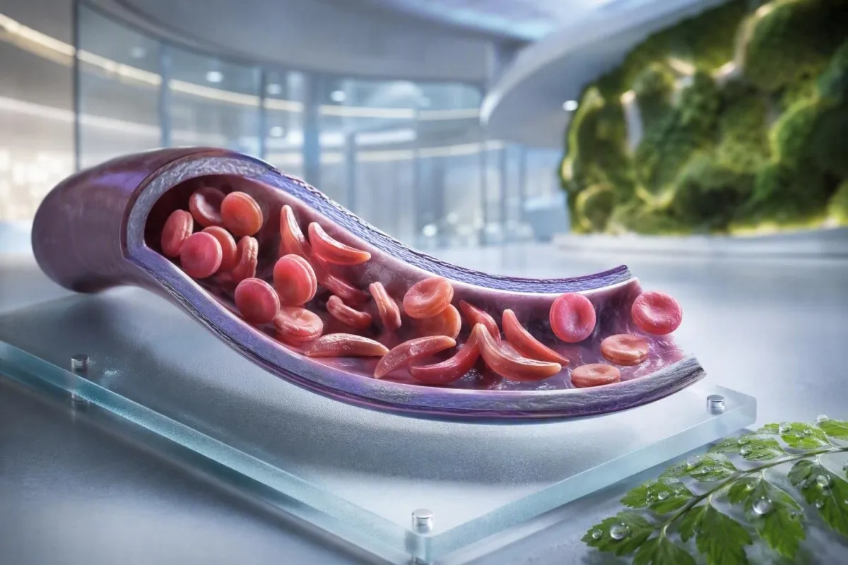
L'anemia falciforme non è un singolo test, un singolo sintomo o un singolo livello di urgenza. Questa guida incentrata sul paziente distingue tra stato di portatore, malattia conclamata, monitoraggio di routine e situazioni che richiedono cure d'emergenza.


- Tratto falciforme significa un gene HbS; di solito non causa anemia cronica o crisi dolorose ricorrenti.
- Screening neonatale identifica la maggior parte dei neonati affetti prima dell'insorgenza dei sintomi, consentendo un inizio precoce della penicillina e del follow-up specialistico.
- Elettroforesi dell’emoglobina separa i tipi di emoglobina e aiuta a confermare HbSS, HbSC, HbS-beta talassemia o stato di portatore.
- Dolore toracico severo, respiro affannoso, febbre di 38,5°C o superiore, confusione o una nuova debolezza richiedono un'urgente valutazione d'emergenza in una persona con malattia falciforme.
- Sindrome toracica acuta può iniziare con tosse, febbre, dolore toracico o bassa ossigenazione e può peggiorare nel corso di ore anziché giorni.
- Una caduta improvvisa dell'emoglobina di 2 g/dL o più da un livello abituale di una persona può segnalare crisi aplastica, sequestro, sanguinamento o emolisi accelerata.
- Conta dei reticolociti aiuta a distinguere una risposta attiva del midollo da una crisi aplastica; una bassa conta durante un peggioramento dell'anemia è un pattern di allarme.
- Idrossiurea è un farmaco modificante la malattia, non semplicemente un antidolorifico; il monitoraggio include comunemente un emocromo completo ogni 4 settimane durante l'aggiustamento della dose.
Cosa significa l'anemia falciforme — e cosa non significa
L'anemia falciforme si riferisce solitamente alla malattia HbSS, in cui una persona eredita due geni che producono emoglobina S; i globuli rossi possono diventare rigidi, rompersi precocemente e ostruire i piccoli vasi. Il tratto falciforme è diverso: un gene HbS è solitamente sufficiente per essere trasmesso geneticamente, ma non sufficiente a causare l'anemia cronica e gli episodi vaso-occlusivi ricorrenti osservati nell'HbSS.
La malattia falciforme è un termine ombrello che include HbSS, HbSC, beta-zero talassemia HbS e beta-plus talassemia HbS. L'HbSS causa spesso l'anemia più pronunciata, ma la gravità varia enormemente anche all'interno della stessa famiglia; un'emoglobina di base di 7,5 g/dL può essere stabile per un adulto e pericolosa per un altro se è scesa rapidamente.
La mutazione dell'emoglobina S modifica un amminoacido nella beta-globina: la valina sostituisce l'acido glutammico in posizione 6. Sotto ipossia, disidratazione, acidosi o febbre, le molecole di HbS possono polimerizzare all'interno della cellula. Quel cambiamento fisico spiega perché una crisi può svilupparsi dopo un lungo volo, una malattia gastrointestinale o una notte di insufficiente apporto di liquidi.
Kantesti AI è un Analizzatore di analisi del sangue AI che colloca CBC, bilirubina, LDH, reticolociti, creatinina e risultati precedenti in un'unica visione longitudinale; non diagnostica la malattia falciforme solo da un CBC. Per un ripasso pratico sulla rendicontazione dell'emoglobina, vedere cosa significa HGB.
Al 4 settembre 2026, vedo ancora pazienti a cui viene detto erroneamente che tratto e malattia sono intercambiabili. Non lo sono. La regola clinica del Dr. Thomas Klein è semplice: chiedere il genotipo esatto dell'emoglobina, non una dichiarazione vaga che un test è stato “positivo”.”
Perché il genotipo cambia la cura
L'HbSC può avere un'emoglobina più alta rispetto all'HbSS e causare comunque complicanze retiniche, dolore o necrosi avascolare. La beta talassemia HbS può assomigliare al tratto o all'HbSS a seconda che la produzione di beta-globina sia parzialmente presente, motivo per cui il test molecolare a volte risolve un quadro elettroforetico ambiguo.
Sintomi della malattia falciforme per età e baseline
I sintomi della malattia falciforme includono comunemente dolore osseo o toracico episodico, affaticamento, ittero, affanno, ritardo della crescita e infezioni frequenti, ma non ci sono due persone che sperimentano lo stesso pattern. I sintomi di solito emergono dopo 4-6 mesi di età perché l'emoglobina fetale protegge inizialmente i neonati dalla polimerizzazione dell'HbS.
La dattilite — gonfiore doloroso di mani o piedi — può essere il primo segno visibile nei neonati. Nei bambini in età scolare, il dolore addominale merita particolare attenzione perché la stitichezza, le malattie della colecisti, l'ingrossamento della milza e la vaso-occlusione possono sembrare sorprendentemente simili a casa.
L'ittero nella malattia falciforme riflette la continua rottura dei globuli rossi e l'aumento della bilirubina indiretta; non significa automaticamente epatite. Urine scure, feci pallide, prurito, febbre o dolore addominale in alto a destra modificano questa valutazione e richiedono una valutazione di cause epatiche e biliari; la nostra guida a pattern di bilirubina diretta spiega la distinzione.
Gli adulti possono normalizzare un dolore che è diventato gradualmente più frequente. Nella mia esperienza, un passaggio da due episodi gestiti a casa all'anno a due episodi al mese è clinicamente significativo anche se le visite al pronto soccorso non sono aumentate. Disturbi del sonno, lavoro perso, sintomi erettili, umore basso e ridotta tolleranza all'esercizio fisico dovrebbero essere documentati perché spesso precedono un cambiamento formale nel trattamento.
Una concentrazione di emoglobina da 6 a 9 g/dL può essere un intervallo di stato stazionario usuale nell'HbSS, mentre un valore inferiore a 10 g/dL sarebbe inaspettato nella maggior parte delle persone senza malattia. Il numero conta, ma il cambiamento rispetto alla baseline personale conta di più.
Chi dovrebbe considerare lo screening per il portatore di anemia falciforme
Lo screening per l'emoglobina falciforme è ragionevole prima della gravidanza o all'inizio della gravidanza per chiunque il cui stato di portatore sia sconosciuto, indipendentemente dall'aspetto, dal cognome o dall'ascendenza presunta. Lo stato di portatore è ereditario e spesso rimane sconosciuto fino a uno screening neonatale o a un test dell'emoglobina inaspettatamente anomalo.
Se entrambi i genitori biologici sono portatori di una variante correlata a HbS o beta-talassemia, ogni gravidanza può avere una probabilità del 25% di ereditare una condizione emoglobinica clinicamente significativa, a seconda delle varianti coinvolte. Un portatore può avere energia normale, risultati normali del CBC e nessuna storia di episodi di dolore.
Lo screening è particolarmente utile quando un partner ha HbAS, tratto HbC, tratto beta-talassemico, microcitosi inspiegata o una storia familiare di anemia falciforme. Un basso volume corpuscolare medio non dovrebbe essere attribuito a carenza di ferro senza controllare la ferritina e considerare la talassemia; confrontare indizi su MCV e MCH.
Un rapporto globale dell'Organizzazione Mondiale della Sanità del 2022 ha stimato che i disturbi dell'emoglobina rimangono un importante onere di malattie ereditarie, in particolare dove l'accesso allo screening neonatale è disomogeneo. L'approccio più rispettoso è l'accesso universale a uno screening informato piuttosto che utilizzare l'etnia come un filtro.
Kantesti è un Piattaforma di interpretazione esami del sangue AI che possono spiegare un risultato HbS riportato in linguaggio semplice, ma la conferma del portatore e la consulenza riproduttiva richiedono un laboratorio accreditato e un medico. Se una coppia sta pianificando una gravidanza, chieda se i test del partner e la consulenza genetica possano avvenire prima del concepimento piuttosto che dopo che un risultato diventi sensibile al tempo.
Come l'elettroforesi dell'emoglobina conferma la diagnosi
L'elettroforesi dell'emoglobina separa le frazioni dell'emoglobina ed è un test fondamentale per confermare l'anemia falciforme o il tratto falciforme, spesso insieme alla cromatografia liquida ad alta prestazione e, quando necessario, ai test genetici. Un CBC di routine non può confermare HbSS o il tratto falciforme perché la carenza di ferro e la talassemia possono produrre indici dei globuli rossi sovrapposti.
In HbSS non trattata, l'elettroforesi mostra tipicamente prevalentemente HbS senza HbA, sebbene la percentuale di HbF vari. HbAS di solito mostra sia HbA che HbS, spesso con HbA più alta di HbS; le percentuali esatte possono variare con la co-ereditarietà dell'alfa-talassemia o trasfusioni recenti.
Una trasfusione recente può rendere un risultato elettroforetico fuorviante per circa 3 mesi perché l'HbA del donatore è ancora in circolo. Questo è anche il motivo per cui l'HbA1c può sottovalutare la glicemia media dopo una trasfusione o in caso di emolisi ad alto turnover; vedere quando HbA1c inganna.
Uno striscio periferico può mostrare cellule bersaglio, policromasia e forme falciformi, ma la microscopia supporta piuttosto che sostituire i test di frazionamento. La Dott.ssa Klein ha visto “normali” strisci d'urgenza ritardare la chiarificazione quando un paziente era stato trasfuso settimane prima: informare sempre il laboratorio e il team di ematologia riguardo alle trasfusioni.
I metodi di screening neonatale identificano l'HbS prima della malattia clinica, ma il risultato necessita comunque di tempestivi test di conferma. La linea guida basata sull'evidenza del 2014 di Yawn et al. raccomanda il coinvolgimento specialistico in una fase precoce della vita, con misure preventive che iniziano prima della prima complicazione maggiore (Yawn et al., 2014).
Monitoraggio ematico di routine: cosa i medici effettivamente monitorano
Il monitoraggio di routine nella drepanocitosi solitamente segue l'andamento dell'emoglobina, dei reticolociti, dei globuli bianchi, delle piastrine, della bilirubina, dell'LDH, della funzionalità renale e delle proteine urinarie, non un singolo risultato isolato. Il confronto più sicuro è con la linea di base stabile del paziente, idealmente prelevata quando sta bene e non è stata trasfusa di recente.
L'emolisi spesso produce LDH elevati, bilirubina indiretta e reticolociti con bassa aptoglobina. L'aptoglobina può essere bassa per altri motivi, tra cui malattie epatiche, quindi il pattern combinato è più forte di qualsiasi singolo marcatore; il nostro guida all’interpretazione dell’aptoglobina mostra perché.
Una percentuale di reticolociti da sola può sovrastimare la risposta del midollo in caso di anemia significativa. I medici calcolano spesso un conteggio assoluto di reticolociti o una risposta corretta dei reticolociti; un conteggio assoluto in calo durante un peggioramento dell'anemia solleva preoccupazioni per la crisi aplastica associata al parvovirus B19.
Kantesti è un Strumento di analisi degli esami del sangue basato su AI che confronta i risultati ematologici ripetuti piuttosto che trattare un LDH elevato come diagnosi autonoma. Un campione segnalato come emolitico nella provetta può aumentare falsamente potassio e LDH, quindi un risultato sorprendente potrebbe richiedere un nuovo prelievo; revisione test ripetuti dopo emolisi.
L'idrossiurea aumenta comunemente MCV e HbF nel tempo, abbassando moderatamente i neutrofili a una dose efficace. Tali variazioni possono essere previste, ma un conteggio assoluto di neutrofili al di sotto della soglia di trattamento individuale o un rapido calo delle piastrine richiede la revisione dello specialista piuttosto che l'autoadjustment del farmaco.
Quando il dolore è un episodio vaso-occlusivo — e quando non lo è
Il dolore vaso-occlusivo è spesso profondo, severo e localizzato nella schiena, nel torace, nei fianchi, negli arti o nell'addome, ma un nuovo dolore non deve essere automaticamente etichettato come crisi falciforme. Febbre, gonfiore focale, lesioni, rigidità addominale o sintomi neurologici unilaterali indicano problemi che richiedono un work-up diverso.
Un piano domiciliare per il dolore include tipicamente idratazione precoce, calore, analgesia prescritta, riposo e una soglia di escalation personale concordata con il team di gestione della drepanocitosi. Bere volumi eccessivi non è più sicuro: le persone con compromissione renale o malattie cardiache possono sviluppare sovraccarico di liquidi, soprattutto durante una malattia acuta.
Il dolore che rimane incontrollato dopo il piano di salvataggio usuale, impedisce di trattenere liquidi o farmaci, o è diverso per qualità o localizzazione dovrebbe indurre una consulenza clinica urgente. Un'articolazione gonfia e calda può rappresentare un'artrite settica o un'infezione ossea piuttosto che un'incontrollata vaso-occlusione; la nostra guida ai test per il dolore inspiegabile delinea le prime indagini utili.
In pronto soccorso, la tempestiva analgesia e la rivalutazione sono più importanti che dimostrare la gravità del dolore attraverso un numero di laboratorio. La linea guida NICE sull'episodio doloroso acuto da falciformazione raccomanda una rapida valutazione analgesica e un frequente riesame, poiché il sottotrattamento stesso contribuisce a un disagio prolungato e a un ritardo nel recupero (NICE, 2012).
Evitare impacchi freddi direttamente su un'area di dolore vaso-occlusivo a meno che il team non li consigli specificamente; il freddo può restringere i vasi periferici. Questo dettaglio pratico sembra minore, ma molti pazienti mi dicono che nessuno glielo ha spiegato fino a dopo diversi episodi difficili.
Sindrome toracica acuta: l'emergenza che molti trascurano
La sindrome toracica acuta è un nuovo infiltrato polmonare all'imaging toracico più sintomi respiratori, febbre, dolore toracico o bassa ossigenazione in una persona con drepanocitosi, ed è un'emergenza. Può deteriorarsi rapidamente, a volte dopo il ricovero per un episodio di dolore piuttosto che al momento in cui iniziano i sintomi toracici.
Chiamare i servizi di emergenza o recarsi al pronto soccorso per nuova dispnea, dolore toracico, labbra blu-grigiastre, svenimento, tosse persistente con febbre o saturazione di ossigeno al di sotto del target prescritto. Una saturazione di 92% può essere un calo importante per qualcuno normalmente al 98%, mentre alcuni pazienti hanno una baseline documentata più bassa; sia il valore assoluto che la variazione sono importanti.
La sindrome toracica acuta può coinvolgere infezioni, embolia grassosa dal midollo osseo, ostruzione vascolare polmonare, atelettasia o diversi processi contemporaneamente. I medici valutano comunemente ossigenazione, CBC, reticolociti, colture quando febbrili, imaging toracico e talvolta emogasanalisi; esami del sangue per l'affanno aggiunge contesto a questi risultati.
Negli adulti, l'eccessiva sedazione da oppioidi, la respirazione superficiale dovuta al dolore e l'eccesso di liquidi endovenosi possono peggiorare lo stato respiratorio. La spirometria incentivata durante il ricovero riduce le complicanze polmonari in episodi dolorosi selezionati, sebbene non sia un sostituto della valutazione urgente quando i sintomi sono già iniziati.
La revisione del 2018 di Kato e colleghi descrive la sindrome toracica acuta come una delle principali cause di morbilità e mortalità nell'arco della vita (Kato et al., 2018). Non guidare se la mancanza di respiro, la sonnolenza o la pressione toracica sono significative.
Febbre, infezione ed emergenze spleniche
Una temperatura di 38,5°C (101,3°F) o superiore in un bambino o adulto con anemia falciforme necessita di una valutazione medica urgente nello stesso giorno, a meno che il loro team specialistico non abbia fornito un piano diverso. La perdita funzionale della funzione immunitaria splenica può far progredire alcune infezioni batteriche molto più velocemente di una comune febbre virale.
Febbre, letargia, eruzione cutanea, ipotensione, confusione o ridotta produzione di urina richiedono cure di emergenza immediate. Un conteggio dei globuli bianchi dall'aspetto normale non esclude in modo affidabile un'infezione grave nell'anemia falciforme, specialmente all'inizio della malattia o durante il trattamento con idrossiurea.
Il sequestro splenico si verifica più frequentemente nei bambini piccoli e causa un improvviso ingrossamento della milza, pallore, pienezza addominale, respiro affannoso e un rapido calo dell'emoglobina. Ai genitori viene talvolta insegnato come palpare la milza del proprio figlio, ma qualsiasi nuovo ingrossamento associato a una malattia dovrebbe innescare una valutazione urgente piuttosto che controlli domestici ripetuti.
Un calo dell'emoglobina di 2 g/dL o più rispetto al valore di base abituale con una milza ingrossata è preoccupante per il sequestro. Al contrario, un'anemia grave con un conteggio molto basso di reticolociti può suggerire una crisi aplastica, frequentemente correlata al parvovirus B19; interpretazione del conteggio dei reticolociti spiega il segnale del midollo osseo.
I laboratori per la sepsi possono includere lattato, colture, CBC, esami renali e marcatori infiammatori, ma le decisioni terapeutiche non dovrebbero mai attendere il ritorno di ogni valore. Per il contesto dei pattern urgenti, leggere indizi sui marcatori della sepsi.
Vaccini e antibiotici preventivi
I vaccini e la profilassi antibiotica con penicillina nell'infanzia riducono il rischio, ma non rendono la febbre a basso rischio. Confermare il proprio piano individualizzato con un team di ematologia o pediatria, in particolare dopo splenectomia, viaggi, un nuovo neonato o un cambiamento nel programma di vaccinazione locale.
Ictus e segnali neurologici d'allarme richiedono un'azione immediata
Qualsiasi improvvisa caduta del volto, debolezza del braccio o della gamba, difficoltà nel parlare, convulsioni, mal di testa insolito e grave, confusione o perdita di equilibrio nell'anemia falciforme deve essere trattata come una possibile emergenza ictus. Chiamare immediatamente i servizi di emergenza; non aspettare a vedere se i sintomi passano o somministrare prima farmaci antidolorifici.
Ai bambini con HbSS o talassemia HbS-beta-zero viene comunemente offerto uno screening ecografico Doppler transcranico dall'età di 2 a 16 anni. Una velocità media massima mediata nel tempo anormale di 200 cm/secondo o superiore identifica un rischio di ictus sostanzialmente aumentato e richiede un'azione specialistica tempestiva.
La terapia trasfusionale cronica può ridurre il rischio di primo ictus nei bambini con risultati Doppler anomali, ma crea esigenze aggiuntive: monitoraggio del sovraccarico di ferro, screening anticorpale e attenta corrispondenza delle cellule del donatore. Questa è una cura specialistica, non un risultato da gestire tramite un'app.
Gli adulti possono presentare sintomi neurologici transitori, cambiamenti cognitivi o lesioni cerebrali silenti senza un classico ictus drammatico. Un episodio di 10 minuti di difficoltà nella ricerca delle parole necessita ancora di una valutazione di emergenza perché i sintomi ischemici transitori possono essere un avvertimento, non una rassicurazione.
Il Dr. Thomas Klein consiglia alle famiglie di salvare il numero di contatto di emergenza per l'anemia falciforme locale accanto al piano per il dolore abituale. Rimuove una decisione durante i pochi minuti in cui un bambino o un adulto potrebbe avere difficoltà a comunicare.
Proteggere reni, occhi e polmoni tra una crisi e l'altra
L'anemia falciforme può danneggiare reni, retina, polmoni e cuore anche quando il dolore è infrequente, quindi la sorveglianza di routine degli organi è parte del trattamento piuttosto che un'aggiunta opzionale. Esami dell'albumina urinaria, controlli della pressione sanguigna, esami oculistici e valutazione polmonare guidata dai sintomi identificano le complicanze prima che diventino evidenti.
L'albuminuria può essere un segno precoce di nefropatia falciforme. Un rapporto albumina urinaria-creatinina di 30 mg/g o superiore è anormale nella maggior parte degli adulti e dovrebbe essere confermato con test ripetuti perché febbre, esercizio fisico, mestruazioni e disidratazione possono aumentarlo transitoriamente.
La creatinina può apparire ingannevolmente normale nell'anemia falciforme perché l'aumentata secrezione tubulare e la ridotta massa muscolare possono mascherare una ridotta filtrazione. La Cistatina C o equazioni combinate possono aggiungere un contesto utile, mentre l'urina persistentemente schiumosa merita una valutazione; vedere guida sulla proteina nelle urine.
La retinopatia è particolarmente importante nella malattia HbSC, che può essere clinicamente più silente per altri aspetti. Nuovi corpi mobili, lampi di luce, un cambiamento visivo a tenda o un'improvvisa visione offuscata richiedono una revisione oftalmica urgente, non un appuntamento di routine settimane dopo.
La gravidanza aggiunge iperfiltrazione fisiologica e un maggior rischio materno-fetale, quindi una valutazione renale e urinaria di base dovrebbe avvenire precocemente. La nostra spiegazione delle modifiche della GFR in gravidanza aiuta a distinguere il cambiamento atteso da una tendenza preoccupante.
Scelte terapeutiche, idrossiclorochina e sicurezza delle trasfusioni
L'idrossiurea riduce gli episodi dolorosi e la sindrome toracica acuta in molte persone con HbSS o beta-zero talassemia S aumentando l'emoglobina fetale, ma richiede un monitoraggio strutturato. La trasfusione può salvare la vita per complicazioni selezionate, eppure trasfusioni ripetute comportano rischi di sovraccarico di ferro e anticorpi anti-globuli rossi.
L'idrossiurea viene solitamente titolata con un CBC e un conteggio dei reticolociti circa ogni 4 settimane fino al raggiungimento di una dose stabile, quindi a intervalli più lunghi determinati dal team curante. Un MCV più alto è spesso un effetto terapeutico atteso, mentre una grave citopenia richiede una revisione del farmaco e talvolta una sospensione temporanea.
La trasfusione semplice aumenta la capacità di trasporto dell'ossigeno, ma un'eccessiva concentrazione di emoglobina può aumentare la viscosità nell'HbSS. Molti protocolli acuti evitano di aumentare l'emoglobina post-trasfusione molto al di sopra di 10 g/dL a meno che uno specialista non stabilisca un obiettivo diverso; la trasfusione a scambio viene utilizzata per complicazioni gravi selezionate.
Il sovraccarico di ferro diventa più probabile dopo trasfusioni ripetute e viene valutato con l'andamento della ferritina più una RM epatica o cardiaca quando appropriato. La ferritina aumenta con l'infiammazione, quindi un singolo valore elevato non è prova di accumulo di ferro nei tessuti; la nostra guida agli studi sul ferro spiega i limiti.
L'evidenza è più forte per l'idrossiurea nelle malattie HbSS e HbS-beta-zero; il beneficio nell'HbSC rimane un'area di minore certezza e di decisione più individualizzata. Un piano terapeutico dovrebbe includere discussioni su contraccezione e gravidanza quando pertinente, barriere all'aderenza e un piano scritto per le dosi saltate.
Gravidanza, fertilità e pianificazione familiare con HbS
La gravidanza con anemia falciforme richiede un'assistenza congiunta precoce da ostetricia ed ematologia perché i rischi di episodi dolorosi, anemia, trombosi, infezione, preeclampsia, restrizione della crescita fetale e parto pretermine sono più elevati. Lo screening dei portatori è importante prima della gravidanza perché il rischio genetico dipende da entrambi i genitori biologici.
Una visita pre-gravidanza dovrebbe rivedere il genotipo, l'emoglobina basale, la funzione renale, l'albumina urinaria, la pressione sanguigna, la storia trasfusionale, gli anticorpi anti-globuli rossi, le vaccinazioni e i farmaci. Le decisioni sull'idrossiurea in gravidanza sono individualizzate e devono essere prese con gli specialisti; non interromperla o riprenderla basandosi solo su consigli online.
Il ferro non deve essere prescritto automaticamente per un'emoglobina bassa nell'HbSS. La carenza di ferro può coesistere, specialmente con la gravidanza o sanguinamento mestruale abbondante, ma la ferritina e la saturazione della transferrina dovrebbero guidare il trattamento perché l'emolisi cronica da sola non equivale a carenza di ferro.
Quando entrambi i genitori portano varianti dell'emoglobina pertinenti, la consulenza genetica può spiegare il concepimento naturale, le scelte diagnostiche prenatali, le opzioni donatore e i test genetici preimpianto senza indirizzare una famiglia verso una decisione. L'obiettivo è la scelta informata, non l'allarme.
Per altre bandierine rosse di laboratorio pratiche in gravidanza, vedere preoccupazioni di laboratorio per la gravidanza in giornata. Il livello basale noto del paziente dovrebbe essere incluso in ogni passaggio di consegne della maternità; modifica il modo in cui viene interpretato un risultato dell'emoglobina.
Utilizzo sicuro dei risultati di laboratorio tra un appuntamento e l'altro
Un risultato di laboratorio per l'anemia falciforme è più utile se confrontato con il proprio genotipo, il livello basale stabile, i sintomi, i trattamenti e le trasfusioni recenti. Una bilirubina alta o un'emoglobina bassa possono essere attesi per una persona con HbSS, ma urgenti per la stessa persona se il risultato cambia bruscamente.
Conservare un registro su una pagina di genotipo, emoglobina usuale, saturazione di ossigeno usuale se nota, gruppo sanguigno e anticorpi, farmaci attuali, date delle trasfusioni e contatti di emergenza. Questo fa risparmiare tempo nei reparti di emergenza non familiari e riduce la possibilità che un valore basale di HbSS venga scambiato per un'anemia di nuova scoperta.
Kantesti è un servizio di interpretazione dei test del laboratorio di IA che possono organizzare i referti di laboratorio caricati e segnalare pattern per la discussione, inclusi marcatori di emolisi, tendenze renali e monitoraggio dei farmaci. Non può vedere il torace, misurare l'ossigenazione o sostituire l'assistenza di emergenza quando i sintomi indicano sindrome toracica acuta, sepsi o ictus.
Un risultato al di fuori dell'intervallo di riferimento di un laboratorio non è automaticamente pericoloso, e un risultato al suo interno può essere comunque pericoloso se è cambiato bruscamente. Per una spiegazione dei contrassegni di segnalazione e degli intervalli di riferimento, leggere significato del risultato fuori intervallo.
L'approccio clinico di Kantesti viene rivisto rispetto a metodi e limitazioni definiti; i lettori che desiderano comprendere le nostre misure di sicurezza possono consultare standard di validazione clinica. Porta il PDF originale al tuo appuntamento di ematologia: l'OCR è utile, ma il referto originale rimane il documento ufficiale.
Un chiaro piano d'azione d'emergenza per la malattia falciforme
Chiedi subito aiuto d'urgenza per dolore al petto o affanno, febbre di 38,5°C o superiore, nuovi sintomi neurologici, svenimento, pallore grave, dolore incontrollabile, addome in rapida espansione o incapacità di bere e assumere farmaci. Questi segnali di allarme possono indicare sindrome toracica acuta, sepsi, ictus, anemia grave o sequestro splenico.
Comunica al personale di emergenza che hai la falcemia, indica il tuo genotipo se noto e descrivi cosa è diverso dal tuo episodio abituale. Menziona trasfusioni recenti, emoglobina basale, farmaci antidolorifici già assunti con dosi e orari, allergie, gravidanza e precedenti episodi di sindrome toracica acuta o ictus.
Non aspettare che il saturimetro domestico diventi anormale se i sintomi toracici peggiorano. I dispositivi di consumo possono leggere erroneamente con dita fredde, prodotti per unghie, movimento, cattiva circolazione e alcuni toni della pelle; i sintomi e la valutazione clinica prevalgono su un display rassicurante.
Se ti prendi cura di un bambino, chiedi al suo team una soglia di febbre scritta, un piano per l'analgesia e istruzioni per il controllo della milza. Se sei un adulto, tieni una copia sul telefono e condividila con una persona fidata; pianificazione della cartella sanitaria familiare può renderlo più facile.
Il team medico di Kantesti, inclusi i nostri Comitato consultivo medico, supporta l'educazione sull'interpretazione di laboratorio, mentre le decisioni di emergenza spettano ai servizi clinici in presenza. Informazioni sulla nostra organizzazione e sulla nostra missione clinica, vedere il nostro team e approccio.
Domande frequenti
Quali sono i primi sintomi dell'anemia falciforme nei neonati?
I primi sintomi dell'anemia falciforme spesso compaiono dopo 4-6 mesi di età, quando l'emoglobina fetale diminuisce naturalmente. Gonfiore doloroso delle mani e dei piedi, chiamato dattilite, febbre, insolita irritabilità, pallore, ittero, scarsa alimentazione e addome gonfio sono comuni segnali di allarme precoci. Una febbre di 38,5°C (101,3°F) o superiore in un neonato con malattia falciforme richiede un'urgente valutazione medica in giornata, poiché un'infezione grave può progredire rapidamente. Lo screening neonatale identifica i neonati affetti prima che inizino i sintomi, ma sono ancora necessari test di conferma e follow-up specialistico.
È possibile avere il tratto falciforme e comunque avere sintomi?
La maggior parte delle persone con tratto falciforme non presenta anemia cronica, crisi di dolore ricorrenti e normale tolleranza all'esercizio fisico quotidiano. Rare complicazioni possono verificarsi in caso di disidratazione estrema, esposizione a calore intenso, altitudine elevata, sforzo molto intenso o ambienti a basso contenuto di ossigeno, e il sangue nelle urine dovrebbe essere valutato anziché dato per scontato come benigno. Tratto significa un gene HbS, mentre l'anemia falciforme di solito significa malattia HbSS con due geni beta-globina colpiti. L'elettroforesi dell'emoglobina o un test di frazionamento equivalente possono chiarire la distinzione.
Quale test conferma la drepanocitosi?
L'elettroforesi dell'emoglobina, la cromatografia liquida ad alte prestazioni o l'elettroforesi capillare confermano le frazioni di emoglobina presenti e sono centrali per la diagnosi dell'anemia falciforme. L'HbSS non trattata mostra comunemente prevalentemente emoglobina S senza emoglobina A, mentre il tratto di solito mostra sia emoglobina A che emoglobina S. Una CBC può identificare l'anemia o globuli rossi piccoli ma non può diagnosticare in modo indipendente HbSS, HbSC o il tratto. Trasfusioni recenti possono distorcere i risultati delle frazioni per circa 3 mesi, quindi potrebbe essere necessario un test molecolare quando il pattern non è chiaro.
Quando una persona con malattia falciforme dovrebbe recarsi al pronto soccorso?
Una persona con malattia falciforme dovrebbe cercare una valutazione di emergenza per febbre di 38,5°C (101,3°F) o superiore, dolore toracico, dispnea, bassa ossigenazione, debolezza improvvisa, difficoltà nel linguaggio, convulsioni, svenimento, pallore in rapido peggioramento, gonfiore addominale severo o dolore non controllato con il piano terapeutico stabilito. Questi sintomi possono indicare sindrome toracica acuta, infezione, ictus, sequestro splenico o un improvviso calo dell'emoglobina. Anche il dolore nuovo o diverso necessita di valutazione perché infezioni ossee, patologie della colecisti, appendicite e coaguli possono mimare il dolore vaso-occlusivo. Non guidare da soli se i sintomi neurologici o respiratori sono significativi.
Qual è il livello di emoglobina pericoloso nella drepanocitosi?
Non esiste un singolo valore di emoglobina pericoloso nella drepanocitosi poiché molte persone con HbSS hanno un livello basale stabile tra circa 6 e 9 g/dL. Una caduta di 2 g/dL o più rispetto all'emoglobina abituale dell'individuo è spesso più preoccupante del valore assoluto e può suggerire crisi aplastica, sequestro splenico, sanguinamento o emolisi accelerata. Sintomi gravi come affanno, svenimento, dolore toracico, battito cardiaco accelerato o confusione richiedono una valutazione urgente a qualsiasi livello di emoglobina. Le decisioni trasfusionali dipendono dai sintomi, dal livello basale, dalla causa dell'anemia, dall'ossigenazione e dal rischio di iperviscosità.
L'idrossiurea cura l'anemia falciforme?
L'idrossiurea non cura la falciformazione, ma può ridurre gli episodi di dolore, la sindrome toracica acuta, la necessità di trasfusioni e l'uso ospedaliero per molte persone con HbSS o talassemia HbS-beta-zero. Agisce in parte aumentando l'emoglobina fetale e di solito richiede un monitoraggio della conta ematica completa (CBC) e dei reticolociti circa ogni 4 settimane mentre la dose viene aggiustata. Un aumento dell'MCV e del livello di HbF può indicare un effetto biologico, sebbene nessuno dei due risultati dimostri una perfetta aderenza. Approcci curativi come il trapianto di cellule staminali e alcune terapie basate sui geni richiedono una valutazione dell'eleggibilità altamente specializzata e un follow-up a lungo termine.
Ottieni oggi l’analisi degli esami del sangue con IA (AI-Powered Blood Test Analysis)
Unisciti a oltre 2 milioni di utenti in tutto il mondo che si fidano di Kantesti per un’analisi istantanea e accurata degli esami di laboratorio. Carica i tuoi risultati analisi del sangue e ricevi un’interpretazione completa dei biomarcatori di 15,000+ in pochi secondi.
📚 Pubblicazioni di ricerca di riferimento
Klein, T., Mitchell, S., & Weber, H. (2026). Guida all’esame del sangue del complemento C3 e C4 e al titolo ANA. Kantesti AI Medical Research.
Klein, T., Mitchell, S., & Weber, H. (2026). Test del sangue per il virus Nipah: guida alla diagnosi e alla diagnosi precoce 2026. Kantesti AI Medical Research.
📖 Riferimenti medici esterni
⚕️ Esclusione di responsabilità medica
Questo articolo ha solo scopo educativo e non costituisce consulenza medica. Consulta sempre un operatore sanitario qualificato per decisioni su diagnosi e trattamento.
Segnali di fiducia E-E-A-T
Esperienza
Revisione clinica guidata da un medico dei flussi di lavoro di interpretazione degli esami.
Competenza
Focus sulla medicina di laboratorio su come i biomarcatori si comportano nel contesto clinico.
autorevolezza
Scritto dal dott. Thomas Klein con revisione della dott.ssa Sarah Mitchell e del Prof. Dr. Hans Weber.
Affidabilità
Interpretazione basata su evidenze, con percorsi di follow-up chiari per ridurre l’allarme.