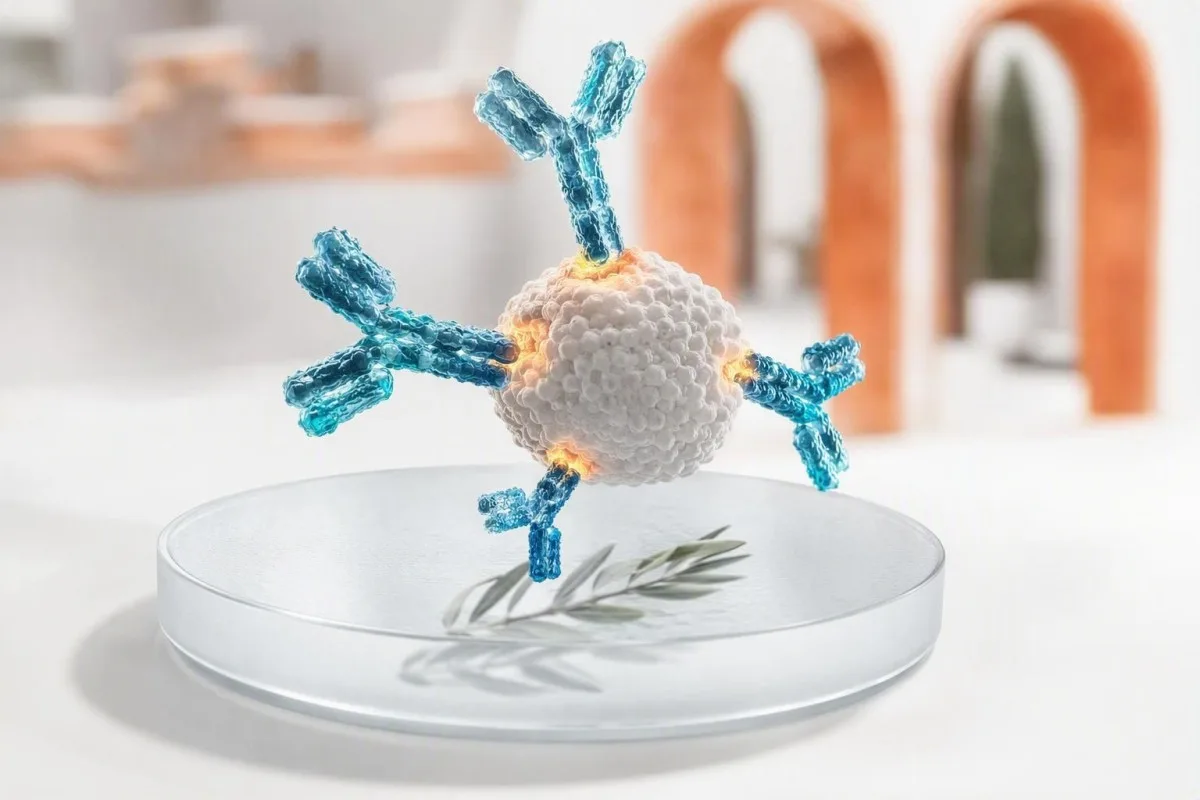
Станоўчы вынік тэсту на антыцелы азначае, што ваша імунная сістэма распазнала пэўную мэту; сам па сабе ён не даказвае актыўную інфекцыю або захворванне. Клас антыцелаў, метад тэсціравання, час, сімптомы і суправаджальныя тэсты вызначаюць, ці патрэбна вам упэўненасць, паўторны тэст, пацвярджэнне або неадкладная медыцынская дапамога.


- Станоўчы вынік тэсту на антыцелы звычайна паказвае на імуннае распазнаванне, а не абавязкова на бягучую хваробу або заразнасць.
- IgM супраць IgG гэта падказка пра час, а не гадзіннік: IgM можа захоўвацца месяцамі, а IgG можа з'явіцца рана.
- Вакцынавыя антыцелы могуць рабіць тэсты на імунітэт станоўчымі, не паказваючы на натуральную інфекцыю.
- Алерген-спецыфічныя IgE больш за 0,35 kUA/л паказвае на сенсібілізацыю, але сімптомы пасля ўздзеяння ўстанаўліваюць клінічную алергію.
- позитивність АНА сустракаецца прыкладна ў 20% здаровых дарослых з нізкімі тытрамі, асабліва з узростам; больш важныя сімптомы і спецыфічныя антыцелы.
- рэактыўнасць на ВІЧ-скрынінг заўсёды патрабуе лабараторнага пацвярджэння перад пастаноўкай любога дыягназу.
- Анты-tTG IgA надзейны для ацэнкі целиакіі толькі тады, калі чалавек спажывае глютен і мае дастатковы ўзровень агульнага IgA.
- Неадкладныя сімптомы такія як дыхавіца, спутанность свядомасці, жаўтуха, боль у грудзях або шырокая бурбалковая сып, перавышаюць вынік руціннага антыцела.
Што насамрэч азначае станоўчы вынік тэсту на антыцелы
A станоўчы тэст на антыцелы азначае, што аналіз выявіў імунныя бялкі, якія звязваюцца з пэўным абетам; гэта не не аўтаматычна азначае, што ў вас ёсць актыўная інфекцыя, аутоіммунное захворванне або алергія, якая патрабуе лячэння. Наступны крок залежыць ад тыпу антыцела, сілы выніку, сімптомаў, часу ўздзеяння і любых парных антыгенаў, ПЦР або тэстаў функцыі органаў.
Антыцелы - гэта B-клетачныя бялкі, якія ўтвараюцца пасля вакцынацыі, інфекцыі, уздзеяння алергена, аутоіммунітэту або часам па прычынах, якія не наносяць клінічнай шкоды. Станоўчы вынік адказвае на адно вузкае аналітычнае пытанне — ці было выяўлена звязванне вышэй за гэты лабараторны ліміт? — а не на больш шырокае клінічнае пытанне аб тым, што адбываецца ў вашым арганізме сёння.
За 15 гадоў маёй клінічнай практыкі найбольш распаўсюджанай памылкай было чытанне “станоўчага” як дыягназу. Практычнае правіла доктара Томаса Кляйна заключаецца ў тым, каб спачатку вызначыць мэта—вірус, вакцынальны антыген, харчовы бялок, шчытападобны фермент, ядзерны антыген або лекі — а затым спытаць, ці выяўляе тэст мінулае ўздзеянне, бягучы арганізм, сенсібілізацыю або імуннае змяненне.
Kantesti AI — гэта Аналізатар крыві са штучным інтэлектам які чытае маркеры антыцелаў побач з датамі, класам імунаглабуліну і звязанымі вынікамі, а не разглядае адну станоўчую лінію як прысуд. Гэты кантэкст асабліва карысны, калі пацыенты параўноўваюць справаздачы з розных лабараторый; якасныя і колькасныя вынікі не з'яўляюцца ўзаемазаменнымі, нават калі абодва кажуць "станоўча".
Станоўчыя IgG супраць IgM: чаму час менш просты, чым здаецца
IgM станоўчы можа сведчыць аб нядаўняй імуннай рэакцыі, у той час як IgG станоўчы часта сведчыць аб мінулым уздзеянні або рэакцыі на вакцыну; ні адзін з гэтых шаблонаў не можа надзейна датаваць захворванне. Некаторыя інфекцыі выпрацоўваюць IgG на працягу 7–14 дзён, а IgM можа заставацца выяўляльным на працягу 3–12 месяцаў ці даўжэй.
IgM - гэта вялікае пяціадзінае антыцела, якое часта выяўляецца рана, але яго памер і шырокае звязванне робяць яго больш схільным да перакрыжаваных рэакцый. Вірус Эпштэйна-Бара, парвавірус B19, рэўматаідны фактар і некаторыя аутоіммунные антыцелы могуць выклікаць памылковыя IgM-рэакцыі; ізаляваны нізкі ўзровень IgM заслугоўвае большага скептыцызму, чым панікі.
IgG звычайна захоўваецца пасля развіцця імуннай памяці, часам на працягу дзесяцігоддзяў. Станоўчы вынік IgG можа адлюстроўваць дзіцячую інфекцыю, нядаўнюю вакцыну, пасіўнае антыцела з нутравеннага імунаглабуліну або мацярынскую перадачу дзіцяці; ён не можа даказаць абарону, калі толькі гэты канкрэтны аналіз не мае пацверджанага ахоўнага парога.
Парныя ўзоры могуць быць больш карыснымі, чым адзін забор: чатырохразовае павышэнне тытра антыцелаў паміж узорамі, узятымі прыкладна з інтэрвалам 2–4 тыдні, пацвярджае нядаўнюю інфекцыю пры некаторых захворваннях. Калі справаздача паказвае агульны IgG, IgA і IgM, разгледзьце шаблон, а не адзін нумар з нашым кіраўніцтва па інтэрпрэтацыі імунаглабулінаў.
Калі антыцелы пры інфекцыях азначаюць, а калі не азначаюць актыўнае захворванне
Антыцелы да інфекцый звычайна паказваюць на ўздзеянне, у той час як ПЦР, тэставанне на антыген, пасеў або павышэнне тытра з'яўляюцца лепшымі інструментамі для выяўлення многіх актыўных інфекцый. Станоўчы вынік на антыцелы без сімптомаў часта з'яўляецца гістарычным, але выключэнне залежыць ад арганізма і вашага імуннага статусу.
Для гепатыту С рэактыўныя антыцелы азначаюць уздзеянне ў нейкі момант, але толькі выяўленая РНК ВГС пацвярджае бягучую вірэмію. Для гепатыту В важна спалучэнне: паверхневы антыген, паверхневыя антыцелы і цэнтральныя антыцелы лепш, чым адзін станоўчы маркер, адрозніваюць актыўную інфекцыю, перанесенае захворванне і вакцынны імунітэт.
ВІЧ - гэта яшчэ адна сітуацыя, дзе формуліроўка надзвычай важная. Рэактыўны скрынінг чацвёртага пакалення не з'яўляецца дыягназам; лабараторыя працягвае з дыферэнцыяльным імунааналізам ВІЧ-1/ВІЧ-2 і, пры неабходнасці, з тэставаннем нуклеінавых кіслот. Чалавек, які прымае постэкспозіцыйную прафілактыку, мае патрэбу ў тэставанні, прывязаным да часу, як тлумачыцца ў нашым кіраўніцтве па тэсціраванні на ВІЧ пасля ПЭП.
Я бачыў здаровага пацыента, якога напалохаў станоўчы трапанемны сіфілітычны антыцел пасля лячэння 18 гадоў таму. Трапанемны тэсты звычайна застаюцца станоўчымі на працягу жыцця, у той час як тытр RPR або VDRL дапамагае ацаніць бягучую актыўнасць і адказ; шаблоны тэсціравання на сіфіліс робяць гэта адрозненне больш ясным.
Чым антыцелы вакцыны адрозніваюцца ад антыцелаў пасля інфекцыі
Антыцелы, звязаныя з вакцынамі, паказваюць, што імунная сістэма адрэагавала на вакцынны антыген; яны не азначаюць, што вакцына выклікала актыўную інфекцыю. Станоўчы вынік можа па-ранейшаму апынуцца ніжэй за вядомы ахоўны карэлят, паколькі звязваючыя антыцелы і функцыянальныя нейтралізуючыя антыцелы не ідэнтычныя.
Канцэнтрацыя паверхневых антыцелаў да гепатыту В 10 мМЕ/мл або вышэй пры вымярэнні праз 1-2 месяцы пасля дакументальна пацверджанай серыі вакцынацыі звычайна лічацца ахоўнымі ў імунакампетэнтных людзей. Анты-HBc, цэнтральныя антыцелы да гепатыту В, не выпрацоўваюцца стандартнай вакцынацыяй супраць гепатыту В; іх наяўнасць сведчыць аб натуральным уздзеянні і павінна падштурхоўваць да поўнай інтэрпрэтацыі панэлі.
IgG да краснухі звычайна правяраецца перад цяжарнасцю, бо ён паказвае на папярэдні імунітэт, а не на актыўнае захворванне на краснуху. Лабараторыі ўсталёўваюць парогі для кожнага аналізу, і памежныя вынікі звычайна разглядаюцца як адсутнасць імунітэту для планавання перад зачаццем, а не як нядаўняя інфекцыя; час падрабязна апісаны ў нашым кіраўніцтве па тэсціраванні на краснуху.
Kantesti AI — гэта Платформа AI для расшыфроўкі аналізу крыві што адрознівае вакцынныя антыцелы ад інфекцыйных пры ўмове, што справаздача ўключае адпаведныя спадарожныя маркеры. Практычнае пытанне часта не “ці станоўчы я?”, а “які антыген вымяраўся, калі я быў вакцынаваны, і ці змяняе гэты вынік сённяшні план абароны?”
Чаму станоўчы вынік тэсту на алергічныя антыцелы можа не азначаць алергію
Станоўчы спецыфічны алерген IgE тэст паказвае на сенсібілізацыю, што азначае, што IgE распазнае алерген; ён не дыягнастуе клінічную алергію без сумяшчальнага анамнезу рэакцыі. Значэнне вышэй 0.35 kUA/L з'яўляецца агульным аналітычным парогам станоўчага выніку, а не універсальным парогам для пазбягання ежы або лекаў.
Анамнез перад тэставаннем мае большае значэнне, чым многія думаюць. Калі чалавек штотыдзень ужывае арахіс без крапіўніцы, хрыпаў, ваніт ці калапсу, адзін толькі нізкі станоўчы IgE да арахісу не павінен прыводзіць да адмовы; непатрэбнае харчовае абмежаванне можа прывесці да харчовай і сацыяльнай шкоды.
Кіраўніцтва NIAID па харчовай алергіі сцвярджае, што тэсты з уколамі на скуру і спецыфічныя IgE выяўляюць магчымыя харчовыя трыгеры, але не могуць самастойна дыягнаставаць харчовую алергію (Sampson et al., 2010). У асобных выпадках алерголаг выкарыстоўвае тэставанне з разбіўкай па кампанентах або медыцынска кантраляваны аральны харчовы выклік, які застаецца залатым стандартам для вырашэння нявызначанасці.
Агульны IgE вымяраецца ў МЕ/мл і можа быць высокім пры экзэме, уздзеянні паразітаў, курэнні і некалькіх імунных станах; ён не можа вызначыць вінаватага алергена. Тэставанне па кампанентах можа адрозніць стабільны запасаючы бялок ад перакрываючагася бялку, звязанага з пылком, што мы даследуем у нашым артыкул пра малекулярнае тэставанне на алергію.
Чаму аўтаімунныя антыцелы патрабуюць сімптомаў і падказак ад органаў
Аўтаіммунныя антыцелы могуць папярэднічаць захворванню, суправаджаць захворванне або прысутнічаць у здаровых людзей; станоўчы вынік сам па сабе рэдка пацвярджае аўтаімунны дыягназ. Клініцысты дзейнічаюць, калі профіль антыцелаў адпавядае сімптомам, дадзеным агляду, маркерам запалення або аб'ектыўным зменам органаў.
Антынуклеарныя антыцелы, або АНА, станоўчыя пры нізкіх тытрах прыкладна ў 13% людзей пры 1:80 і каля 20% пры 1:40 у папуляцыйных даследаваннях, з больш высокімі паказчыкамі ў жанчын і пажылых людзей. Аднастайны або плямісты малюнак не дыягнастуе сістэмны чырвоны ваўканок; ANA лепш за ўсё выкарыстоўваць як тэст-шлюз, калі клінічная карціна робіць захворванне злучальнай тканіны праўдападобным.
Крытэрыі класіфікацыі сістэмнага чырвонага ваўчанкі EULAR/ACR 2019 года выкарыстоўваюць ANA ў тытры не менш за 1:80 як крытэрый уключэння, а затым патрабуюць дадатковых абцяжараных клінічных і імуналагічных прыкмет (Aringer et al., 2019). Новы бялок у мачы, нізкі C3/C4, цитопении, запаленчыя ацёкі суставаў або пераканаўчы фотаадчувальны сып змяняюць тэрміновасць значна больш, чым слабы ANA сам па сабе.
Anti-dsDNA набывае большае значэнне ў спалучэнні з нізкім кампанентам і нырачнымі дадзенымі, хоць метады аналізу істотна адрозніваюцца. Наш кіраўніцтва па наступным назіранні за anti-dsDNA і агляд тэставання кампаненту тлумачаць, чаму спецыяліст можа паўтарыць тэставанне на іншай платформе.
Прыклады органаспецыфічных антыцелаў: шчытападобная залоза, целиакия і страўнік
Органоспецифические антыцелы вызначаюць імунную атаку на тканіну, але яны не заўсёды паказваюць, ці пашкоджаны орган у цяперашні час ці мае патрэбу ў лячэнні. Антыцелы да тиреоидной пероксидазы, антыцелы да тканіннай трансглутаміназы і антыцелы да ўнутранага фактару патрабуюць розных спадарожных тэстаў.
Антыцелы TPO часта сустракаюцца пры тырэяідзіце Хашымота і могуць прысутнічаць гадамі, пакуль TSH і свабодны T4 застаюцца нармальнымі. У дарослага чалавека, які не цяжарны, з нармальнай функцыяй шчытападобнай залозы, вынік звычайна падтрымлівае перыядычны маніторынг TSH, а не лячэнне гармонамі шчытападобнай залозы; цяжарнасць, планы фертыльнасці і сімптомы могуць змяніць гэты падыход.
Поўстаноўчы tTG-IgA прапануе целиакию толькі ў тым выпадку, калі чалавек усё яшчэ спажывае глютен, а агульны IgA дастатковы. Селектыўны недахоп IgA можа зрабіць tTG-IgA памылкова адмоўным, таму клініцысты выкарыстоўваюць тэсты на дэамідаваны пептыд гліядыну на аснове IgG або тэсты tTG, калі агульны IgA нізкі; гл. наша тлумачэнне памылкі пры нізкім IgA.
Антыцелы да ўнутранага фактару адносна спецыфічныя, але не вельмі адчувальныя для перніцыёзнай анеміі. Калі яны станоўчыя разам з нізкім B12, павышанай метилмалоновой кіслатой і макроцитозом, профіль пераканаўчы; без гэтага профілю я пазбягаю лячэння лабараторнага пазначэння, а не пацыента. тэставанне на перніцыёзную анемію паказвае звычайную паслядоўнасць пацвярджэння.
Што выклікае ілжыва-станоўчы вынік тэсту на антыцелы
Памылкова станоўчыя вынікі аналізу антыцелаў узнікаюць, калі аналіз выяўляе ненаўмыснае звязванне, а не калі лабараторыя проста “памылілася”. Крыжаваныя антыцелы, рэўматоідны фактар, нядаўняя імунная стымуляцыя, пасіўны імунаглабулін і нізкая перадтэставая верагоднасць з'яўляюцца распаўсюджанымі прычынамі.
Спецыфічнасць - гэта не дасканаласць. Тэст з 99% спецыфічнасцю дае прыкладна 10 памылкова станоўчых вынікаў на кожныя 1000 сапраўды адмоўных людзей; калі захворванне рэдкае, гэтыя вынікі могуць перавышаць колькасць сапраўдных станоўчых. Вось чаму шырокае скрынінг у чалавека без сімптомаў часта стварае неадназначнасць, а не супакойванне.
Біятын вядомы сваім умяшаннем у некаторыя аналізы гармонаў і сардэчных маркераў, у той час як гетерофільные антыцелы і рэўматоідны фактар могуць уплываць на некаторыя імунааналізы. Лабараторыя часам можа разводзіць узор, выкарыстоўваць блокіруючыя рэагенты, паўтараць вынік на іншым аналізатары або выконваць больш спецыфічны пацвярджаючы метад.
Станоўчы вынік, які супярэчыць цалкам непрыкметнай клінічнай карціне, не варта ігнараваць; гэта прычына для праверкі. Kantesti AI параўноўвае несумяшчальныя маркеры і даты тэстаў, а наша чэк-ліст дакладнасці лабараторных вынікаў можа дапамагчы вам запісаць харчовыя дабаўкі, нядаўнія інфузіі і хваробы перад размовай з які прызначыў лекарам.
Станоўчы вынік тэсту на антыцелы, але без сімптомаў: план практычнага сартавання
Калі аналіз антыцелаў станоўчы, але вы адчуваеце сябе добра, большасці людзей патрэбна яснасць, а не неадкладная дапамога. Самыя бяспечныя першыя крокі - гэта праверыць дакладны аналіз, праверыць даты ўздзеяння і вакцынацыі, а таксама знайсці спадарожны тэст, які пацвярджае бягучае захворванне або ўцягванне органаў.
Пачніце са справаздачы: запішыце назву аналіта, клас антыцелаў, лікавы індэкс або тытр, рэферэнтны інтэрвал, лабараторны метад і дату збору. “Рэактыўны” можа азначаць суадносіны сігналу да парога, ледзь вышэйшае за 1,0, або значна больш моцны вынік; лабараторыі не выкарыстоўваюць аднолькавыя парогі, і велічыня толькі часам адпавядае клінічнай верагоднасці.
Далей спытайце, ці існуе тэст на актыўнае захворванне. Прыклады ўключаюць ПЦР на гепатыт C, серыйныя RPR на сіфіліс, аналіз калу або выдыху на актыўны Helicobacter pylori, TSH/свабодны T4 на антыцелы да шчытападобнай залозы і аналіз мачы плюс крэатынін для праблем з ныркамі, звязаных з воўчанкай.
Па стане на 8 верасня 2026 г. я б не рэкамендаваў самастойна пачынаць прыём антыбіётыкаў, супрацьвірусных прэпаратаў, безглютеновой дыеты, лекаў для шчытападобнай залозы або пазбягання прадуктаў харчавання выключна на аснове адзінкавага выніку аналізу антыцелаў. Захавайце арыгінальны PDF, адзначце сваю зыходную кропку з падоўжальным назіранні за лабараторнымі паказчыкамі, і арганізуйце звычайны агляд, калі няма трывожных сімптомаў.
Пацвярджальныя тэсты, якія змяняюць наступны крок
Найлепшы пацвярджаючы тэст залежыць ад таго, што з'яўляецца мішэнню для антыцелаў: малекулярнае выяўленне пацвярджае многія інфекцыі, функцыянальныя аналізы ўдакладняюць алергію, а тэсціраванне функцый органаў ацэньвае аутоіммунны уплыў. Паўторнае выкананне таго ж тэсту на антыцелы карысна толькі тады, калі чаканая змена з цягам часу мае клінічнае значэнне.
Для станоўчага тэсту на ВІЧ шлях пацвярджэння – гэта дыферэнцыяльны аналіз з наступным тэставаннем нуклеінавых кіслот на ВІЧ, калі вынікі неадназначныя. Для антыцелаў да гепатыту C, РНК HCV з'яўляецца вырашальным наступным крокам; для станоўчага антыцела да ядра гепатыту B, паверхневы антыген, паверхневыя антыцелы і часам ДНК HBV удакладняюць карціну.
Для падазронай целиакии рашэнні аб біяпсіі павінны прымацца, калі ўздзеянне глютена дастаткова для тэсціравання. Для магчымай харчовай алергіі алерголаг улічвае гісторыю ўздзеяння, узровень спецыфічнага IgE, профіль кампанентаў і рызыку правядзення праверкі — а не панэлі незвязаных станоўчых вынікаў, прызначаных без клінічнага пытання.
Kantesti AI — гэта Інструмент аналізу вынікаў аналізу крыві з падтрымкай AI што можа арганізаваць гэтыя звязаныя маркеры ў спіс пытанняў, гатовых для клініцыста, але гэта не замяняе пацвярджальнае тэсціраванне або дыягностыку. Калі вы не ўпэўненыя, наколькі давяраць выніку, які выходзіць за рамкі нормы, наша стандарты медыцынскай валідацыі апісвае, чаму праверка і клінічны нагляд маюць значэнне.
Сімптомы, якія робяць станоўчы вынік тэсту на антыцелы больш тэрміновым
Станоўчы вынік аналізу на антыцелы патрабуе тэрміновай ацэнкі, калі ён суправаджаецца цяжкасцямі з дыханнем, ацёкам твару або горла, стратай прытомнасці, спутанностью свядомасці, болем у грудзях, жаўтухай, хутка распаўсюджваецца сыпам, моцным абязводжваннем або новай неўралагічнай слабасцю. Сімптомы — а не сам лік антыцелаў — вызначаюць тэрміновасць.
Магчымы анафілактычны шок – гэта надзвычайная сітуацыя: раптоўная распаўсюджаная крапіўніца з хрыпамі, сцісканнем у горле, устойлівай ванітамі, слабасцю або нізкім крывяным ціскам пасля ўздзеяння патрабуе экстранных службаў і прызначанага адрэналіну, калі ён даступны. Станоўчы тэст на харчовы алерген або яд IgE ад некалькіх месяцаў таму не прагназуе і не выключае гэтую неадкладную рызыку.
Новая жаўтуха з цёмнай мочой, выяўлены боль у жываце, ліхаманка або спутанность свядомасці павінны выклікаць ацэнку ў той жа дзень, незалежна ад таго, станоўчыя антыцелы да віруснага гепатыту ці не. Перапонныя ферменты, білірубін, INR і прамое тэставанне на патаген дапамагаюць адрозніць імунную гісторыю ад актыўнага гепатабіліярнага захворвання; кампанентах печаночнай панэлі з'яўляюцца карысным кантэкстам.
Аутоіммунная заклапочанасць становіцца тэрміновай пры кашлі з крывёй, дыхавіцы, хутка растучым крэатынінам, крывавым дыярэяй, зменамі зроку, курчамі або новай аднабаковай слабасцю. Я навучыўся не супакойваць кагосьці на аснове мяжуючага тытра антыцелаў, калі траекторыя іх сімптомаў відавочна пагаршаецца.
Ці азначае большы лік антыцелаў больш цяжкае захворванне?
Больш высокі індэкс або тытр антыцелаў часам павялічвае верагоднасць таго, што вынік сапраўдны, але ён звычайна не вымярае цяжкасць захворвання. Значэнні аналізаў спецыфічныя для метаду, і вынік 80 AU/mL нельга параўноўваць непасрэдна з 80 U/mL з іншай лабараторыі.
Пры аутоіммунных захворваннях тэндэнцыі anti-dsDNA часам могуць адпавядаць актыўнасці воўчанкі, асабліва калі яны вымяраюцца паслядоўна адным і тым жа метадам і інтэрпрэтуюцца разам з комплементам і бялком у мачы. Аднак тытр ANA з'яўляецца дрэнным паказчыкам цяжкасці воўчанкі і не павінен праследжвацца паўторна ў стабільнага чалавека.
Для інфекцый моцны сігнал антыцелаў можа падтрымліваць папярэдняе ўздзеянне, але рэдка сведчыць аб заразлівасці. Тэсты на нейтралізуючыя антыцелы з'яўляюцца спецыялізаванымі, і іх ахоўныя парогі могуць адрознівацца ў залежнасці ад узбуджальніка, узросту, вакцыннага прадукту і імунасупрэсіі; просты камерцыйны аналіз IgG не з'яўляецца асабістым пашпартам імунітэту.
Пры параўнанні справаздач трымайце лабараторыю, аналіз і адзінкі бачнымі. Рэальная біялагічная змена павінна перавышаць звычайныя аналітычныя і штодзённыя варыяцыі, таму наша даведнік па параўнанні аналізаў крыві засяроджваецца на датах і метадах, а не толькі на каляровых пазначках.
Цяжарнасць, дзеці і імунасупрэсія змяняюць інтэрпрэтацыю
Цяжарнасць, дзіцячы ўзрост і імунасупрэсіўная тэрапія могуць змяняць як выпрацоўку антыцелаў, так і бяспеку чакання пацвярджэння. У гэтых групах станоўчы вынік можа адлюстроўваць мацярынскія антыцелы, зніжаную рэакцыю на вакцыну, рызыку рэактывацыі або неабходнасць больш ранняй кансультацыі спецыяліста.
Материнскія IgG перасякаюць плацэнту і могуць выяўляцца ў дзіцяці на працягу некалькіх месяцаў, тады як матчыны IgM звычайна не перасякаюць. Такім чынам, ізаляваная IgG-позитивность дзіцяці часта адлюстроўвае мацярынскі імунітэт, у той час як арганізмова-спецыфічныя IgM або прамое выяўленне могуць патрабаваць больш мэтанакіраванай ацэнкі ў залежнасці ад інфекцыі і ўзросту.
Падчас цяжарнасці вынікі выяўлення антыцелаў да краснухі або парвавіруса трактуюцца з улікам дат уздзеяння, тэсту на авіднасць і паўторных проб, а не толькі аднаразовага IgM. Фальшыва-пазітыўны IgM з'яўляецца вядомай праблемай, і пазбегнуць трывогі можна, каб пазбегнуць непатрэбных інвазівных даследаванняў; узгадненне з акушэрскім або інфекцыйным спецыялістам разумна, калі ўздзеянне імаверна.
Людзі, якія атрымліваюць рытуксімаб, высокія дозы кортікостэроідаў, хіміётэрапію або трансплантацыйныя лекі, могуць мець слабыя або адсутнічаюць адказы антыцелаў, нягледзячы на інфекцыю або вакцынацыю. Для сямейных рашэнняў, абмежаванні інтэрпрэтацыі AI для дзяцей і Kantesti conseil médical падкрэсліваюць неабходнасць разгляду клініцыстам.
Як падрыхтавацца да карыснага агляду станоўчага выніку клініцыстам
Прынясіце поўны лабараторны справаздачу, прычыну прызначэння тэсту, даты сімптомаў або ўздзеяння, гісторыю вакцынацыі, лекі і папярэднія вынікі. Гэтая інфармацыя можа ператварыць, здавалася б, невыразны пазітыўны вынік антыцелаў у яснае наступнае дзеянне на працягу аднаго прыёму.
Запішыце пяць фактаў перад візітам: першы дзень сімптомаў, апошняе імавернае ўздзеянне, даты вакцынацыі, лекі, якія падаўляюць імунітэт, або інфузіі імунаглабуліну, і ці паляпшаюцца ці пагаршаюцца сімптомы. Ліхаманка, якая пачалася за 3 дні да ўзяцця пробы, трактуецца вельмі адрозніваецца ад стомленасці, якая пачалася 6 месяцаў таму.
Задайце тры мэтанакіраваныя пытанні: “Што выяўляе гэты тэст?”, “Які тэст вызначае актыўнае захворванне або паражэнне органа?”, і “Які вынік зменіць лячэнне?” Доктар Томас Кляйн лічыць, што гэтыя пытанні змяншаюць непатрэбныя паўторныя панэлі і дапамагаюць пацыентам сысці з вызначаным часам - сёння, 1-2 тыдні, або звычайны маніторынг.
Kantesti AI падтрымлівае бяспечны агляд справаздач на больш чым 75 мовах і можа выяўляць прапушчаныя дадатковыя тэсты з фатаграфаванага або загружанага PDF прыкладна за 60 секунд. Наш кіраўніцтва па тэхналогіі AI тлумачыць падыход супастаўлення кантэксту, у той час як наша клінічная каманда забяспечвае медыцынскі нагляд за інтэрпрэтацыяй для пацыента.
Часта задаваныя пытанні
Што азначае станоўчы вынік на антыцелы, калі ў мяне няма сімптомаў?
Станоўчы вынік на антыцелы без сімптомаў часцей за ўсё сведчыць аб папярэднім кантакце, вакцынацыі, сенсібілізацыі або аб выніку, які патрабуе пацвярджэння, а не аб актыўным захворванні. Наступны крок залежыць ад мэты: антыцелы да гепатыту патрабуюць РНК ВГС, рэактыўны скрынінг на ВІЧ патрабуе алгарытму лабараторнага пацвярджэння, а тырэоідныя антыцелы патрабуюць TSH і вольнага Т4. Ізаляваны нізка-станоўчы вынік менш пераканаўчы пры адсутнасці сумяшчальнага анамнезу ўздзеяння або сімптомаў. Звярніцеся за тэрміновай дапамогай, калі з'явіцца новая жаўтуха, дыхавіца, спутанность свядомасці, боль у грудзях або хуткае пагаршэнне сыпу.
Ці заўсёды IgM станоўчы і IgG адмоўны азначае новую інфекцыю?
IgM станоўчы пры адмоўным IgG можа з'явіцца на ранніх стадыях інфекцыі, але гэта не заўсёды сведчыць аб новай інфекцыі. IgM можа перакрыжавана рэагаваць з незвязанымі імуннымі бялкамі, а некаторыя аналізы даюць ілжыва-станоўчыя вынікі IgM часцей, чым вынікі IgG. Клініцысты могуць паўтарыць сералагічныя даследаванні праз 1–2 тыдні, запытаць ПЦР або антыгеннае тэсціраванне, або выкарыстоўваць больш спецыфічны пацвярджальны аналіз. Правільная інтэрпрэтацыя таксама залежыць ад храналагіі захворвання і канкрэтнага выяўленага арганізма.
Ці можа вакцына зрабіць тэст на антыцелы станоўчым?
Так, вакцыны звычайна робяць тэсты на антыцелы станоўчымі на антыген, які змяшчаецца ў вакцыне. Для гепатыту В канцэнтрацыя анты-HBs не менш за 10 мМЕ/мл, вымераная праз 1–2 месяцы пасля поўнага курса, звычайна сведчыць аб абарончай рэакцыі на вакцыну ў імунакампетэнтных людзей. Стандартная вакцынацыя супраць гепатыту В не выпрацоўвае анты-HBc, таму пазітыўны вынік на антыцелы да ядра сведчыць аб натуральным уздзеянні, а не толькі аб вакцынацыі. Пазітыўны вынік, звязаны з вакцынай, не азначае, што ў вас актыўная інфекцыя.
Ці можа станоўчы тэст на аутоіммунныя антыцелы быць пры адсутнасці захворвання?
Так, у здаровых людзей могуць быць станоўчыя аутоіммунныя антыцелы, асабліва вынікі АНА з нізкім тытрам. АНА выяўляецца прыкладна ў 13% людзей пры тытры 1:80 і каля 20% пры тытры 1:40, таму гэта не самастойны тэст на ваўчанку. Такія сімптомы, як запаленчы ацёк суставаў, язвы ў роце, фотаадчувальная сып, нізкі ўзровень комплемента, бялок у мачы або анамальныя паказчыкі крыві, вызначаюць, ці патрэбна кансультацыя спецыяліста. Станоўчы вынік павінен інтэрпрэтавацца ў кантэксце прычыны прызначэння аналізу.
Ці азначае станоўчы тэст на IgE у крыві, што я павінен пазбягаць гэтай ежы?
Не, станоўчы вынік IgE да пэўнага прадукту азначае сенсібілізацыю і не даказвае, што ўжыванне прадукту выклікае алергічную рэакцыю. Многія лабараторыі называюць вынікі вышэй за 0,35 kUA/L станоўчымі, але гэты парог прызначаны для выяўлення, а не для універсальнага рашэння аб пазбяганні. Чалавек, які рэгулярна ўжывае прадукт без крапіўніцы, хрыпаў, паўторнага ванітавання або слабасці, не павінен аўтаматычна выключаць яго з рацыёну. Алерголаг можа выкарыстоўваць гісторыю рэакцый, тэсціраванне кампанентаў і часам наглядны пероральны харчовы тэст для ўдакладнення рызыкі.
Як доўга антыцелы застаюцца пазітыўнымі пасля інфекцыі?
Працягласць антыцелаў моцна вар'іруецца ў залежнасці ад інфекцыі і класа антыцелаў. IgM можа знікаць на працягу некалькіх тыдняў, але можа захоўвацца на працягу 3-12 месяцаў ці даўжэй, у той час як IgG можа заставацца вымяральным на працягу некалькіх гадоў ці на працягу ўсяго жыцця. Сіфілітычныя антыцелы да трэпонемы часта застаюцца станоўчымі на працягу ўсяго жыцця пасля паспяховага лячэння, у той час як тытры RPR або VDRL выкарыстоўваюцца для адсочвання актыўнасці. Такім чынам, станоўчы вынік аналізу на антыцелы не можа надзейна паведаміць вам, калі адбылася інфекцыя, без іншай клінічнай і лабараторнай інфармацыі.
Атрымаць аналіз крыві з AI сёння
Далучайцеся больш чым да 2 мільёнаў карыстальнікаў па ўсім свеце, якія давяраюць Kantesti для імгненнага, дакладнага аналізу лабараторных тэстаў. Загрузіце вынікі аналізу крыві і атрымайце поўную інтэрпрэтацыю біямаркераў 15,000+ за лічаныя секунды.
📚 Публікацыі даследаванняў са спасылкамі
Кляйн, Т., Мітчэл, С., і Вебер, Г. (2026). Шматмоўная AI-падтрымка прыняцця клінічных рашэнняў для ранняй трыяж хантавіруса: дызайн, інжынерная валідацыя і рэальнае разгортванне на 50 000 прааналізаваных справаздач аб аналізах крыві. Figshare. https://www.researchgate.net/. Kantesti AI Medical Research.
Кляйн, Т., Мітчэл, С., і Вебер, Г. (2026). Зарання зарэгістраваны аўтаматызаваны тэхнічны бенчмарк, заснаваны на рубрыках, рухавіка інтэрпрэтацыі аналізаў крыві Kantesti на 100 000 сінтэтычных выпадкаў тэсціравання. Figshare. https://www.academia.edu/. Kantesti AI Medical Research.
📖 Знешнія медыцынскія спасылкі
⚕️ Медыцынская адмова ад адказнасці
Гэты артыкул прызначаны толькі для адукацыйных мэт і не з’яўляецца медычнай парадай. Заўсёды кансультуйцеся з кваліфікаваным спецыялістам аховы здароўя для прыняцця рашэнняў па дыягностыцы і лячэнні.
Сігналы даверу E-E-A-T
Вопыт
Клінічны агляд, які вядзе лекар, працэсаў інтэрпрэтацыі лабараторных паказчыкаў.
Экспертыза
Фокус лабараторнай медыцыны на тым, як біямаркеры паводзяць сябе ў клінічным кантэксце.
Аўтарытэтнасць
Напісана доктарам Томасам Кляйнам, з аглядам доктара Сара Мітчэл і праф. доктара Ганса Вебера.
Надзейнасць
Інтэрпрэтацыя на аснове доказаў з выразнымі шляхамі далейшых дзеянняў, каб паменшыць трывогу.