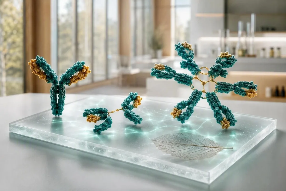
Аналіз на імунаглабуліны найбольш карысны, калі IgG, IgA і IgM разглядаюцца як заканамернасць, а не тры асобныя паказчыкі. Высокія значэнні могуць адлюстроўваць шырокую імунную актывацыю або адзін анамальны бялок; нізкія значэнні могуць сведчыць пра лекі, страту бялку або парушэнне выпрацоўкі антыцелаў.


- IgG звычайна складае 700-1600 мг/дл у дарослых; Устойлівае павышэнне некалькіх класаў часта адлюстроўвае хранічнае запаленне або захворванне печані.
- IgA звычайна складае 70-400 мг/дл у дарослых; селектыўны дэфіцыт можа зрабіць ненадзейным адмоўны тэст на целиакию на аснове IgA.
- антыцелы IgM звычайна складае 40-230 мг/дл у дарослых; ізаляванае, устойлівае павышэнне патрабуе электрафарэзу бялкоў сыроваткі для выключэння моноклональнага бялку.
- Усе тры нізкія выклікае больш занепакоенасць, чым адзін нізкі вынік, і патрабуе аналізу інфекцый, лекаў, страты бялку і адказаў антыцелаў на вакцыну.
- Полікланальная гіперакліў азначае, што некалькі класаў імунаглабулінаў павышаны; гэта часта суправаджае аутоіммуннае захворванне, хранічнае захворванне печані або працяглую інфекцыю.
- Моноклональны ўзор азначае, што адзін клас непрапарцыйна павышаны або з'яўляецца вузкая паласа на электрафарэзе; гэта патрабуе пацвярджэння імунафіксацыі і тэсціравання свабодных легкіх ланцужкоў.
- Неадкладны агляд мае сэнс пры ліхаманцы з паўторнымі сур'ёзнымі інфекцыямі, новай блытанінай свядомасці, дыхавіцай, жаўтухай, стратай вагі, болем у костках або хутка растучым агульным бялком.
- Важны трэнд таму што розніца ў 10-20% можа ўзнікнуць з-за гідратацыі, варыяцыі аналізу, нядаўняй хваробы або іншага лабараторнага метаду.
Як чытаць аналіз на імунаглабуліны як адну заканамернасць
Вынікі IgG, IgA і IgM разам азначаюць рознае, чым паасобку. Шырока павышаная панэль звычайна сведчыць аб поліклональнай імуннай стымуляцыі, у той час як адзін дамінантны клас — асабліва з высокім агульным бялком — ставіць асобнае пытанне аб манакланальным бялку. Па стане на 5 верасня 2026 года гэта адрозненне застаецца практычным першым крокам перад прысваеннем дыягназу аналізу крыві на імунаглабуліны.
Дарослыя рэферэнтныя інтэрвалы вар'іруюцца ў залежнасці ад метаду, але многія лабараторыі выкарыстоўваюць IgG 700-1600 мг/дл, IgA 70-400 мг/дл і IgM 40-230 мг/дл. Значэнне 5% па-за межамі дыяпазону не з'яўляецца аўтаматычным захворваннем; спачатку я правяраю лабараторны інтэрвал, узрост, статус цяжарнасці, нядаўнюю інфекцыю, страту нырак і ці быў узор узяты пасля нутравеннага увядзення імунаглабуліна.
Кантэсці — гэта Аналізатар крыві са штучным інтэлектам які паказвае IgG, IgA і IgM побач з агульным бялком, альбумінам, маркёрамі печані, крэацінінам і CBC, а не разглядае чырвоны сігнал як дыягназ. Гэтая скрыжаваная праверка карысная, таму што нізкі альбумін з павышанымі глобулінамі распавядае зусім іншую гісторыю, чым абязводжванне з абодвума канцэнтраванымі значэннямі; наша кіраўніцтва па біямаркерах тлумачыць звязаныя вымярэнні.
У маёй клінічнай практыцы найбольш супакойваючая панэль часта не ідэальна сярэдняя, а стабільная: напрыклад, IgG 1690 мг/дл пры трох аналізах на працягу 18 месяцаў з нармальным электрафарэзам і без сімптомаў. Доктар Томас Кляйн усё яшчэ пытаўся б аб сухіх вачах, ацёку суставаў, паўторных інфекцыях і леках, але стабільная слабая варыяцыя рэдка мае такую вагу, як уздым на 600 мг/дл за шэсць месяцаў.
Тры імунаглабуліны простымі словамі
антыцелы IgM з'яўляецца раннім шырокім адказнікам, IgA абараняе слізістыя паверхні ў дыхальных шляхах і кішачніку, і IgG забяспечвае найбольш працяглую цыркуляцыйную актыўнасць антыцелаў. Іх біялагічны перыяд паўраспаду значна адрозніваецца: IgG жыве каля 21 дня, IgA каля 6 дзён у сыроватцы крыві, а IgM каля 5 дзён, таму серыйныя змены не адбываюцца з аднолькавай хуткасцю.
Звычайныя нормы для дарослых і чаму ваша лабараторыя можа адрознівацца
Рэферэнтны дыяпазон, надрукаваны побач з вашым уласным вынікам, мае прыярытэт перад любым онлайн-дыяпазонам. Нефеламетрыя, турбідыметрыя і папуляцыі, адкалібраваныя па ўзросце, даюць сціпла розныя межы, асабліва для IgA і IgM.
Звычайныя дарослыя інтэрвалы IgG 700-1600 мг/дл, IgA 70-400 мг/дл і IgM 40-230 мг/дл пералічваюцца прыкладна ў 7-16 г/л, 0,7-4,0 г/л і 0,4-2,3 г/л. Дзеці маюць істотна залежныя ад узросту значэнні, і дарослы дыяпазон ніколі не павінен выкарыстоўвацца для ацэнкі немаўляці або падлетка.
Вынік, блізкі да парога, асабліва ўразлівы да звычайнай біялагічнай варыяцыі. Калі IgM складае 238 мг/дл пры верхняй мяжы 230 мг/дл пасля рэспіраторнай інфекцыі, паўторны аналіз праз 8-12 тыдняў часта больш паказальны, чым неадкладнае прызначэнне вялікай колькасці тэстаў; гл. наша кіраўніцтва па рэальных змяненняў паміж аналізамі крыві.
Галаданне звычайна не патрабуецца для колькасных імунаглабулінаў, але моцнае абязводжванне можа канцэнтраваць бялкі, а нутравенны імунаглабулін можа павышаць вымераны IgG на тыдні. Спытайце ў лекара, які прызначыў панэль, ці атрымлівалі вы замяшчэнне антыцелаў, рытуксімаб, кортікастэроіды, супрацьсутаргавыя сродкі або лячэнне супраць CD20 за папярэднія 6 месяцаў.
Калі ўсе тры класы імунаглабулінаў высокія
Высокія ўзроўні IgG, IgA і IgM разам звычайна сведчаць аб паліклональнай імуннай актывацыі, а не аб адным анамальным клоне. Хранічныя захворванні печані, аутоіммунныя станы, працяглыя інфекцыі і некаторыя запаленчыя захворванні кішачніка з'яўляюцца распаўсюджанымі клінічнымі сітуацыямі.
Паліклональнае павышэнне часта пашырае гама-рэгіён пры электрофарэзе бялкоў сыроваткі замест стварэння рэзкага вузкага піка. На практыцы, IgG 2,100 мг/дл, IgA 520 мг/дл і IgM 310 мг/дл з павышаным CRP сведчыць аб іншым шляху, чым IgG 2,400 мг/дл з двума іншымі класамі, якія зніжаны.
аутоіммуннае захворванне можа выклікаць такую карціну, але сімптомы накіроўваюць наступнае тэставанне. Устойлівая сухасць вачэй і рота, ацёк суставаў, сып, дыярэя або нейропатия могуць апраўдаць накіраваныя аутоантитела і тэставанне на кампаненты сістэмы дапаўнення; наша даведнік па C3 і C4 тлумачыць, чаму нізкія кампаненты сістэмы дапаўнення змяняюць гэту інтэрпрэтацыю.
Хранічныя інфекцыі таксама могуць павышаць некалькі класаў на працягу некалькіх месяцаў, хоць імунаглабуліны не ідэнтыфікуюць арганізм і не пацвярджаюць актыўную інфекцыю. Высокі ESR з агульнымі сімптомамі заслугоўвае клінічнай ацэнкі, і наша тлумачэнне пра высокія прычыны ESR паказвае, чаму неспецыфічны запаленчы маркер не можа вызначыць прычыну.
Што можа азначаць асобна павышаны IgM
Ізаляванае высокае значэнне IgM можа быць вынікам нядаўняй інфекцыі, сустракацца пры холестатычным аутоіммунным захворванні печані або прадстаўляць сабой моноклональны бялок IgM. Устойлівасць, ступень павышэння, сімптомы і электрофарэз маюць большае значэнне, чым адзінкавае мяжавае значэнне.
IgM - гэта вялікае пентамернае антыцела, і ўзровень крыху вышэй 230 мг/дл часта нармалізуецца пасля імуннага выкліку. Паўторны ўзровень IgM 800 мг/дл або 1,200 мг/дл, асабліва пры нармальным або нізкім IgG і IgA, павінен паслужыць падставай для электрофарэзу бялкоў сыроваткі, імунафіксацыі і тэставання на свабодныя лёгкія ланцугі сыроваткі, а не для здагадак.
Першасны біліярны халангіт класічна асацыюецца з павышаным IgM, асабліва калі шчолачная фасфатаза і гама-глутамілтрансфераза таксама высокія. Кіраўніцтва EASL рэкамендуе ацэнку холестатычнага захворвання печані пры ўстойлівым павышэнні шчолачнай фасфатазы і наяўнасці антымітахандрыяльных антыцелаў (EASL, 2017); наша даведнік па ўзору халестазу ахоплівае суправаджальныя маркеры.
моноклональны бялок IgM адрозніваецца ад хваробы Вальденстрэма, і ў большасці людзей з невялікай моноклональнай гамапатыяй няма неадкладнага дыягназу раку. Тым не менш, сімптомы, такія як затуманенасць зроку, галаўны боль, насавыя крывацёкі, новая нейропатия або неабгрунтаваная стомленасць, патрабуюць тэрміновай ацэнкі, бо вельмі высокі IgM можа змяніць глейкасць сыроваткі; чытайце больш пра высокі IgM выклікае.
Нізкі IgA: пастка тэсту на целиакию і прыкметы інфекцыі
Нізкі IgA можа зрабіць коакс-скрынінг з дапамогай IgA-тканіннай трансглютаміназы ілжыва адмоўным. Селектыўны недахоп IgA звычайна вызначаецца як IgA сыроваткі ніжэй 7 мг/дл у чалавека старэй 4 гадоў з нармальным IgG і IgM, хоць лакальныя вызначэнні і паўторнае тэставанне маюць значэнне.
Калі агульны IgA нізкі, клініцысты звычайна выкарыстоўваюць IgG-базіраваны тэст на дэамідаваны глютенавы пептыд або IgG-тканінавую трансглютаміназу разам з анамнезам. Не пачынайце дыету безглютеновую перад адпаведным тэставаннем, калі іншае не рэкамендавана, бо ўзровень антыцелаў можа знізіцца і затушаваць вынік; наша кіраўніцтва па глютенавай нагрузцы апісвае праблему з таймінгам.
У многіх людзей з частковым дэфіцытам IgA няма сур'ёзных праблем са здароўем, у той час як іншыя паведамляюць аб паўторных інфекцыях сінусаў, грудной клеткі або страўнікава-кішачнага тракту. Частата, цяжар і дакументальна пацверджаныя бактэрыяльныя прычыны маюць большае значэнне, чым наяўнасць двух звычайных прастуд у зімовы перыяд; гісторыя вакцынацыі і адказ на антыцелы супраць пнеўмакока могуць быць больш інфарматыўнымі, чым толькі агульны IgA.
Рэакцыі на прадукты крыві рэдкія, але значныя: людзям з глыбокім дэфіцытам IgA і антыцеламі супраць IgA могуць спатрэбіцца спецыяльна падабраныя кампаненты, калі патрабуецца пераліванне. Гэта прычына паведамляць камандам бальніцы аб пацверджаным цяжкім дэфіцыце, а не прычына пазбягаць медыцынска неабходнай дапамогі; гл. наш больш падрабязны разгляд нізкіх вынікаў IgA.
Нізкі IgG і паўторныя інфекцыі: калі вынік мае значэнне
Нізкі IgG становіцца клінічна значным, калі ён суправаджаецца паўторнымі, незвычайна цяжкімі або дрэнна якія праходзяць інфекцыямі. Узровень IgG у дарослых ніжэй за 400 мг/дл звычайна выклікае больш турботы, чым значэнне 650 мг/дл, але функцыянальныя вакцынна-антыцелавыя адказы могуць адмяніць высновы, заснаваныя толькі на колькасці.
Інфекцыі, якія прымушаюць імунолагаў задумацца, - гэта паўторная пнеўманія, бронхаэктазы, паўторныя бактэрыяльныя сінусавыя інфекцыі, якія патрабуюць некалькіх антыбіётыкаў, інвазівныя бактэрыяльныя захворванні або хранічная гиардыяз. Чатыры лёгкія вірусныя прастуды ў работніка дашкольнага ўстановы не эквівалентныя тром пнеўманіям, пацверджаным рэнтгеналагічна, за 24 месяцы.
Другасныя прычыны распаўсюджаныя: рытуксімаб і іншыя тэрапеўтычныя сродкі, накіраваныя на B-клеткі, працяглы прыём кортікостэроідаў унутр, некаторыя супрацьсутаргавыя сродкі, страта бялку ў нефротычным дыяпазоне, энтэрапатыя з стратай бялку і лімфоідная пухліна могуць знізіць IgG. Бялок у мачы, альбумін, колькасць лімфацытаў і гісторыя прыёму лекаў часта даюць больш інфармацыі, чым іншая выпадковая панэль антыцелаў; наша гід па бялку ў мачы тлумачыць ключавы шлях страты.
Баніла і інш. (2015) рэкамендуюць ацэньваць колькасныя імунаглабуліны разам са спецыфічнымі адказамі антыцелаў пры падазрэнні на першасны імунадэфіцыт. На маю думку, нармальны адказ пасля вакцынацыі можа супакойваць, нават калі IgG злёгку паніжаны, тады як дрэнныя адказы пры IgG ніжэй за 500 мг/дл заслугоўваюць увагі спецыяліста.
Калі IgG, IgA і IgM нізкія
Нізкія ўзроўні IgG, IgA і IgM разам называюцца гіпагамаглабулінеміяй і патрабуюць структураванага даследавання, асабліва пры інфекцыях. Такая карціна можа адлюстроўваць парушэнне выпрацоўкі антыцелаў, эфекты лекаў, страту бялку, запушчанае лімфоіднае захворванне або, радзей, часовую змену пасля хваробы.
Панэль IgG 360 мг/дл, IgA 28 мг/дл і IgM 18 мг/дл не з'яўляецца вынікам, які варта чакаць, калі ў чалавека ёсць ліхаманка, паўторныя інфекцыі грудной клеткі або страта вагі. Клініцыст можа паўтарыць панэль, праверыць электрафарэз, CBC з дыферэнцыялам, аналізы нырак і печані, бялок у мачы і вакцынна-спецыфічныя антыцелы, а затым накіраваць да клінічнага імунолага або гематолага.
Частая зменлівая імунадэфіцыт часта праяўляецца ў дарослым узросце, але яе дыягностыка патрабуе больш, чым нізкіх лічбаў: сімптомы, выключаныя другасныя прычыны і зніжаная функцыянальная выпрацоўка антыцелаў з'яўляюцца цэнтральнымі. Kantesti AI - гэта сервісі інтерпретації тестів ШІ , які можа арганізаваць гэтыя суседнія высновы ў рэзюмэ прыёму ў лекара, але ён не можа дыягнаставаць імунадэфіцыт або прыняць рашэнне аб замяшчальнай тэрапіі імунаглабулінамі.
Замяшчальная тэрапія імунаглабулінамі прызначаецца для прадухілення сур'ёзных інфекцый у асобных пацыентаў, а не проста для нармалізацыі лабараторнага выніку. Тыповыя рэжымы замены індывідуальныя і звычайна пачынаюцца прыблізна з 400-600 мг/кг у месяц нутравенна або падскурна, з карэкціроўкай дозы ў залежнасці ад кантролю інфекцыі і мінімальнага ўзроўню IgG, а не універсальнай мэты; наша кіраўніцтва па аналізаў крыві на імунную сістэму дадае карысны кантэкст.
Як аналіз на імунаглабуліны можа ўказваць на моноклональны бялок
Монакланальны бялок мяркуецца, калі адзін клас імунаглабуліна непрапарцыйна высокі, іншыя класы падушаны, або электрафарэз паказвае вузкую паласу. Колькасныя імунаглабуліны самі па сабе не могуць даказаць кланальнасць; серумны электрафарэз бялкоў і імунафіксацыя з'яўляюцца пацвярджаючымі інструментамі.
Разгледзьце IgG 2900 мг/дл з IgA 38 мг/дл і IgM 21 мг/дл: такая карціна больш падазроная, чым калі ўсе тры паказчыкі ўмерана павышаны. Высокі агульны бялок, нізкае суадносіны альбумін/глабулін, анемія, зніжэнне eGFR, высокі ўзровень кальцыя, боль у костках або паўторныя інфекцыі дадаюць клінічнай тэрміновасці; азнаёмцеся з нашым кіраўніцтвам па высокаму агульнаму бялку.
Міжнародная рабочая група па міеломе адрознівае MGUS ад актыўнай міеломы на падставе колькасці бялку, вынікаў даследавання касцявога мозгу і ўплыву на органы, а не толькі выніку на імунаглабуліны (Rajkumar et al., 2014). Узровень M-бялку ніжэйшы за 3 г/дл усё яшчэ можа патрабаваць назірання, а больш нізкае значэнне пры анеміі або пашкоджанні нырак можа патрабаваць больш хуткай ацэнкі гематолагам.
Не дазваляйце ўвесці сябе ў зман нармальнаму ўзроўню агульнага бялку. Захворванні, пры якіх выпрацоўваюцца толькі лёгкія ланцугі, могуць не выклікаць відавочнага павышэння бялку, таму пры адпаведнай клінічнай карціне лекары спалучаюць даследаванне свабодных лёгкіх ланцугоў у сыроватцы, імунафіксацыю і даследаванні мачы; наша шлях тэсціравання на рак крыві тлумачэнне апісвае, што адбываецца далей.
Заканамернасці захворванняў печані: падказкі па IgA, IgG і IgM
Захворванні печані могуць павышаць узровень імунаглабулінаў, але пераважны клас дае толькі падказку, а не дыягназ. IgA часта павышаецца пры алкагольна-асацыяваным пашкоджанні печані, IgG — пры аўтаімунным гепатыце, а IgM — пры першасным біліярным халангіце; перакрыцце бывае часта.
Павышэнне IgG больш чым у 1,1 раза ад верхняй мяжы нормы падтрымлівае дыягназ аўтаімуннага гепатыту, калі вынікі трансаміназ і аўтаантыцелаў адпавядаюць, але само па сабе яно недастаткова адчувальнае і спецыфічнае для пастаноўкі дыягназу. Вострае віруснае захворванне, хранічнае пашкоджанне печані і сістэмнае аўтаімуннае захворванне могуць даваць падобны сігнал, таму лекары часта выкарыстоўваюць візуалізацыйныя даследаванні і абследаванні, прызначаныя спецыялістам.
Павышэнне IgA разам з павышанымі ўзроўнямі GGT, AST і макрацытозам можа адпавядаць алкагольна-асацыяванаму пашкоджанню печані, аднак нельга ніколі меркаваць пра ўжыванне алкаголю толькі на падставе панэлі аналізаў. Частымі прычынамі таксама з'яўляюцца ўплыў лекаў, метабалічная тлушчавая хвароба печані і іншыя фактары; наша тлумачэнне вынікаў ALT дапамагае разглядаць паказчыкі ферментаў разам з імунаглабулінамі.
Важна памятаць пра бяспеку: жаўтуха, цёмная мача, светлы кал, спутанасць свядомасці, павелічэнне жывата або ваніты крывёй патрабуюць неадкладнай медыцынскай дапамогі незалежна ад узроўняў імунаглабулінаў. Стабільнае ізаляванае павышэнне IgA да 430 мг/дл не выклікае гэтых сімптомаў і не павінна адцягваць увагу ад неадкладнага абследавання.
Аддзяленне інфекцыі, запалення і аутоіммунной актыўнасці
Узровень імунаглабулінаў павышаецца занадта павольна і неспецыфічна, каб сам па сабе дыягнаставаць вострую інфекцыю. CRP, агульны аналіз крыві, пасевы або мэтанакіраваныя аналізы на ўзбуджальнікаў лепш адказваюць на непасрэдныя пытанні аб інфекцыі, тады як імунаглабуліны апісваюць больш шырокі імунны фон.
IgM часта апісваюць як антыцела ранняй фазы інфекцыі, але агульны сыроватачны IgM — гэта не тое самае, што тэст на IgM, спецыфічны да пэўнага ўзбуджальніка. Агульны ўзровень IgM 290 мг/дл не можа пацвердзіць грып, інфекцыю, выкліканую вірусам Эпштэйна—Бара, або інфекцыю мачавых шляхоў; неабходнасць лячэння вызначаецца сімптомамі і непасрэдным тэставаннем.
Аўтаімуннае запаленне можа павышаць IgG на працягу многіх гадоў да таго, як дыягназ стане відавочным, асабліва пры гіперγ-глабулінеміі і шырокім профілі электрафарэзу. Панэлі ANA, рэўматоіднага фактару і ENA варта прызначаць для адказу на канкрэтнае клінічнае пытанне, а не для пошуку чаго-небудзь выпадковага; напрыклад, наша кіраўніцтва па anti-dsDNA тлумачыць, чаму станоўчы вынік неабходна ацэньваць з улікам стану нырак і ўзроўню кампанентаў камплементу.
Доктар Thomas Klein назіраў пацыентаў, напалоханых высокім узроўнем IgG пасля віруснага захворвання, які нармалізаваўся пры паўторным даследаванні праз 3 месяцы. І наадварот, устойлівы ўзровень IgG вышэйшы за 2 000 мг/дл у спалучэнні з нізкім C3, пратэінурыяй і сустаўнымі сімптомамі — не тое, з чым я адклаў бы абследаванне на год; такое спалучэнне вынікаў патрабуе своечасовай клінічнай ацэнкі.
Лекі, страта бялку і эфекты часу, якія змяняюць вынікі
Лекі і страта бялку могуць зніжаць узровень імунаглабулінаў без першаснага імуннага захворвання. Прэпараты anti-CD20 могуць спачатку зніжаць IgM, а пазней IgG, тады як страта бялку праз ныркі або кішэчнік можа зніжаць узровень некалькіх класаў адначасова з альбумінам.
Гіпагамаглабулінемія, звязаная з рытаксімабам, можа развіцца праз некалькі месяцаў пасля апошняй інфузіі, паколькі аднаўленне B-клетак і аднаўленне ўзроўню антыцелаў адбываюцца неаднолькава. Вазьміце на прыём поўны спіс лекаў, уключаючы біялагічныя прэпараты, дозу стэроідаў, супрацьсутаргавыя прэпараты і хіміятэрапію; наша кіраўніцтва па тэндэнцыях бяспекі лекаў тлумачыць, чаму важныя даты.
Станы, якія суправаджаюцца стратай бялку, часта пакідаюць дадатковыя прыкметы: нізкі альбумін — ніжэй за прыкладна 3,5 г/дл, ацёкі, дыярэю, павышанае суадносіны альбуміну і крэатыніну ў мачы або зніжаны агульны ўзровень бялку. Наадварот, абязводжванне можа ствараць уражанне павышанага ўзроўню імунаглабулінаў, у той час як альбумін і гематакрыт павышаюцца паралельна.
Паўторнае даследаванне звычайна лепш праводзіць, калі ваш стан клінічна стабільны, па магчымасці ў той жа лабараторыі. Kantesti можа параўнаць датаваны PDF-файл або фатаграфію з папярэднімі вынікамі і адзначыць значны зрух, але дбайная праверка загружаных матэрыялаў будзе разумнай, бо адзінкі вымярэння і рэферэнтныя дыяпазоны могуць быць распазнаныя няправільна.
Дадатковыя аналізы, якія ўдакладняюць анамальную панэль
Правільны наступны аналіз залежыць ад характару змяненняў: электрафарэз — пры непрапарцыйным павышэнні, аналіз антыцелаў пасля вакцынацыі — пры падазрэнні на дэфіцыт, а даследаванні мачы або печані — пры страце бялку ці прыкметах захворвання печані. Паўторнае вымярэнне тых жа трох паказчыкаў без канкрэтнага пытання часта мала што дадае.
Пры высокіх або нераўнамерных выніках лекары звычайна прызначаюць электрафарэз бялкоў сыроваткі, імунафіксацыю, аналіз свабодных лёгкіх ланцугоў у сыроватцы, агульны аналіз крыві, кальцый, крэатынін, альбумін і ферменты печані. Вузкую паласу на электрафарэзаграме неабходна тыпаваць з дапамогай імунафіксацыі, бо клас — IgG, IgA або IgM — змяняе дыферэнцыяльную дыягностыку і план далейшага назірання.
Пры нізкіх выніках і інфекцыях даследаванне можа ўключаць падкласы IgG і тытры антыцелаў да слупняка, Haemophilus influenzae тыпу b або сератыпаў пнеўмакока да і пасля вакцынацыі. Не існуе адзінага ахоўнага парога для пнеўмакокавых антыцелаў, які падыходзіў бы для кожнай лабараторыі і ўзроставай групы; гэта адна з прычын, чаму спецыялізаваная інтэрпрэтацыя важнейшая за онлайн-парог.
Кантэсці — гэта Інструмент аналізу вынікаў аналізу крыві з падтрымкай AI якая можа сабраць гэтыя вынікі ў храналагічную паслядоўнасць і выявіць адсутны кантэкст, напрыклад адсутнасць паказчыкаў альбуміну або электрафарэзу, для разгляду вашым лекарам. Наша гід па тэхналогіі тлумачыць, як фарміруюцца кантэкстуальныя папярэджанні, не замяняючы прыняцця медыцынскіх рашэнняў.
Сімптомы, якія не павінны чакаць звычайнага паўторнага аналізу
Адзін вынік даследавання імунаглабулінаў рэдка сам па сабе стварае надзвычайную сітуацыю, але пэўныя сімптомы — так. Неадкладна звярніцеся па медыцынскую дапамогу ў той жа дзень пры высокай тэмпературы з цяжкім агульным станам, дыхавіцы, спутанасці свядомасці, новай слабасці, жаўтусе, рэзкім памяншэнні колькасці мачы, моцным галаўным болі са зменамі зроку або некантралюемым крывацёку.
Нізкія ўзроўні імунаглабулінаў разам з тэмпературай 38,0 °C або вышэй выклікаюць большую занепакоенасць у чалавека, які атрымлівае хіміятэрапію, высокія дозы стэроідаў або тэрапію, накіраваную супраць B-клетак, ці мае анамнез інвазіўных інфекцый. Не чакайце паведамлення ў медыцынскім партале, калі дыханне абцяжарана, узровень кіслароду нізкі або чалавек выглядае спутаным ці незвычайна санлівым.
Магчымы монакланальны бялок патрабуе больш хуткай ацэнкі, калі ён спалучаецца з новай болем у касцях, невытлумачальнай анеміяй, павышэннем крэатыніну, узроўнем кальцыю вышэй за мясцовы верхні рэферэнтны дыяпазон, неўрапатыяй або ненаўмыснай стратай вагі. Гэта не з'яўляецца доказам захворвання плазматычных клетак, але менавіта гэтыя прыкметы лекары выкарыстоўваюць для вызначэння прыярытэтнасці абследавання; наша даведнік па другім меркаванні можа дапамагчы падрыхтаваць пытанні.
Пры лёгкіх стабільных адхіленнях без трывожных прыкмет звычайна разумна запланаваць прыём на працягу некалькіх тыдняў. Вазьміце з сабой папярэднія вынікі, даты інфекцый, змены ў прыёме лекаў і сямейны анамнез; графік за 2 гады часта больш карысны з клінічнага пункту гледжання, чым адзін вылучаны паказчык.
Бяспечнае выкарыстанне ІІ-інтэрпрэтацыі для вынікаў імунаглабулінаў
ШІ можа сістэматызаваць характар змяненняў у аналізе імунаглабулінаў, але не можа вызначыць, ці ёсць у чалавека імунны дэфіцыт, захворванне печані або монакланальнае парушэнне. Бяспечная інтэрпрэтацыя ўлічвае рэферэнтны дыяпазон лабараторыі, сімптомы, лекі, дынаміку паказчыкаў і пацвярджальныя даследаванні.
Kantesti AI аналізуе IgG, IgA і IgM разам з паказчыкамі бялкоў і органаў, якія змяняюць іх значэнне, а затым прапануе пытанні для далейшага абследавання, а не абвяшчае дыягназ. У нашым аналітычным працэсе асобна паніжаны IgA выклікае рэкамендацыю ўлічыць неабходнасць абследавання на целиакию, тады як паніжэнне ўсіх трох паказчыкаў разам з нізкім альбумінам прымушае разгледзець страту бялку, уплыў лекаў і кансультацыю спецыяліста па клінічнай імуналогіі.
Канфідэнцыяльнасць мае значэнне пры выніках, звязаных з імуннай сістэмай і анкалагічнымі захворваннямі. Kantesti Ltd выкарыстоўвае апрацоўку даных у адпаведнасці з GDPR, а наша старонка медыцынскай валідацыі апісвае клінічны нагляд і межы аўтаматызаванай інтэрпрэтацыі лабараторных вынікаў.
Добрая справаздача павінна дапамагчы вам спытаць: “Ці з'яўляецца гэта шырокай актывацыяй імуннай сістэмы, стратай бялку, зніжэннем яго выпрацоўкі або змяненнем аднаго бялку?” Гэта належны ўзровень упэўненасці да таго, як электрафарэз або спецыялізаванае абследаванне дадуць адказ.
Пытанні, якія трэба задаць пасля анамальнай панэлі імунаглабулінаў
Наступнае пытанне звычайна павінна гучаць: “Якая тут карціна?” — а не: “Як мне знізіць або павысіць гэты паказчык?” Спытайце, ці з'яўляецца вынік устойлівым, поліклональным або моноклональным, ці сведчаць інфекцыі аб парушэнні функцыі і якія другасныя прычыны ўжо былі правераны.
Карысныя пытанні ўключаюць: ці праводзілася электрафарэза сыроватачных бялкоў? Ці адпавядаюць узроўні альбуміну, бялку ў мачы і пячоначных ферментаў страце бялку або захворванню печані? Ці варта мне прайсці аналіз на антыцелы да вакцын? Ці патрэбна паўторнае даследаванне праз 8–12 тыдняў? Гэтыя пытанні больш прадуктыўныя, чым купля харчовых дабавак, якія не выпраўляюць надзейным чынам клінічна значны дэфіцыт антыцелаў.
Калі плануецца накіраванне да спецыяліста, зафіксуйце колькасць інфекцый, якія лячыліся антыбіётыкамі, за апошнія 12 месяцаў, наведванні бальніцы, пнеўманію, пацверджаную метадамі візуалізацыі, і прыём лекаў. Дакладны запіс — “тры курсы антыбіётыкаў і адна пнеўманія” — дапамагае больш, чым фраза “я часта хварэю”; наш трэкер лабараторных вынікаў прапануе практычны фармат вядзення запісаў.
Наш Медыцынская кансультатыўная рада падтрымлівае стандарты клінічнага агляду Kantesti, але ваш уласны ўрач застаецца тым, хто можа вас агледзець, прызначыць пацвярджальныя даследаванні і адрэагаваць на тэрміновыя вынікі. Большасць пацыентаў лічыць, што спакойная размова, заснаваная на аналізе карціны, ператварае трывожны сігнал у кіраваны наступны крок.
Часта задаваныя пытанні
Які нармальныя дыяпазоны імунаглабулінаў для дарослых?
Звычайныя дыяпазоны імунаглабулінаў для дарослых: IgG 700-1600 мг/дл, IgA 70-400 мг/дл і IgM 40-230 мг/дл, хаця для інтэрпрэтацыі трэба выкарыстоўваць прыведзеныя межы кожнай лабараторыі. У многіх краінах вынікі таксама паведамляюцца ў г/л: 7-16 г/л для IgG, 0,7-4,0 г/л для IgA і 0,4-2,3 г/л для IgM. Вынік, які крыху выходзіць за межы дыяпазону, можа адлюстроўваць нядаўнюю хваробу, узровень гідратацыі або варыяцыю аналізу. Устойлівыя анамаліі і сімптомы вызначаюць, ці патрэбна наступнае назіранне.
Што азначае высокі ўзровень IgG, IgA і IgM разам?
Высокі ўзровень IgG, IgA і IgM найчасцей сведчыць аб полікланальнай актывацыі імунітэту з-за хранічнага запалення, аутоіммуннага захворвання, пастаяннай інфекцыі або захворвання печані. Шырокае павышэнне ўсіх трох класаў адрозніваецца ад монокланольнага бялку, які звычайна стварае адзін непрапарцыйна высокі клас або вузкую паласу электрафарэзу. Значэнні, такія як IgG 2000 мг/дл, IgA 500 мг/дл і IgM 300 мг/дл, трэба інтэрпрэтаваць разам з CRP, печеночнымі пробамі, альбумінам і электрофарэзам сыроватачных бялкоў. Сам па сабе такі паказчык не з'яўляецца дыягназам аутоіммуннага захворвання або інфекцыі.
Ці могуць нізкія ўзроўні імунаглабулінаў выклікаць частыя інфекцыі?
Нізкі ўзровень імунаглабулінаў можа павялічыць рызыку рэцыдывавальных бактэрыяльных рэспіраторных або страўнікава-кішачных інфекцый, асабліва калі IgG ніжэй за 400-500 мг/дл або адказ антыцелаў на вакцыну слабы. Найбольш трывожным з'яўляецца паўторная пнеўманія, бронхаэктатычная хвароба, інвазівная бактэрыяльная інфекцыя або некалькі сінусітаў, якія лячыліся антыбіётыкамі, а не аднадзённыя вірусныя прастуды. Нізкі ўзровень IgG, IgA і IgM разам павінен падштурхнуць да агляду на прадмет лекаў, страты бялку ныркамі або кішкамі, лімфоідных станаў і першаснага імунадэфіцыту. Функцыянальнае тэставанне антыцелаў часта лепш удакладняе рызыку, чым адно значэнне агульнага IgG.
Ці ўплывае нізкі ўзровень IgA на тэставанне на целиакию?
Нізкі ўзровень IgA можа прывесці да ілжыва-адмоўнага выніку тэсту на целиакію з выкарыстаннем IgA-антыцелаў да тканіннай трансглютаміназы, паколькі тэст залежыць ад выпрацоўкі антыцелаў IgA. Селектыўны дэфіцыт IgA часта вызначаецца як узровень IgA ніжэй за 7 мг/дл пасля 4 гадоў пры нармальных узроўнях IgG і IgM. Калі агульны ўзровень IgA нізкі, лекары звычайна выкарыстоўваюць замест гэтага тэставанне на IgG-дэамідаваны пептыд гліядыну або IgG-антыцелы да тканіннай трансглютаміназы. Працягвайце ўжываць глютен да ўзгаднення плана тэсціравання, паколькі пазбяганне глютена можа знізіць узровень антыцелаў і зменшыць адчувальнасць тэсту.
Які аналіз пацвярджае наяўнасць манакланальнага імунаглабулінавага бялку?
Электрофарэз сыварачных бялкоў выяўляе анамальны профіль бялку, а імунафіксацыя пацвярджае наяўнасць манакланальнага імунаглабуліну і вызначае яго клас. Звычайна дадаецца тэст на свабодныя лёгкія ланцугі сыроваткі, паколькі некаторыя плазматкалетные парушэнні вырабляюць у асноўным лёгкія ланцугі і могуць не выклікаць значнага павышэння агульнага бялку. Манакланальны вынік патрабуе ацэнкі канцэнтрацыі M-бялку, агульнага аналізу крыві, кальцыя, функцыі нырак і сімптомаў; адзін M-бялок аўтаматычна не азначае рак. У некаторых выпадках таксама могуць спатрэбіцца даследаванні бялку ў мачы.
Ці можа стрэс павысіць узровень IgG, IgA або IgM?
Звычайны псіхалагічны стрэс звычайна не выклікае значнага ізаляванага павелічэння IgG, IgA або IgM па выніках аналізу крыві на імунаглабуліны. Стрэс можа ўплываць на сон, успрыимлівасць да інфекцый і запаленчае сігналаванне, але пастаянныя значэнні вышэй верхняй мяжы лабараторыі не варта спісваць на стрэс без агляду печаначных маркераў, CRP, лекаў і бялковай электрафарэзу, калі гэта неабходна. Сціплае змяненне на 10-20% можа адбыцца паміж узяццем проб з-за нармальных біялагічных і аналітычных варыяцый. Паўторнае правядзенне аналізу праз 8-12 тыдняў, калі пацыент клінічна здаровы, часта з'яўляецца разумным першым крокам пры невялікім невытлумачальным павышэнні.
Атрымаць аналіз крыві з AI сёння
Далучайцеся больш чым да 2 мільёнаў карыстальнікаў па ўсім свеце, якія давяраюць Kantesti для імгненнага, дакладнага аналізу лабараторных тэстаў. Загрузіце вынікі аналізу крыві і атрымайце поўную інтэрпрэтацыю біямаркераў 15,000+ за лічаныя секунды.
📚 Публікацыі даследаванняў са спасылкамі
Кляйн, Т., Мітчэл, С., і Вебер, Г. (2026). Kantesti LTD. (2026). AI Blood Test Analyzer: 2,5M Tests Analyzed | Global Health Report 2026. Zenodo.. Kantesti AI Medical Research.
Кляйн, Т., Мітчэл, С., і Вебер, Г. (2026). Kantesti LTD. (2026). Аналіз крыві RDW: поўны гід па RDW-CV, MCV і MCHC. Zenodo.. Kantesti AI Medical Research.
📖 Знешнія медыцынскія спасылкі
⚕️ Медыцынская адмова ад адказнасці
Гэты артыкул прызначаны толькі для адукацыйных мэт і не з’яўляецца медычнай парадай. Заўсёды кансультуйцеся з кваліфікаваным спецыялістам аховы здароўя для прыняцця рашэнняў па дыягностыцы і лячэнні.
Сігналы даверу E-E-A-T
Вопыт
Клінічны агляд, які вядзе лекар, працэсаў інтэрпрэтацыі лабараторных паказчыкаў.
Экспертыза
Фокус лабараторнай медыцыны на тым, як біямаркеры паводзяць сябе ў клінічным кантэксце.
Аўтарытэтнасць
Напісана доктарам Томасам Кляйнам, з аглядам доктара Сара Мітчэл і праф. доктара Ганса Вебера.
Надзейнасць
Інтэрпрэтацыя на аснове доказаў з выразнымі шляхамі далейшых дзеянняў, каб паменшыць трывогу.