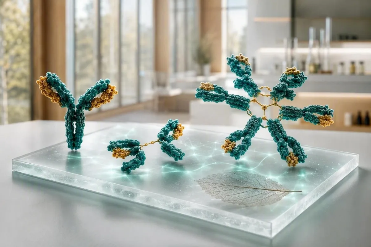
Иммуноглобулиныйн шинжилгээ нь IgG, IgA болон IgM-ийг гурван тусдаа үзүүлэлт биш, харин нэгэн цогц байдлаар авч үзэхэд хамгийн их ач холбогдолтой байдаг. Өндөр утга нь дархлааны системийн өргөн хүрээтэй идэвхжилт эсвэл ганцхан хэвийн бус уураг илэрч болно; бага утга нь эм, уургийн алдагдал эсвэл дархлааны үйлдвэрлэл буурсантай холбоотой байж болно.


- IgG-ийн өсөлттэй хавсарч илэрдэг нь насанд хүрэгчдэд ихэвчлэн 700-1,600 мг/dL байдаг; хэд хэдэн ангилалд тогтмол өсөх нь архаг үрэвсэл эсвэл элэгний өвчнийг илтгэдэг.
- IgA нь насанд хүрэгчдэд ихэвчлэн 70-400 мг/dL байдаг; сонгомол дутагдал нь IgA-д суурилсан целиакийн сөрөг шинжилгээг найдвартай бус болгож болно.
- IgM нь насанд хүрэгчдэд ихэвчлэн 40-230 мг/dL байдаг; моноклонал уургийг үгүйсгэхийн тулд тусгаарлагдсан, тогтмол өсөлт нь ийлдэсний уургийн электрофорезыг шаарддаг.
- Гурав гурвуулаа бага нь нэг бага үр дүнгээс илүү санаа зовоосон байдаг ба халдвар, эм, уургийн алдагдал, вакцинжуулалтын дархлааны хариу урвалыг хянах шаардлагатай.
- Поликлональ өсөлт нь олон иммуноглобулины ангилал өндөр байхыг илтгэнэ; энэ нь ихэвчлэн аутоиммун өвчин, архаг элэгний өвчин эсвэл удаан хугацааны халдвартай холбоотой байдаг.
- Моноклональ хэв шинж нь нэг ангилал нь хэт их нэмэгдсэн эсвэл электрофорезод нарийн зурвас гарч ирснийг илтгэнэ; энэ нь баталгаажуулах иммунофиксаци болон чөлөөт хөнгөн гинжин шинжилгээг шаарддаг.
- Яаралтай хяналт нь халуурах, ойртох ноцтой халдвар, шинээр хэлэхэд ухаан алдах, амьсгал давхцах, шарлах, жин алдах, яс өвдөх эсвэл цусны ерөнхий уураг хурдан нэмэгдэх үед бодолтой байх нь зүйтэй.
- Хандлага (trend) чухал учир нь 10-20% зөрүү нь шингэн алдалт, шинжилгээний өөрчлөлт, саяхны өвчин эсвэл өөр лабораторийн аргаас үүсч болно.
Иммуноглобулины шинжилгээг нэгэн цогц байдлаар хэрхэн унших вэ
IgG, IgA болон IgM үр дүн нь дангаараа болон хамтдаа өөр өөр утгатай. Өргөн хүрээтэй нэмэгдсэн багц нь ихэвчлэн поликлональ дархлааны өдөөлтийг илтгэнэ, харин нэг давамгайлсан анги - ялангуяа өндөр цусны ерөнхий уурагтай хамт - моноклональ уургийн тусдаа асуудлыг хөндөнө. 9-р сарын 5, 2026 байдлаар, энэ ялгаа нь иммуноглобулины цусны шинжилгээнд онош тавихын өмнө практик эхний алхам хэвээр байна.
Насанд хүрэгчдийн хэвийн хэмжээ нь аргаас хамаардаг боловч олон лаборатори ашигладаг IgG 700-1,600 мг/дл, IgA 70-400 мг/дл, IgM 40-230 мг/дл. Хэвийн хязгаараас гадуурх үнэ цэнэ нь автоматаар өвчин болохгүй; Би эхлээд лабораторийн хязгаар, нас, жирэмсний байдал, саяхны халдвар, бөөрний алдагдал, багц нь суд V immunoglobulin авсны дараа авсан эсэхийг шалгадаг.
Кантести бол AI цусны шинжилгээний анализатор нь уургийн ерөнхий хэмжээ, альбумин, элэгний маркер, креатинин ба CBC-г онош болгон авч үзэхээс илүүтэйгээр IgG, IgA, IgM-г харуулдаг. Энэ давхар шалгалт нь шингэн алдалтын улмаас хоёр үнэ цэнэ нэмэгдсэнээс тэс өөр түүхийг хэлж өгдөг тул бага альбумин нь өндөр глобулинтой байх нь ашигтай байдаг; бидний биомаркерийн гарын авчим нь холбогдох хэмжилтийг тайлбарладаг.
Миний эмнэлзүйн ажилд хамгийн тайвшруулдаг багц нь ихэвчлэн төгс дундаж биш харин тогтвортой байдаг: жишээлбэл, 18 сарын хугацаанд гурван удаа татахад IgG 1,690 мг/дл, хэвийн электрофорез болон ямар нэгэн шинж тэмдэггүй. Томас Клейн эмч нүд хуурайших, үе мөч хавагнах, ойртох халдвар, эм тарианы талаар асуух боловч тогтвортой бага зэргийн өөрчлөлт нь зургаан сарын дотор 600 мг/дл-ийн өсөлттэй ижил жинтэй байх нь ховор.
Энгийн хэлээр гурван иммуноглобулин
IgM нь анхдагч өргөн хариу үйлдэгч юм, IgA нь амьсгалын зам, гэдэсний салст бүрхэвчийг хамгаалдаг, ба IgG-ийн өсөлттэй хавсарч илэрдэг нь хамгийн удаан эдэлгээтэй эргэлдэх антибиотикийн идэвхийг хангадаг. Тэдний биологийн хагас хугацаа нь ихээхэн ялгаатай: IgG ойролцоогоор 21 хоног, IgA нь цусанд ойролцоогоор 6 хоног, IgM нь ойролцоогоор 5 хоног үргэлжилдэг тул цуврал өөрчлөлтүүд ижил хурдтай явахгүй.
Насанд хүрэгчдийн ердийн хэмжээ ба танай лаборатори яагаад ялгаатай байж болох вэ
Өөрийнхөө үр дүнгийн хажууд байрлах хэвийн хязгаар нь онлайн хязгаараас давуу талтай. Нефелометр, турбидиметр ба наснаас хамааралтай лабораторийн популяци нь бага зэргийн өөрчлөлттэй хязгаар, ялангуяа IgA ба IgM-д бий болгодог.
IgG 700-1,600 мг/дл, IgA 70-400 мг/дл, IgM 40-230 мг/дл-ийн нийтлэг насанд хүрэгчдийн хязгаар нь ойролцоогоор 7-16 г/л, 0.7-4.0 г/л, 0.4-2.3 г/л болж хувирдаг. Хүүхдүүд нь наснаас хамааралтай их хэмжээний утгатай байдаг бөгөөд насанд хүрэгчдийн хязгаарыг нярай эсвэл өсвөр насныханыг шүүхэд хэзээ ч ашиглаж болохгүй.
Хязгаарын ойролцоох үр дүн нь ердийн биологийн өөрчлөлтөд онцгой эмзэг байдаг. Хэрэв IgM нь 230 мг/дл-ийн дээд хязгаарт 238 мг/дл байвал амьсгалын замын өвчний дараа 8-12 долоо хоногийн дараа дахин хийлгэх нь шууд том багц захиалахаас илүү их зүйлийг илчлэх нь ихэвчлэн илүү үр дүнтэй байдаг; манай гарын авлагыг үзнэ үү. цусны шинжилгээний хоорондох бодит өөрчлөлтүүд.
Ерөнхийдөө шингэнийг хэмжихэд өлсөх шаардлагагүй, гэхдээ хүнд хэлбэрийн шингэн алдалт нь уургийг өтгөрүүлж, суд V immunoglobulin нь хэдэн долоо хоногийн турш IgG-г нэмэгдүүлж болно. Багц захиалсан эмчээс та өмнөх 6 сард антибиотикийн нөхөн сэргээлт, ритуксимаб, кортикостероид, антиконвульсант эсвэл anti-CD20 эмчилгээ хийлгэсэн эсэхийг асуугаарай.
Гурав гурван иммуноглобулины ангилал бүгд өндөр байх үед
IgG, IgA болон IgM зэрэг өндөр байх нь ихэвчлэн нэг хэвийн бус клоноос илүүтэй поликлональ дархлааны идэвхжлийг илтгэнэ. Элэгний архаг өвчин, аутоиммун эмгэгүүд, удаан үргэлжилсэн халдвар болон гэдэсний үрэвслийн зарим эмгэг нь түгээмэл тохиолддог эмнэлзүйн нөхцөлүүд юм.
Поликлональ өсөлтийн үед сийвэнгийн уургийн электрофорезээр хурц, нарийн оргил үүсэхийн оронд гамма бүс ихэвчлэн өргөсөн харагдана. Практик талаас нь авч үзвэл, CRP ихэссэн үед IgG 2,100 мг/дл, IgA 520 мг/дл, IgM 310 мг/дл байх нь бусад хоёр ангилал дарангуйлагдсан IgG 2,400 мг/дл байхаас өөр эмгэг жамыг示нэ.
Аутоиммун өвчин ийм хэв шинж үүсгэж болох боловч дараагийн шинжилгээг шинж тэмдэгт үндэслэн сонгоно. Нүд, ам байнга хуурайших, үе мөч хавдах, тууралт гарах, суулгалт эсвэл мэдрэлийн эмгэг илэрвэл чиглэсэн аутоэсрэгбие болон комплементийн шинжилгээ хийх шаардлагатай байж болно; манай C3 ба C4-ийн гарын авлага тайлбар нь комплемент багассан үед энэ тайлбар хэрхэн өөрчлөгддөгийг тайлбарлана.
Архаг халдварын үед мөн хэд хэдэн ангиллын иммуноглобулин хэдэн сарын турш ихэсч болох боловч иммуноглобулин нь үүсгэгчийг тодорхойлохгүй, идэвхтэй халдварыг батлахгүй. Биеийн ерөнхий шинж тэмдэгтэй хавсарсан ESR өндөр байх нь эмнэлзүйн үнэлгээ шаарддаг бөгөөд өндөр ESR-ийн шалтгаанууд талаарх бидний тайлбар нь өвөрмөц бус үрэвслийн маркер шалтгааныг дангаараа тогтоож чадахгүйг харуулна.
Тусгаарлагдсан өндөр IgM юуг илтгэж болох вэ
IgM дангаараа өндөр байх нь саяхан болсон халдварын дараа илэрч болохоос гадна холестазтай аутоиммун элэгний өвчний үед тохиолдож, эсвэл IgM моноклональ уургийг төлөөлж болно. Нэг удаагийн хязгаарын ойролцоох тооноос илүүтэй үзүүлэлтийн тогтвортой байдал, ихсэлтийн хэмжээ, шинж тэмдэг болон электрофорезийн үр дүн чухал.
IgM нь том хэмжээтэй пентамер эсрэгбие бөгөөд 230 мг/дл-ээс ялимгүй өндөр түвшин нь дархлааны өдөөлтийн дараа ихэвчлэн хэвийн болдог. IgG болон IgA хэвийн эсвэл бага байхад IgM давтан шинжилгээгээр 800 мг/дл буюу 1,200 мг/дл гарвал таамгаар шийдэхийн оронд сийвэнгийн уургийн электрофорез, иммунофиксаци болон сийвэнгийн чөлөөт хөнгөн гинжийн шинжилгээ хийх хэрэгтэй.
Анхдагч билиар холангит нь IgM ихсэлттэй, ялангуяа шүлтлэг фосфатаза болон гамма-глутамилтрансфераза мөн өндөр байх үед сонгодог байдлаар хамааралтай байдаг. Шүлтлэг фосфатаза тогтвортой өндөр, антимитохондрийн эсрэгбие илэрсэн тохиолдолд холестазын элэгний өвчнийг үнэлэхийг EASL-ийн удирдамж зөвлөдөг (EASL, 2017); дагалдах маркеруудын талаар манай холестазын хэв шинжийн гарын авлага тайлбарт авч үзсэн.
Моноклональ IgM уураг нь Вальденстрёмийн макроглобулинемитэй адил зүйл биш бөгөөд бага хэмжээний моноклональ гаммопати бүхий ихэнх хүнд шууд хорт хавдрын онош тавигддаггүй. Гэсэн хэдий ч хараа бүрэлзэх, толгой өвдөх, хамраас цус гарах, шинээр үүссэн мэдрэлийн эмгэг эсвэл шалтгаан тодорхойгүй ядрах зэрэг шинж тэмдэг илэрвэл яаралтай үнэлгээ хийлгэх шаардлагатай. Учир нь IgM маш өндөр үед сийвэнгийн зуурамтгай чанар өөрчлөгдөж болно; дэлгэрэнгүйг өндөр IgM нь.
Бага IgA: целиакийн шинжилгээний алдаа ба халдварын шинж тэмдэг
IgA бага байх нь IgA-ийн эдийн трансглутаминазын целиакийн скринингийн хариуг хуурамч сөрөг болгож болно. Сонгомол IgA дутагдлыг ихэвчлэн 4-өөс дээш настай, IgG болон IgM хэвийн хүний сийвэнгийн IgA 7 мг/дл-ээс бага байх гэж тодорхойлдог боловч тухайн газрын тодорхойлолт болон давтан шинжилгээ чухал.
Нийт IgA бага байвал эмч нар ихэвчлэн дараах шинжилгээг ашигладаг. IgG-д суурилсан деамиджүүлсэн глиадины пептид эсвэл IgG эдийн трансглутаминазын шинжилгээ эмнэлзүйн түүхийн хамт авч үзнэ. Тохирох шинжилгээ хийлгэхээс өмнө эмч зөвлөөгүй л бол глютенгүй хоолны дэглэм бүү эхлүүл, учир нь эсрэгбиеийн түвшин буурч, шинжилгээний хариуг далдлах боломжтой; манай глютений өдөөлтийн заавар хугацаатай холбоотой асуудлыг тайлбарласан.
IgA-ийн хэсэгчилсэн дутагдалтай олон хүнд эрүүл мэндийн ноцтой асуудал байдаггүй бол зарим нь давтан синус, цээжний хөндий эсвэл ходоод гэдэсний халдварын талаар мэдээлдэг. Нэг өвөлд хоёр удаа энгийн ханиад хүрсэн эсэхээс илүү халдварын давтамж, хүндийн зэрэг болон баримтаар тогтоогдсон бактерийн шалтгаан чухал; вакцин хийлгэсэн түүх болон пневмококкийн эсрэгбиеийн хариу нь нийт IgA-ийн түвшнээс илүү мэдээлэлтэй байж болно.
Цусны бүтээгдэхүүнд үзүүлэх урвал ховор боловч анхаарах шаардлагатай: хүнд хэлбэрийн IgA дутагдалтай, anti-IgA эсрэгбиетэй хүмүүст цус сэлбэх шаардлагатай бол тусгайлан сонгосон цусны бүрэлдэхүүн хэрэгтэй байж болно. Энэ нь батлагдсан хүнд хэлбэрийн дутагдлын талаар эмнэлгийн багт мэдэгдэх шалтгаан болохоос эмнэлгийн зайлшгүй шаардлагатай тусламжаас зайлсхийх шалтгаан биш; манай дэлгэрэнгүй тайлбарыг үзнэ үү IgA-ийн бага түвшний хариу.
Бага IgG ба дахилтат халдвар: үр дүн нь чухал байх үед
IgG-ийн бага түвшин давтан, ер бусын хүнд эсвэл удаан арилах халдваруудтай хавсарсан үед эмнэлзүйн ач холбогдолтой болдог. Насанд хүрсэн хүний IgG 400 мг/дл-ээс бага байх нь 650 мг/дл байхаас ерөнхийдөө илүү анхаарал татна, гэхдээ вакцины эсрэгбиеийн үйл ажиллагааны хариу нь зөвхөн тоон үзүүлэлтэд тулгуурласан таамгийг өөрчилж болно.
Дархлаа судлаачдыг анхаарахад хүргэдэг халдварууд нь давтан уушгины хатгалгаа, бронхоэктаз, хэд хэдэн антибиотик шаардсан давтан бактерийн синусын халдвар, инвазив бактерийн өвчин эсвэл архаг гиардиаз юм. Хүүхэд асрах ажилтанд тохиолдсон дөрвөн удаагийн хөнгөн вирусийн ханиад нь 24 сарын дотор дүрс оношилгоогоор батлагдсан гурван удаагийн уушгины хатгалгаатай адил биш.
Хоёрдогч шалтгаан түгээмэл: ритуксимаб болон В-эсэд чиглэсэн бусад эмчилгээ, амаар удаан хугацаагаар хэрэглэсэн кортикостероид, зарим таталтын эсрэг эм, нефротик түвшний уургийн алдагдал, уураг алдалттай энтеропати болон лимфоидын хорт хавдар нь IgG-ийг бууруулж болно. Шээсний уураг, альбумин, лимфоцитын тоо болон эмийн түүх нь өөр нэг санамсаргүй эсрэгбиеийн багцаас илүү их мэдээлэл өгөх нь олонтаа; манай шээсний уургийн заавар алдагдлын нэг чухал замыг тайлбарласан.
Bonilla нар (2015) анхдагч дархлал хомсдол сэжиглэгдсэн үед тоон иммуноглобулиныг өвөрмөц эсрэгбиеийн хариутай хамт үнэлэхийг зөвлөсөн. Миний туршлагаар вакцины дараах хэвийн хариу нь IgG бага зэрэг буурсан байсан ч тайвшруулах шинж байж болно, харин IgG 500 мг/дл-ээс бага бөгөөд хариу сул байвал нарийн мэргэжлийн эмчийн зөвлөгөө шаардлагатай.
IgG, IgA болон IgM бүгд бага байх үед
IgG, IgA болон IgM гурвуулаа бага байхыг гипогаммаглобулинеми гэдэг бөгөөд ялангуяа халдвар хавсарсан тохиолдолд шат дараалсан оношилгоо шаарддаг. Энэ хэв шинж нь эсрэгбиеийн нийлэгжилт алдагдах, эмийн нөлөө, уураг алдагдах, лимфоидын өвчин хүндэрсэн байх эсвэл ховор тохиолдолд өвчний дараах түр зуурын өөрчлөлтийг илэрхийлж болно.
IgG 360 мг/дл, IgA 28 мг/дл, IgM 18 мг/дл гэсэн үзүүлэлт нь тухайн хүн халуурах, цээжний хөндийн давтан халдвар эсвэл жин бууралттай бол ажиглаад хүлээх үр дүн биш. Эмч шинжилгээг давтан хийж, электрофорез, ялгавартай тоолол бүхий CBC, бөөр ба элэгний шинжилгээ, шээсний уураг болон вакцины өвөрмөц эсрэгбиесийг шалгаад эмнэлзүйн дархлаа судлалын эсвэл гематологийн эмчид илгээж болно.
Түгээмэл хувьсамтгай дархлал хомсдол нь ихэвчлэн насанд хүрсэн үед илэрдэг боловч оношлоход бага тоон үзүүлэлтээс илүү зүйл шаардлагатай: шинж тэмдэг, хоёрдогч шалтгааныг үгүйсгэх болон үйл ажиллагааны хувьд буурсан эсрэгбиеийн нийлэгжилт нь гол ач холбогдолтой. Kantesti AI нь AI лабораторийн шинжилгээний тайлбар үйлчилгээ орчны эдгээр ололтыг эмчийн үзлэгийн хураангуй болгон зохион байгуулахад тусалж болох боловч дархлал хомсдолыг оношлох эсвэл иммуноглобулин орлуулах эмчилгээ хийх эсэхийг шийдэх боломжгүй.
Иммуноглобулин орлуулах эмчилгээг лабораторийн үзүүлэлтийг хэвийн болгохын тулд биш, сонгогдсон өвчтөнд ноцтой халдвараас сэргийлэх зорилгоор бичиж өгдөг. Ердийн орлуулах эмчилгээний схемийг хүн бүрт тохируулдаг бөгөөд ихэвчлэн судсаар эсвэл арьсан дор сар бүр ойролцоогоор 400–600 мг/кг-аар эхлүүлж, тунг бүх нийтэд адил зорилтот түвшнээр бус халдварын хяналт болон доод IgG түвшинд үндэслэн тохируулна; манай зааврыг үзнэ үү дархлааны тогтолцооны цусны шинжилгээнүүд хэрэгтэй нэмэлт ойлголт өгдөг.
Иммуноглобулины шинжилгээ нь моноклонал уургийг хэрхэн илрүүлж болох вэ
Нэг иммуноглобулины ангилал харьцангуй өндөр, бусад ангилал дарангуйлагдсан эсвэл электрофорезоор нарийн зурвас илэрвэл моноклональ уураг сэжиглэнэ. Тоон иммуноглобулины шинжилгээ дангаараа клональ шинжийг баталж чадахгүй; ийлдсийн уургийн электрофорез болон иммунофиксаци нь баталгаажуулах шинжилгээ юм.
IgG 2,900 мг/дл, IgA 38 мг/дл, IgM 21 мг/дл гэсэн үзүүлэлтийг авч үзье: гурвуулаа бага зэрэг нэмэгдсэн байхаас илүү энэ хэв шинж сэжигтэй. Нийт уураг ихсэх, альбумин-глобулины харьцаа буурах, цус багадалт, eGFR буурах, кальци ихсэх, яс өвдөх эсвэл давтан халдвар нь эмнэлзүйн яаралтай байдлыг нэмэгдүүлнэ; манай зааврыг үзнэ үү нийт уураг ихсэх.
Олон улсын миеломын ажлын хэсэг нь MGUS-ийг идэвхтэй миеломоос ялгахдаа зөвхөн иммуноглобулины хариунд бус, уургийн хэмжээ, ясны чөмөгний өөрчлөлт болон эрхтэнд үзүүлэх нөлөөнд тулгуурладаг (Rajkumar нар, 2014). М-уургийн хэмжээ 3 г/дл-ээс бага байсан ч хяналт шаардлагатай байж болох бөгөөд цус багадалт эсвэл бөөрний гэмтэл хавсарсан бага хэмжээний үзүүлэлт нь гематологийн нарийн үнэлгээг яаравчлуулах шаардлагатай байж болно.
Нийт уураг хэвийн байна гэж төөрөгдөж болохгүй. Зөвхөн хөнгөн гинж үүсгэдэг эмгэгийн үед уураг мэдэгдэхүйц ихсэхгүй байж болох тул тохирох эмнэлзүйн нөхцөлд эмч нар ийлдсийн чөлөөт хөнгөн гинж, иммунофиксаци болон шээсний шинжилгээг хамтатган үнэлдэг; манай цусны хавдрын шинжилгээний замнал дараагийн алхам юу болохыг тайлбарласан болно.
Элэгний өвчний хэв шинж: IgA, IgG болон IgM-ийн шинж тэмдэг
Элэгний өвчин иммуноглобулиныг ихэсгэж болох ч давамгайлж буй ангилал нь зөвхөн сэжүүр болохоос онош биш. Архитай холбоотой элэгний гэмтлийн үед IgA, аутоиммун гепатитын үед IgG, анхдагч билиар холангитын үед IgM ихсэх нь элбэг боловч давхцал түгээмэл тохиолддог.
Трансаминаз болон аутоэсрэгбиеийн үзүүлэлтүүд тохирч байвал хэвийн дээд хязгаараас 1.1 дахин их IgG нь аутоиммун гепатитыг дэмжих боловч дангаараа оношлоход хангалттай мэдрэг, өвөрмөц биш. Цочмог вирусын өвчин, элэгний архаг гэмтэл болон системийн аутоиммун өвчин ижил төстэй өөрчлөлт үүсгэж болох тул эмч нар дүрс оношилгоо болон мэргэжлийн эмчийн заавраар хийх шинжилгээг ихэвчлэн ашигладаг.
GGT, AST ихсэх болон макроцитозтой зэрэгцэн IgA нэмэгдэх нь архитай холбоотой элэгний гэмтэлтэй нийцэж болох ч шинжилгээний багцын үр дүнд үндэслэн архины хэрэглээг хэзээ ч таамаглаж болохгүй. Эмийн нөлөө, бодисын солилцоотой холбоотой өөхлөг элэгний өвчин болон бусад шалтгаан түгээмэл байдаг; манай ALT-ийн үр дүн тайлбар нь ферментийн үзүүлэлтүүдийг иммуноглобулины үзүүлэлтүүдтэй хамтатган ойлгоход тусална.
Анхаарах чухал зүйл бол иммуноглобулины хэмжээ ямар байхаас үл хамааран шарлалт, бараан шээс, цайвар өтгөн, будилах, хэвлий хөөх эсвэл цусаар бөөлжих үед яаралтай эмнэлгийн тусламж авах шаардлагатай. Тогтвортой, дангаар илэрсэн 430 мг/дл хэмжээний IgA нь эдгээр шинж тэмдгийг үүсгэдэггүй бөгөөд яаралтай үнэлгээ хийлгэхээс анхаарлыг сарниулах ёсгүй.
Халдвар, үрэвсэл, аутоиммун байдлыг ялгах нь
Иммуноглобулины хэмжээ дангаараа цочмог халдварыг оношлоход хэт удаан нэмэгддэг бөгөөд өвөрмөц бус байдаг. CRP, цусны дэлгэрэнгүй шинжилгээ, өсгөвөр болон чиглэсэн үүсгэгчийн шинжилгээ нь халдвартай холбоотой яаралтай асуултад илүү сайн хариулдаг бол иммуноглобулин нь дархлааны ерөнхий байдлыг илэрхийлдэг.
IgM-ийг ихэвчлэн халдварын эхэн үеийн эсрэгбие гэж тайлбарладаг боловч ийлдсийн нийт IgM нь үүсгэгчид өвөрмөц IgM-ийн шинжилгээтэй адилгүй. Нийт IgM 290 мг/дл байгаа нь томуу, Эпштейн–Баррын вирусын халдвар эсвэл шээсний замын халдварыг баталж чадахгүй; эмчилгээ шаардлагатай эсэхийг шинж тэмдэг болон шууд шинжилгээ тодорхойлно.
Аутоиммун үрэвслийн үед онош тодорхой болохоос хэдэн жилийн өмнө IgG ихсэж болно. Ялангуяа гипергаммаглобулинеми болон электрофорезийн өргөн хүрээний хэв шинж хавсарсан үед ANA, ревматоид хүчин зүйл болон ENA-ийн багц шинжилгээг зүгээр нэг таамгаар бус, эмнэлзүйн тодорхой асуултад хариулах зорилгоор захиалах ёстой; жишээлбэл, манай anti-dsDNA-ийн гарын авлага эерэг хариуг бөөрний үйл ажиллагаа болон комплементийн үзүүлэлтүүдийн хүрээнд тайлбарлах шаардлагатайг тайлбарласан.
Доктор Thomas Klein вирусын халдварын дараа IgG нь ихэссэн боловч 3 сарын дараах давтан шинжилгээгээр хэвийн болсон тул айж сандарсан өвчтөнүүдтэй уулзаж байсан. Харин IgG 2,000 мг/дл-ээс дээш хэвээр байх, C3 буурах, протеинури болон үе мөчний шинж тэмдэг хавсрах нь нэг жил хүлээж болох зүйл биш—эдгээр хавсарсан үзүүлэлт нь цаг алдалгүй эмнэлзүйн үнэлгээ хийх үндэслэл болно.
Эм, уургийн алдагдал, цаг хугацааны нөлөө нь үр дүнг өөрчилдөг
Эмийн хэрэглээ болон уураг алдагдал нь анхдагч дархлааны эмгэггүй байсан ч иммуноглобулиныг бууруулж болно. Anti-CD20 эмүүд эхлээд IgM-ийг, дараа нь IgG-ийг бууруулж болох бол бөөрөөр эсвэл гэдсээр уураг алдагдах үед альбуминтай хамт хэд хэдэн ангиллын иммуноглобулин буурч болно.
Ритуксимабтай холбоотой гипогаммаглобулинеми нь сүүлийн дуслаас хойш хэдэн сарын дараа илэрч болно. Учир нь В-эсийн нөхөн сэргээлт болон эсрэгбиеийн нөхөн сэргээлт ижил хугацаанд явагддаггүй. Эмчийн үзлэгт биологийн эм, стероидын тун, таталтын эсрэг эм болон хими эмчилгээ зэрэг бүх эмийн бүрэн жагсаалтыг авч очоорой; манай эмийн аюулгүй байдлын чиг хандлагын гарын авлага огноо яагаад чухал болохыг тайлбарлана.
Уураг алдагддаг эмгэгийн үед ихэвчлэн нэмэлт шинжүүд илэрдэг: альбумин ойролцоогоор 3.5 г/дл-ээс бага байх, хаван, суулгалт, шээсний альбумин-креатинины харьцаа ихсэх эсвэл нийт уураг буурах. Харин шингэн алдалтын үед иммуноглобулины хэмжээ өндөр мэт харагдаж болох бөгөөд альбумин болон гематокрит зэрэгцэн нэмэгддэг.
Ихэвчлэн биеийн байдал тогтвортой үед, боломжтой бол нэг лабораторид шинжилгээг давтан өгөх нь хамгийн зөв. Kantesti нь огноотой PDF файл эсвэл зургийг өмнөх хариутай харьцуулж, их хэмжээний өөрчлөлтийг тэмдэглэж чадна. Гэхдээ сайтар нягталж байршуулах нь зүйтэй. Учир нь нэгж болон лавлах хэмжээг буруу таньж авах боломжтой.
Хэвийн бус бүлгийг тодруулах дараахь шинжилгээнүүд
Зөв дараагийн шинжилгээ нь илэрсэн хэв шинжээс хамаарна: харьцангуй ихэссэн үзүүлэлтийн үед электрофорез, дутагдал сэжиглэгдсэн үед вакцины эсрэгбие, уураг алдагдсан эсвэл элэгний өөрчлөлт сэжиглэгдсэн үед шээсний болон элэгний шинжилгээ хийнэ. Тодорхой асуултгүйгээр ижил гурван үзүүлэлтийг давтан шинжлэх нь ихэвчлэн бага мэдээлэл нэмдэг.
Үзүүлэлт өндөр эсвэл жигд бус гарсан үед эмч нар ихэвчлэн сийвэнгийн уургийн электрофорез, иммунофиксаци, сийвэнгийн чөлөөт хөнгөн гинж, цусны ерөнхий шинжилгээ, кальци, креатинин, альбумин болон элэгний ферментийн шинжилгээ захиалдаг. Электрофорезийн үед нарийн зурвас илэрвэл иммунофиксациар төрлийг тогтоох шаардлагатай. Учир нь IgG, IgA эсвэл IgM-ийн аль нь болохоос ялган оношлох хүрээ болон цаашдын хяналтын төлөвлөгөө хамаарна.
Халдварын хамт бага үзүүлэлт гарсан тохиолдолд вакцин хийлгэхийн өмнө болон дараа IgG-ийн дэд бүлгүүд, татран, Haemophilus influenzae type b эсвэл пневмококкийн серотипүүдийн эсрэгбиеийн титрийг шинжилж болно. Бүх лаборатори болон насны бүлэгт тохирох пневмококкийн хамгаалах нэг л босго үзүүлэлт байдаггүй. Иймээс энэ нь мэргэжлийн эмчийн тайлбар онлайн босго үзүүлэлтээс илүү чухал байдаг салбарын нэг юм.
Кантести бол AI-аар ажилладаг цусны шинжилгээний үр дүнгийн шинжилгээний хэрэгсэл эдгээр үр дүнг цаг хугацааны дарааллаар нэгтгэж, альбумин эсвэл электрофорезийн хариу байхгүй зэрэг эмчийн анхаарах шаардлагатай дутуу мэдээллийг илрүүлж чадна. Манай технологийн гарын авлага тайлбар нь эмнэлгийн шийдвэр гаргалтыг орлохгүйгээр нөхцөл байдлын тэмдэглэгээг хэрхэн үүсгэдгийг тайлбарладаг.
Ердийн давталтыг хүлээхгүйгээр шууд үзүүлэх шинж тэмдэг
Иммуноглобулины дан хариу нь ховор тохиолдолд яаралтай байдал үүсгэдэг боловч зарим шинж тэмдэг үүсгэж болно. Өндөр халууралт хүнд биеийн байдалтай хавсрах, амьсгал давчдах, будилах, шинээр сулрах, шарлах, шээсний хэмжээ мэдэгдэхүйц буурах, хараа өөрчлөгдөхтэй хавсарсан хүчтэй толгой өвдөх, эсвэл цус алдалт тогтохгүй байх үед тэр өдөртөө яаралтай үзүүлээрэй.
Хими эмчилгээ, өндөр тунгийн стероид, В-эсэд чиглэсэн эмчилгээ хийлгэж байгаа эсвэл хүнд хэлбэрийн халдварын түүхтэй хүнд иммуноглобулины түвшин бага, халуун 38.0°C буюу түүнээс дээш байх нь илүү анхаарал татна. Амьсгалахад хүндрэлтэй, хүчилтөрөгчийн түвшин бага эсвэл хүн будилсан, ер бусын нойрмог байвал порталын мессежийг хүлээж болохгүй.
Шинээр яс өвдөх, шалтгаан тодорхойгүй цус багадалт, креатинины хэмжээ нэмэгдэх, кальци тухайн лабораторийн дээд хязгаараас давах, мэдрэлийн эмгэг эсвэл санаандгүй жин хасагдахтай хавсарсан моноклональ уураг сэжиглэгдсэн тохиолдолд илүү шуурхай үнэлгээ шаардлагатай. Эдгээр нь плазмын эсийн эмгэгийн баталгаа биш боловч эмч нар шинжилгээний яаралтай дарааллыг тогтооход ашигладаг шинжүүд юм; манай хоёр дахь санал (second-opinion) өгөх гарын авлага нь асуух асуултаа бэлтгэхэд тусална.
Сэрэмжлүүлэх шинжгүй, хөнгөн бөгөөд тогтвортой өөрчлөлтийн үед хэдэн долоо хоногийн дотор төлөвлөсөн үзлэгт орох нь ихэвчлэн тохиромжтой. Өмнөх шинжилгээний хариу, халдварын огноо, эмийн өөрчлөлт болон гэр бүлийн өвчний түүхээ авч очоорой; 2 жилийн график нь ганц тодруулсан тооноос эмнэлзүйн хувьд илүү ашигтай байдаг.
Иммуноглобулины үр дүнг аюулгүйгээр CE ашиглан тайлбарлах нь
AI нь иммуноглобулины шинжилгээний хэв шинжийг эмхэлж чаддаг боловч тухайн хүнд дархлааны дутагдал, элэгний өвчин эсвэл моноклональ эмгэг байгаа эсэхийг тогтоож чадахгүй. Аюулгүй тайлбарлахын тулд лабораторийн лавлах хэмжээ, шинж тэмдэг, эмүүд, явцын өөрчлөлт болон баталгаажуулах шинжилгээнүүдийг хамтад нь авч үзэх шаардлагатай.
Kantesti AI нь IgG, IgA болон IgM-ийг тэдгээрийн утгыг өөрчилдөг уураг, эрхтний үзүүлэлтүүдийн хамт хянаж, онош зарлахын оронд дараагийн асуултуудыг танилцуулдаг. Манай шинжилгээний ажлын урсгалд дангаар илэрсэн IgA-ийн бууралт нь целиакийн өвчний шинжилгээг анхаарах шаардлагатайг сануулдаг бол гурван үзүүлэлт бүгд бага, альбумин мөн бага байх нь уураг алдагдах, эмийн нөлөө болон эмнэлзүйн дархлаа судлалын үнэлгээг авч үзэхэд хүргэдэг.
Дархлаа болон хавдартай холбоотой шинжилгээний хариунд нууцлал чухал. Kantesti Ltd нь GDPR-д нийцсэн мэдээлэлтэй харьцах зарчим ашигладаг бөгөөд манай эмнэлзүйн баталгаажуулалтын хуудас автоматжуулсан лабораторийн тайлбарын цаадах эмнэлзүйн хяналт болон хязгаарлалтыг тайлбарладаг.
Сайн тайлан нь “Энэ нь өргөн хүрээний дархлааны идэвхжил үү, уураг алдагдал уу, нийлэгжил буурсан уу, эсвэл ганц уурагтай холбоотой юу?” гэж асуухад туслах ёстой. Электрофорез эсвэл мэргэжлийн эмчийн үнэлгээгээр хариу гарахаас өмнө баттай хэлж болох түвшин нь энэ юм.
Хэвийн бус иммуноглобулины бүлгээс хойш асуух асуултууд
Дараагийн хамгийн зөв асуулт нь ихэвчлэн “Энэ ямар хэв шинжтэй вэ?” болохоос “Энэ тоог хэрхэн бууруулах эсвэл нэмэгдүүлэх вэ?” биш байдаг.” Үр дүн тогтвортой эсэх, поликлональ эсвэл моноклональ эсэх, халдварууд дархлааны үйл ажиллагаа алдагдсаныг示ж байгаа эсэх, мөн хоёрдогч шалтгаануудын алийг нь шалгасан талаар асуугаарай.
Хэрэгтэй асуултуудад дараах зүйлс орно: Сийвэнгийн уургийн электрофорез хийсэн үү? Альбумин, шээсний уураг болон элэгний ферментүүд уураг алдагдал эсвэл элэгний өвчинтэй нийцэж байна уу? Вакцины эсрэгбиеийн шинжилгээ хийлгэх шаардлагатай юу? 8–12 долоо хоногийн дараа давтан шинжилгээ хэрэгтэй юу? Эдгээр асуулт нь эмнэлзүйн ач холбогдолтой эсрэгбиеийн дутагдлыг найдвартай засдаггүй нэмэлт бүтээгдэхүүн худалдан авахаас илүү үр дүнтэй.
Мэргэжлийн эмчид үзүүлэхээр төлөвлөж байгаа бол сүүлийн 12 сарын хугацаанд антибиотикоор эмчилсэн халдварын тоо, эмнэлэгт хандсан тохиолдол, дүрс оношилгоогоор батлагдсан хатгалгаа болон эмийн хэрэглээг тэмдэглээрэй. “Би байнга өвддөг” гэхээсээ илүү “антибиотикийн гурван курс хэрэглэж, нэг удаа хатгалгаа туссан” гэх мэт нарийн бүртгэл илүү тустай; манай лабораторийн үр дүн хянагч практик бүртгэлийн загварыг санал болгодог.
Манай Эмнэлгийн зөвлөх зөвлөл Kantesti-ийн эмнэлзүйн үнэлгээний стандартыг дэмждэг боловч таныг үзэж, баталгаажуулах шинжилгээ захиалж, яаралтай илэрсэн үр дүнгийн дагуу арга хэмжээ авах хүн нь таны эмчлэгч эмч хэвээр байна. Ихэнх өвчтөнүүд тайван, хэв шинжид тулгуурласан ярилцлага нь санаа зовоосон үзүүлэлтийг хэрэгжүүлэхэд хялбар дараагийн алхам болгон хувиргадаг гэж үздэг.
Байнга асуудаг асуултууд
Насанд хүрэгчдэд зориулсан ердийн иммуноглобулины шинжилгээний хязгаар нь хэд вэ?
Насанд хүрсэн хүний дархлааны глобулины ердийн хэмжээ нь IgG 700-1,600 мг/дл, IgA 70-400 мг/дл, IgM 40-230 мг/дл байдаг боловч тайлбар хийхдээ тухайн лабораторийн хэвлэсэн хязгаарыг ашиглах ёстой. Олон оронд үр дүнг г/л-ээр мөн мэдүүлдэг: IgG-ийн хувьд 7-16 г/л, IgA-ийн хувьд 0.7-4.0 г/л, IgM-ийн хувьд 0.4-2.3 г/л. Хязгаараас арай гадуурх үр дүн нь саяхан өвдсөн, усны тэнцвэр эсвэл шинжилгээний хувьсах байдлыг тусгаж болно. Байнга тохиолддог хэвийн бус байдал, шинж тэмдгүүд нь дараачийн хяналт шалгалт хийх шаардлагатай эсэхийг тодорхойлно.
Өндөр IgG, IgA болон IgM хамтдаа юу гэсэн үг вэ?
IgG, IgA, болон IgM-ийн хэмжээ ихсэх нь ихэвчлэн архаг үрэвсэл, аутоиммун өвчин, удаан үргэлжилсэн халдвар эсвэл элэгний өвчнөөс шалтгаалсан поликлональ дархлааны идэвхжилийг илтгэнэ. Эдгээр гурван ангиллын хэмжээ ихсэх нь ихэвчлэн нэг ангиллын хэмжээг огцом нэмэгдүүлдэг эсвэл нарийн электрофорезийн бүс үүсгэдэг моноклональ уургийн нөлөөнөөс ялгаатай. IgG 2,000 мг/дл, IgA 500 мг/дл, IgM 300 мг/дл зэрэг утгуудыг CRP, элэгний шинжилгээ, альбумин болон ийлдэсний уургийн электрофорезтой хамт авч үзэх хэрэгтэй. Зөвхнөө энэ хэлбэр нь аутоиммун өвчин эсвэл халдварыг оношлох боломжгүй.
Өндөр бус иммуноглобулины түвшин нь байнга халдвар авахад хүргэж болох уу?
Иммуноглобулины (ИГ) багасах нь бактерийн шалтгаант амьсгалын замын болон ходоод гэдэсний замын халдварын эрсдэлийг нэмэгдүүлдэг, ялангуяа ИГG нь 400-500 мг/дл-ээс бага эсвэл вакцины эсрэгбие урвал муу байвал. Хамгийн санаа зовоосон зүйл бол ойр ойрхон үүсдэг уушгины хатгалга, гуурванцгийн өргөсөх, инвазив бактерийн халдвар эсвэл антибиотикоор эмчилсэн хэд хэдэн синусит юм. ИГG, ИГA, ИГM-ийн түвшин бага байвал эмийн хэрэглээ, бөөр эсвэл гэдэсний уургийн алдагдал, тунгалгийн зангилааны өвчин, анхдагч дархлааны дутагдал зэргийг шалгах хэрэгтэй. Функциональ эсрэгбиеийн шинжилгээ нь заримдаа нэгэн ИГG-ийн нийт утгаас илүү эрсдлийг тодорхойлох боломжтой.
IgA-гийн бага түвшин нь целиак өвчнийг илрүүлэхэд нөлөөлдөг үү?
IgA-ийн бага хэмжээ нь IgA-ийн дутагдлаас шалтгаалдаг тул IgA-ийн түвшин бага байх нь цөмийн шинжилгээнд буруу сөрөг үр дүнг өгч болно, учир нь энэ шинжилгээ нь IgA эсрэг биеийн үйлдвэрлэлээс хамаардаг. Сонгомол IgA дутагдал нь ихэвчлэн 4 наснаас хойш IgG болон IgM хэвийн байх үед IgA-ийн хэмжээ 7 мг/дл-ээс бага байх гэж тодорхойлогддог. Нийт IgA бага байх үед эмч нар ихэвчлэн оронд нь IgG деамид hóa gliadin peptide эсвэл IgG tissue-transglutaminase шинжилгээг ашигладаг. Шинжилгээний төлөвлөгөөг тохиролцон шийдэх хүртэл цавуулаг хэрэглэж байгаарай, учир нь цавуулаг зайлуулах нь эсрэг биеийн түвшинг бууруулж, шинжилгээний мэдрэмжийг бууруулж болзошгүй юм.
Моноклоныг иммуноглобулиныг баталгаажуулдаг шинжилгээ юу вэ?
Цусны уургийн электрофорез нь уургийн хэвийн бус байдлыг илрүүлдэг бөгөөд дархлааны фиксаци нь моноклональ иммуноглобулин байгаа эсэхийг баталж, түүний ангиллыг тодорхойлдог. Цусны чөлөөт гинжин хөнгөн цагааны шинжилгээ нь ихэвчлэн нэмж хийгддэг, учир нь зарим плазмын эсийн эмгэг нь голчлон хөнгөн цагааныг үүсгэдэг бөгөөд нийт уургийг ихээхэн нэмэгдүүлдэггүй байж болно. Моноклональ үр дүн нь М-уургийн концентраци, цусны тоолол, кальци, бөөрний үйл ажиллагаа, шинж тэмдгүүдийг үнэлэхийг шаарддаг; М-уураг дангаараа хорт хавдар гэсэн үг биш юм. Зарим тохиолдолд шээсээр уургийг судлах шаардлагатай байж болно.
Стресс IgG, IgA эсвэл IgM-ийг нэмэгдүүлж чадах уу?
Өдөр тутмын сэтгэлзүйн стресс нь ихэвчлэн дархлаа шинжилгээнд IgG, IgA эсвэл IgM-ийн их хэмжээний өсөлтийг үүсгэдэггүй. Стресс нь нойр, халдварт өртөмтгий байдал, үрэвслийн дохиололд нөлөөлж болно. Гэхдээ лабораторийн дээд хязгаараас давсан байнгын үр дүнг стресс гэж үзэхээсээ өмнө шаардлагатай бол элэгний маркер, CRP, эм, уургийн электрофорезийг хянах хэрэгтэй. Хэвийн биологийн болон шинжилгээний хувиралтай холбоотойгоор дээж авах хооронд 10-20%-ийн дунд зэргийн өөрчлөлт гарч болно. Эмнэлзүйн хувьд сайн байвал 8-12 долоо хоногийн дараа мөчлөгийг давтах нь учир шалтгаангүй бага зэргийн өсөлтийг арилгах анхны ухаалаг алхам болдог.
Өнөөдөр AI-аар дэмжигдсэн цусны шинжилгээний тайлал аваарай
Лабораторийн шинжилгээний агшин зуур, үнэн зөв тайлалд итгэдэг дэлхийн 2 сая гаруй хэрэглэгчтэй нэгдээрэй. Цусны шинжилгээний хариугаа байршуулж, 15,000+ биомаркеруудын цогц тайллыг хэдхэн секундын дотор аваарай.
📚 Иш татсан судалгааны нийтлэлүүд
Клейн, Т., Митчелл, С., ба Вебер, Х. (2026). Kantesti LTD. (2026). AI Цусны шинжилгээний анализатор: 2.5M шинжилгээ хийгдсэн | Дэлхийн эрүүл мэндийн тайлан 2026. Zenodo.. Kantesti AI Медикл Рисерч.
Клейн, Т., Митчелл, С., ба Вебер, Х. (2026). Kantesti LTD. (2026). RDW цусны шинжилгээ: RDW-CV, MCV ба MCHC-ийн иж бүрэн гарын авлага. Zenodo.. Kantesti AI Медикл Рисерч.
📖 Гадаад эмнэлгийн лавлагаа
⚕️ Эмнэлгийн мэдэгдэл
Энэ нийтлэл зөвхөн боловсролын зорилготой бөгөөд эмнэлгийн зөвлөгөө биш болно. Оношилгоо, эмчилгээний шийдвэр гаргахдаа мэргэшсэн эрүүл мэндийн үйлчилгээ үзүүлэгчтэй заавал зөвлөлдөнө.
E-E-A-T итгэлийн дохио
Туршлага
Эмчийн удирдлагатай лабораторийн тайлалын ажлын урсгалын клиник хяналт.
Мэргэшсэн байдал
Биомаркерууд клиникийн нөхцөлд хэрхэн ажиллаж/өөрчлөгдөж байгаад чиглэсэн лабораторийн анагаах ухаан.
Эрх мэдэл
Доктор Томас Клейн бичсэн, Доктор Сара Митчелл, Проф. Доктор Ханс Вебер нар хянан тохиолдуулсан.
Найдвартай байдал
Сэрэмжлүүлгийг бууруулахын тулд тодорхой дараагийн алхамтай, нотолгоонд суурилсан тайлал.