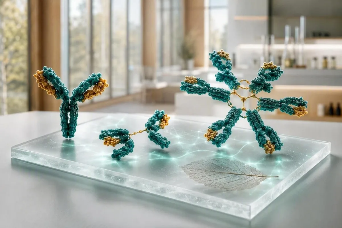
Ang pagsulay sa immunoglobulin labi nga mapuslanon kung ang IgG, IgA ug IgM basahon ingon usa ka sumbanan, dili tulo nga nahimulag nga mga bandila. Ang taas nga mga kantidad mahimong magpakita sa lapad nga pagpaaktibo sa immune o usa ka talagsaon nga abnormal nga protina; ang ubos nga mga kantidad mahimong magpunting sa mga tambal, pagkawala sa protina, o nadaot nga produksiyon sa antibody.


- IgG kasagaran 700-1,600 mg/dL sa mga hamtong; ang nagpadayon nga pagsaka sa daghang klase kanunay nagpakita sa laygay nga paghubag o sakit sa atay.
- IgA kasagaran 70-400 mg/dL sa mga hamtong; ang pinili nga kakulangan mahimong maghimo sa usa ka negatibo nga IgA-based nga pagsulay sa coeliac dili kasaligan.
- ang mga antibody nga IgM kasagaran 40-230 mg/dL sa mga hamtong; ang usa ka nahimulag, nagpadayon nga pagsaka nagkinahanglan og serum protein electrophoresis aron dili iapil ang usa ka monoclonal nga protina.
- Tulo ka ubos tanan labi ka makapabalaka kaysa usa ka ubos nga resulta ug nagkinahanglan og pagrepaso sa mga impeksyon, mga tambal, pagkawala sa protina ug mga tubag sa antibody sa bakuna.
- Polyclonal nga pagtaas nagpasabut nga daghang mga klase sa immunoglobulin ang taas; kini kasagarang nag-uban sa autoimmune nga sakit, laygay nga sakit sa atay o dugay nga impeksyon.
- Monoclonal nga sumbanan nagpasabut nga ang usa ka klase labaw pa sa kasarangan nga pagtaas o usa ka pig-ot nga banda ang makita sa electrophoresis; kini nagkinahanglan og pagpamatuod nga immunofixation ug libre-light-chain testing.
- dali nga pagrepaso makatarunganon alang sa hilanat nga adunay balik-balik nga grabe nga mga impeksyon, bag-ong kalibog, kakulang sa gininhawa, jaundice, pagkawala sa timbang, kasakit sa bukog o kusog nga pagsaka sa kinatibuk-ang protina.
- Importante ang trend tungod kay ang kalainan nga 10-20% mahimong gikan sa hydration, kausaban sa assay, bag-o nga sakit o lain nga pamaagi sa laboratoryo.
Unsaon pagbasa ang pagsulay sa immunoglobulin ingon usa ka sumbanan
Ang mga resulta sa IgG, IgA ug IgM nagkahulugan og lainlain nga magkauban kaysa mag-inusara. Ang usa ka halapad nga gipataas nga panel kasagarang nagpaila sa polyclonal immune stimulation, samtang ang usa ka dominanteng klase—labi na sa taas nga kinatibuk-ang protina—nagpataas sa lahi nga pangutana sa monoclonal protein. Sukad niadtong Septiyembre 5, 2026, kana nga kalainan nagpabilin nga praktikal nga unang lakang sa wala pa idugang ang usa ka diagnosis sa usa ka immunoglobulin blood test.
Ang mga reperensya nga agwat sa mga hamtong managlahi sa matag pamaagi, apan daghang mga laboratoryo ang naggamit IgG 700-1,600 mg/dL, IgA 70-400 mg/dL, ug IgM 40-230 mg/dL. Ang bili nga 5% sa gawas sa sakup dili awtomatikong sakit; Una nakong susihon ang agwat sa laboratoryo, edad, kahimtang sa pagmabdos, bag-o nga impeksyon, pagkawala sa amimislon ug kung ang sampol gikuha pagkahuman sa intravenous immunoglobulin.
Si Kantesti usa ka AI blood test analyzer nga nagbasa sa IgG, IgA ug IgM tupad sa kinatibuk-ang protina, albumin, mga marka sa atay, creatinine ug CBC kaysa pagtratar sa usa ka pulang bandila ingon usa ka diagnosis. Kana nga cross-check mapuslanon tungod kay ang ubos nga albumin nga adunay taas nga globulins nagsulti usa ka lainlaing istorya kaysa dehydration nga adunay pareho nga mga kantidad nga konsentrado; among biomarker guide nagpatin-aw sa mga may kalabutan nga pagsukod.
Sa akong klinikal nga trabaho, ang labing makapadasig nga panel kasagaran dili perpekto apan lig-on: pananglitan, IgG 1,690 mg/dL sa tulo ka pagkuha sulod sa 18 ka bulan nga adunay normal nga electrophoresis ug walay mga sintomas. Si Dr. Thomas Klein mangutana gihapon bahin sa uga nga mga mata, paghubag sa lutahan, balik-balik nga mga impeksyon ug mga tambal, apan ang lig-on nga gamay nga pagkalainlain panagsa ra magdala sa parehas nga gibug-aton sama sa 600 mg/dL nga pagsaka sa unom ka bulan.
Ang tulo ka immunoglobulins sa yano nga pinulongan
ang mga antibody nga IgM mao ang sayo nga halapad nga tigtubag, IgA nanalipod sa mga mucosal nga ibabaw sa agianan sa hangin ug tinai, ug IgG naghatag sa labing malungtaron nga circulating antibody activity. Ang ilang biyolohikal nga katunga sa kinabuhi managlahi pag-ayo: ang IgG molungtad mga 21 ka adlaw, ang IgA mga 6 ka adlaw sa serum, ug ang IgM mga 5 ka adlaw, mao nga ang sunod-sunod nga mga pagbag-o dili managsama ug katulin.
Kasagaran nga mga sakup sa hamtong ug ngano nga ang imong laboratoryo mahimong magkalainlain
Ang reperensya nga sakop nga gipatik tupad sa imong kaugalingon nga resulta maoy prayoridad labaw sa bisan unsang online nga sakop. Ang Nephelometry, turbidimetry ug mga populasyon sa laboratoryo nga gitakda sa edad makagama og gamay nga managlahi nga mga limitasyon, labi na alang sa IgA ug IgM.
Ang komon nga mga agwat sa hamtong sa IgG 700-1,600 mg/dL, IgA 70-400 mg/dL ug IgM 40-230 mg/dL nag-convert sa gibana-bana nga 7-16 g/L, 0.7-4.0 g/L ug 0.4-2.3 g/L. Ang mga bata adunay daghan kaayo nga mga kantidad nga nagdepende sa edad, ug ang agwat sa hamtong dili kinahanglan gamiton sa paghukom sa usa ka masuso o tin-edyer.
Ang usa ka resulta nga duol sa usa ka cutoff huyang kaayo sa ordinaryong biyolohikal nga kausaban. Kung ang IgM kay 238 mg/dL batok sa taas nga limitasyon nga 230 mg/dL pagkahuman sa sakit sa respiratoryo, ang pag-usab sa 8-12 ka semana kasagaran mas nagpadayag kaysa pag-order dayon og dako nga baterya; tan-awa ang among giya sa tinuod nga mga kausaban tali sa mga blood test.
Ang pagpuasa dili kasagaran gikinahanglan alang sa quantitative immunoglobulins, apan ang grabeng dehydration makakonsentra sa mga protina ug ang IV immunoglobulin makapataas sa gisukod nga IgG sulod sa mga semana. Pangutan-a ang clinician nga nag-order sa panel kung nakadawat ka ug antibody replacement, rituximab, corticosteroids, anticonvulsants o usa ka anti-CD20 treatment sa miaging 6 ka bulan.
Kung ang tanan nga tulo nga mga klase sa immunoglobulin taas
Ang taas nga IgG, IgA, ug IgM nga dungan kasagaran nagpakita sa polyclonal immune activation kaysa usa ka abnormal nga clone. Ang laygay nga sakit sa atay, mga kondisyon sa autoimmune, padayon nga impeksyon, ug pipila ka mga sakit sa panghubag sa tinai mga kasagaran nga klinikal nga kahimtang.
Ang usa ka polyclonal nga pagsaka kanunay nagpalapad sa rehiyon sa gamma sa serum protein electrophoresis imbes nga maghimo usa ka hait ug pig-ot nga spike. Sa praktikal nga mga termino, ang IgG 2,100 mg/dL, IgA 520 mg/dL, ug IgM 310 mg/dL nga adunay taas nga CRP nagsugyot og lahi nga agianan kaysa IgG 2,400 mg/dL nga adunay ubang duha ka klase nga gipugngan.
Ang sakit nga autoimmune mahimong makamugna niini nga sumbanan, apan ang mga sintomas naggiya sa sunod nga pagsulay. Ang padayon nga uga nga mga mata ug baba, nanghubag nga mga lutahan, pantal, kalibanga, o neuropathy mahimong maghatag ug katarungan sa gipunting nga mga autoantibodies ug pagsulay sa complement; ang among C3 ug C4 guide nagpatin-aw ngano nga ang ubos nga complement nagbag-o sa interpretasyon.
Ang laygay nga impeksyon mahimo usab nga makapataas sa daghang mga klase sulod sa mga bulan, bisan kung ang mga immunoglobulin wala mag-ila sa organismo o nagpamatuod sa aktibo nga impeksyon. Ang taas nga ESR nga adunay constitutional symptoms angay sa klinikal nga pagrepaso, ug ang among pagpatin-aw sa hinungdan sa taas nga ESR nagpakita ngano nga ang usa ka nonspecific inflammatory marker dili makasulbad sa hinungdan.
Unsa ang mahimong ipasabut sa nahimulag nga taas nga IgM
Ang usa ka nahilit nga taas nga resulta sa IgM mahimong mosunod sa bag-o nga impeksyon, mahitabo sa cholestatic autoimmune liver disease, o magrepresentar sa usa ka IgM monoclonal protein. Ang pagpadayon, ang gidak-on sa pagsaka, ang mga sintomas, ug ang electrophoresis mas importante kaysa usa ka kinutuban nga numero.
Ang IgM usa ka dako nga pentameric antibody, ug ang mga lebel nga gamay ra sa 230 mg/dL kasagaran molig-on human sa usa ka immune challenge. Ang gisubli nga IgM nga 800 mg/dL o 1,200 mg/dL, labi na kung normal o ubos ang IgG ug IgA, kinahanglan magpalihok sa serum protein electrophoresis, immunofixation, ug serum free-light-chain testing imbes nga mangagapulo.
Ang primary biliary cholangitis klasiko nga nag-uban sa taas nga IgM, labi na kung ang alkaline phosphatase ug gamma-glutamyl transferase usab taas. Ang EASL guideline nagtambag sa pagtimbang-timbang alang sa cholestatic liver disease kung ang alkaline phosphatase padayon nga taas ug ang antimitochondrial antibody anaa (EASL, 2017); ang among giya sa cholestasis pattern naglangkob sa nag-uban nga mga marker.
Ang usa ka monoclonal IgM protein dili parehas sa Waldenström macroglobulinaemia, ug kadaghanan sa mga tawo nga adunay gamay nga monoclonal gammopathy dili dayon adunay diagnosis sa kanser. Bisan pa, ang mga sintomas sama sa pagblur sa panan-aw, sakit sa ulo, sipon sa ilong, bag-ong neuropathy, o dili masabtan nga kakapoy nagkinahanglan ug dinalian nga pagtimbang-timbang tungod kay ang taas kaayo nga IgM mahimong makabag-o sa viscosity sa serum; basaha ang dugang bahin sa taas nga IgM hinungdan sa.
Ubos nga IgA: ang sayup sa pagsulay sa coeliac ug mga timailhan sa impeksyon
Ang ubos nga IgA mahimong makapahimo sa usa ka IgA tissue-transglutaminase coeliac screen nga sayop nga negatibo. Ang selective IgA deficiency kasagaran gihubit ingon nga serum IgA nga ubos sa 7 mg/dL sa usa ka tawo nga kapin sa 4 ka tuig ang edad nga adunay normal nga IgG ug IgM, bisan kung ang lokal nga mga depinisyon ug gisubli nga pagsulay importante.
Kung ang kinatibuk-ang IgA ubos, ang mga clinician kasagaran mogamit og IgG-based deamidated gliadin peptide o IgG tissue-transglutaminase test kauban sa kaagi sa klinika. Ayaw pagsugod sa usa ka gluten-free diet sa wala pa ang angay nga pagsulay gawas kon tambagan, tungod kay ang lebel sa antibody mahimong mokunhod ug makatabon sa resulta; ang among gabay sa gluten challenge naghulagway sa problema sa pag-timing.
Daghang mga tawo nga adunay partial IgA deficiency walay dagkong problema sa panglawas, samtang ang uban nagreport sa balik-balik nga sinus, dughan, o gastrointestinal infections. Ang kasubsob, kabug-at, ug dokumentado nga mga hinungdan sa bakterya mas hinungdanon kaysa sa pagbaton og duha ka ordinaryong sip-on sa usa ka tingtugnaw; ang kasaysayan sa pagbakuna ug tubag sa pneumococcal antibody mahimong mas makapatin-aw kay sa kinatibuk-ang IgA lamang.
Ang mga reaksyon sa produkto sa dugo dili kasagaran apan importante: ang mga tawo nga adunay lawom nga IgA deficiency ug anti-IgA antibodies mahimong magkinahanglan og espesyal nga gipili nga mga sangkap kung gikinahanglan ang transfusion. Kini usa ka rason sa pagpahibalo sa mga team sa ospital mahitungod sa usa ka napamatud-an nga grabe nga kakulangan, dili usa ka rason sa paglikay sa medikal nga gikinahanglan nga pag-atiman; tan-awa ang among mas bug-os nga diskusyon sa ubos nga IgA results.
Ubos nga IgG ug balik-balik nga mga impeksyon: kanus-a hinungdanon ang resulta
Ang ubos nga IgG mahimong klinikal nga importante kung kini nag-uban sa balik-balik, dili kasagaran nga grabe, o dili maayo nga pag-ayo sa mga impeksyon. Ang usa ka hamtong nga IgG ubos sa 400 mg/dL kasagaran usa ka mas lig-on nga kabalaka kaysa sa bili nga 650 mg/dL, apan ang mga functional vaccine-antibody responses mahimong makapausab sa mga pangagpas base lamang sa gidaghanon.
Ang mga impeksyon nga makapahunong sa mga immunologist mao ang balik-balik nga pneumonia, bronchiectasis, balik-balik nga bacterial sinus infections nga nanginahanglan og daghang antibiotics, invasive bacterial disease, o chronic giardiasis. Upat ka minor viral colds sa usa ka childcare worker dili katumbas sa tulo ka radiological confirmed pneumonias sa 24 ka bulan.
Ang secondary causes komon: rituximab ug uban pang B-cell-directed therapies, dugay nga oral corticosteroids, pipila ka anticonvulsants, nephrotic-range protein loss, protein-losing enteropathy, ug lymphoid malignancy mahimong makapaubos sa IgG. Ang urine protein, albumin, lymphocyte count, ug kasaysayan sa tambal kasagaran makahatag ug labaw pa kay sa laing random antibody panel; among protein sa ihi nga giya nagpatin-aw sa usa ka yawe nga agianan sa pagkawala.
Si Bonilla et al. (2015) nagrekomendar sa pagtimbang-timbang sa quantitative immunoglobulins kauban ang mga specific antibody responses kung ang primary immunodeficiency gisuspetsahan. Sa akong kasinatian, ang normal nga post-vaccine response mahimong makahatag og kasegurohan bisan kung ang IgG gamay ra ang ubos, samtang ang dili maayo nga mga tubag nga adunay IgG ubos sa 500 mg/dL nagkinahanglan og espesyalista nga input.
Kung ang IgG, IgA ug IgM tanan ubos
Ang ubos nga IgG, IgA, ug IgM nga magkauban gitawag nga hypogammaglobulinaemia ug nagkinahanglan og structured work-up, ilabi na sa mga impeksyon. Ang pattern mahimong magpakita sa dili maayo nga paghimo sa antibody, mga epekto sa tambal, pagkawala sa protina, advanced lymphoid disease, o, dili kaayo sagad, usa ka temporaryo nga pagbag-o pagkahuman sa sakit.
Ang usa ka panel sa IgG 360 mg/dL, IgA 28 mg/dL, ug IgM 18 mg/dL dili usa ka resulta nga hulatan ug tan-awon kung ang tawo adunay hilanat, balik-balik nga mga impeksyon sa dughan, o pagkawala sa timbang. Ang usa ka clinician mahimong mag-usab sa panel, susihon ang electrophoresis, CBC nga adunay differential, kidney ug liver tests, urine protein, ug vaccine-specific antibodies, dayon modangop sa clinical immunology o haematology.
Ang common variable immunodeficiency sagad nagpakita sa pagkatigulang, apan ang pagdayagnos niini nagkinahanglan ug labaw pa sa ubos nga mga numero: mga sintomas, gipagawas nga secondary causes, ug nabawasan nga functional antibody production mao ang sentro. Kantesti AI usa ka AI lab test interpretation service nga makahan-ay niining mga naglibot nga mga nakaplagan ngadto sa usa ka summary sa pagbisita sa doktor, apan dili kini makadiskubre sa immune deficiency o makadesisyon sa immunoglobulin replacement.
Ang immunoglobulin replacement gireseta aron mapugngan ang grabe nga mga impeksyon sa mga pinili nga pasyente, dili lamang aron i-normalize ang usa ka resulta sa laboratoryo. Ang tipikal nga mga regimen sa pagpuli gihimo matag tawo ug kasagarang magsugod sa mga 400-600 mg/kg matag bulan pinaagi sa intravenous o subcutaneous nga mga agianan, nga adunay dosing nga gi-adjust sa pagkontrol sa impeksyon ug trough IgG imbes nga usa ka universal target; ang among giya sa mga blood test sa immune-system naghatag ug mapuslanong konteksto.
Unsaon ang pagsulay sa immunoglobulin nga makatudlo sa usa ka monoclonal nga protina
Ang monoclonal protein gisugyot kung ang usa ka immunoglobulin class labi nga taas, ang ubang mga klase gipugngan, o ang electrophoresis nagpakita og pig-ot nga banda. Ang Quantitative immunoglobulins lamang dili makapamatud-og sa clonality; ang serum protein electrophoresis ug immunofixation mao ang mga confirmatory tools.
Ikonsiderar ang IgG 2,900 mg/dL nga adunay IgA 38 mg/dL ug IgM 21 mg/dL: kana nga pattern mas kaduda-dudahan kay sa tanan nga tulo nga kasarangan nga gipataas. Ang taas nga kinatibuk-ang protina, ubos nga albumin-to-globulin ratio, anemia, nabugnaw nga eGFR, taas nga calcium, sakit sa bukog, o balik-balik nga impeksyon makadugang sa klinikal nga pagkadinalian; ribyuha ang among giya sa taas nga kinatibuk-ang protina.
Ang International Myeloma Working Group nagpalahi sa MGUS gikan sa aktibong myeloma pinaagi sa gidaghanon sa protina, mga kaplag sa utok sa bukog (marrow findings) ug mga epekto sa organo—dili lang sa resulta sa immunoglobulin (Rajkumar et al., 2014). Ang M-protein nga ubos sa 3 g/dL mahimo gihapon nga nagkinahanglan og pag-monitor, samtang ang mas ubos nga kantidad uban sa anemia o kadaot sa kidney mahimong magkinahanglan og mas paspas nga pagtimbang-timbang sa haematology.
Ayaw palimbong sa normal nga total protein. Ang mga disorder nga light-chain-only mahimong dili makapataas og klaro sa protina, mao nga ang mga clinician naghiusa sa serum free light chains, immunofixation ug mga pagtuon sa ihi sa usa ka angay nga presentasyon; ang among nagpasabot nganong ang CBC usa ka sinyales nga magsugod imbes nga diagnosis sa cancer. naglatid kon unsay sunod nga mahitabo.
Mga sumbanan sa sakit sa atay: mga timailhan sa IgA, IgG ug IgM
Ang sakit sa atay makapataas sa immunoglobulins, apan ang dominanteng klase naghatag lamang og timailhan, dili usa ka diagnosis. Ang IgA kanunay nga motaas uban sa kadaot sa atay nga may kalabotan sa alkohol, IgG uban sa autoimmune hepatitis, ug IgM uban sa primary biliary cholangitis; kasagaran ang pag-overlap.
Ang pagtaas sa IgG nga labaw sa 1.1 ka pilo sa upper limit of normal nagsuporta sa autoimmune hepatitis kon ang transaminases ug autoantibodies mohaom, apan kini dili sensitibo o espesipiko nga igo aron ma-diagnose kini nga mag-inusara. Ang acute viral illness, chronic liver injury ug systemic autoimmune disease ang tanan makahimo og susamang signal, busa ang mga clinician kasagarang mogamit og imaging ug pagsulay nga gidumala sa espesyalista.
Ang pagtaas sa IgA uban sa taas nga GGT, AST ug macrocytosis mahimong mohaom sa alcohol-associated liver injury, apan ang paggamit sa alkohol dili angay nga ipangagpas base lang sa usa ka panel. Ang mga epekto sa tambal, metabolic fatty liver disease ug uban pang mga hinungdan kay kanunay; ang among katin-awan sa ALT nga mga resulta makatabang sa pagbutang sa mga enzyme tupad sa immunoglobulins.
Ang mapuslanong mensahe sa kaluwasan mao nga ang jaundice, itom nga ihi, luspad nga hugaw, kalibog, paghubag sa tiyan o pagsuka og dugo nagkinahanglan og dinalian nga medikal nga pagtagad bisan unsa pa ang mga kantidad sa immunoglobulin. Ang lig-on ug nag-inusarang pagtaas sa IgA nga 430 mg/dL dili makapahinabo sa maong mga simtomas ug dili angay nga makabalda sa dinalian nga ebalwasyon.
Pagbulag sa impeksyon, paghubag ug autoimmune nga kalihokan
Ang mga lebel sa immunoglobulin motaas og hinay kaayo ug dili espesipiko aron ma-diagnose ang usa ka acute infection sa ilang kaugalingon. Ang CRP, full blood count, cultures o targeted pathogen tests mas maayo nga makatubag sa mga pangutana mahitungod sa dinalian nga impeksyon, samtang ang immunoglobulins naghulagway sa mas lapad nga background sa immune system.
Ang IgM kanunay nga gihulagway nga antibody sa sayong impeksyon, apan ang total serum IgM dili sama sa pathogen-specific IgM test. Ang total IgM nga 290 mg/dL dili makakumpirma sa trangkaso, Epstein-Barr virus o impeksyon sa ihi; ang mga simtomas ug direkta nga pagsulay ang magdeterminar kon gikinahanglan ba ang pagtambal.
Ang autoimmune inflammation mahimong makapataas sa IgG sulod sa daghang katuigan sa dili pa mahimong klaro ang diagnosis, ilabi na sa hypergammaglobulinaemia ug lapad nga electrophoresis pattern. Ang mga panel sa ANA, rheumatoid factor ug ENA angay nga i-order aron sa pagtubag sa usa ka klinikal nga pangutana, dili isip usa ka fishing expedition; pananglitan, ang among anti-dsDNA nga giya nagpatin-aw kon nganong ang positibong resulta nagkinahanglan og konteksto sa kidney ug complement.
Si Dr. Thomas Klein nakakita og mga pasyente nga nalisang sa taas nga IgG human sa usa ka viral illness nga mibalik sa normal sa pag-usab sa pagsulay human sa 3 ka bulan. Sa laing bahin, ang padayon nga IgG nga labaw sa 2,000 mg/dL uban sa ubos nga C3, proteinuria ug mga simtomas sa lutahan dili usa ka butang nga akong i-defer sulod sa usa ka tuig—ang maong hiniusa nga mga kaplag nagpakamatarong sa tukma sa panahon nga klinikal nga ebalwasyon.
Mga tambal, pagkawala sa protina ug mga epekto sa oras nga nagbag-o sa mga resulta
Ang mga tambal ug pagkawala sa protina makapaubos sa immunoglobulins nga walay nag-unang immune disorder. Ang mga tambal nga Anti-CD20 makapaminus sa IgM una ug sa IgG sa ulahi, samtang ang pagkawala sa protina sa kidney o tinai mahimong makapaubos sa pipila ka mga klase uban sa albumin.
Ang hypogammaglobulinaemia nga may kalabotan sa Rituximab mahimong motungha pipila ka bulan human sa kataposang infusion tungod kay ang pagkaayo sa B-cell ug pagkaayo sa antibody dili managsama. Pagdala og kompleto nga listahan sa tambal—lakip ang biologics, dosis sa steroid, tambal batok sa kombulsyon ug chemotherapy—ngadto sa appointment; ang among giya sa mga uso sa kaluwasan sa tambal nagpatin-aw kon nganong importante ang mga petsa.
Ang mga kahimtang nga mawad-an og protina sagad magbilin og dugang nga mga timailhan: ubos nga albumin nga ubos sa mga 3.5 g/dL, edema, kalibanga, taas nga urine albumin-creatinine ratio o pagkunhod sa kinatibuk-ang protina. Sa laing bahin, ang dehydration makapahimo sa mga immunoglobulin nga tan-awon nga taas samtang ang albumin ug haematocrit motaas dungan niini.
Ang balik-balik nga pagsulay sagad nga labing maayo kung ikaw klinikal nga lig-on, gamit ang parehas nga laboratoryo kung mahimo. Ang AI makatandi sa usa ka karaan nga PDF o litrato batok sa mga miaging resulta ug makamatikod og dako nga kausaban, apan ang usa ka mabinantayon nga pagsusi sa gi-upload makatarunganon tungod kay ang mga yunit ug mga reference range mahimong sayop nga makuha.
Mga pagsulay sa pag follow-up nga maklaro ang usa ka abnormal nga panel
Ang husto nga follow-up nga pagsulay nagdepende sa sumbanan: electrophoresis alang sa dili timbang nga pagtaas, mga antibody sa bakuna alang sa gidudahang kakulangan, ug mga pagsulay sa ihi o atay alang sa pagkawala sa protina o mga timailhan sa atay. Ang pagsubli sa parehas nga tulo ka numero nga walay pangutana sagad makadugang og gamay ra nga impormasyon.
Alang sa taas o dili parehas nga mga resulta, ang mga clinician sagad mangayo og serum protein electrophoresis, immunofixation, serum free light chains, kompletong blood count, calcium, creatinine, albumin ug mga enzyme sa atay. Ang usa ka pig-ot nga banda sa electrophoresis kinahanglan nga i-type pinaagi sa immunofixation tungod kay ang klase—IgG, IgA o IgM—makapausab sa differential ug plano sa follow-up.
Alang sa ubos nga mga resulta nga adunay mga impeksyon, ang pagsulay mahimong maglakip sa mga IgG subclass ug antibody titre alang sa tetanus, Haemophilus influenzae type b o pneumococcal serotypes sa wala pa ug pagkahuman sa pagbakuna. Walay usa ka protective pneumococcal threshold nga mohaom sa matag laboratoryo ug grupo sa edad, nga usa ka dapit diin ang paghubad sa espesyalista mas importante kaysa sa usa ka online cutoff.
Si Kantesti usa ka AI-powered blood test analysis tool nga makahimo sa pagtigom niini nga mga resulta ngadto sa usa ka timeline ug pag-ila sa nawala nga konteksto, sama sa absent nga albumin o electrophoresis, aron ikonsiderar sa imong clinician. Ang among giya sa teknolohiya nagpatin-aw kung giunsa ang paghimo sa mga contextual flag nga dili mopuli sa medikal nga paghimog desisyon.
Mga simtomas nga dili angay maghulat sa usa ka routine repeat
Ang usa ka resulta sa immunoglobulin lamang panagsa ra magmugna og emerhensya, apan ang pipila ka mga sintomas makahimo. Pangita og dinalian nga assessment sa samang adlaw alang sa taas nga hilanat nga adunay grabeng sakit, kalisud sa pagginhawa, kalibog, bag-ong kahuyang, jaundice, mamatikdan nga pagkunhod sa ihi, grabeng labad sa ulo nga adunay kausaban sa panan-aw, o dili mapugngan nga pagdugo.
Ang ubos nga lebel sa immunoglobulin dugang pa sa hilanat nga 38.0°C o mas taas pa mas makapabalaka sa usa ka tawo nga nagpa-chemotherapy, naggamit og taas nga dosis sa steroids, B-cell-directed therapy o adunay kasaysayan sa invasive nga mga impeksyon. Ayaw paghulat og mensahe sa portal kung lisud ang pagginhawa, ubos ang lebel sa oxygen, o ang tawo morag naglibog o dili kasagaran nga katulgon.
Ang usa ka posible nga monoclonal protein nagkinahanglan og mas paspas nga pagrepaso kung kini mahitabo uban sa bag-ong kasakit sa bukog, dili masabtan nga anemia, pagtaas sa creatinine, calcium nga labaw sa lokal nga upper range, neuropathy o wala tuyoa nga pagkawala sa timbang. Dili kini pamatuod sa usa ka sakit sa plasma-cell, apan kini ang mga bahin nga gigamit sa mga clinician aron unahon ang pagsulay; ang among second-opinion guide makatabang sa pag-andam og mga pangutana.
Alang sa malumo ug lig-on nga mga abnormalidad nga walay red flags, ang usa ka giplano nga appointment sulod sa pipila ka mga semana sagad nga makatarunganon. Dad-a ang mga miaging resulta, mga petsa sa impeksyon, mga kausaban sa tambal ug family history; ang usa ka 2-tuig nga graph sagad mas mapuslanon sa klinikal kaysa sa usa ka gi-highlight nga numero.
Paggamit sa interpretasyon sa AI sa luwas nga paagi alang sa mga resulta sa immunoglobulin
Ang AI makahimo sa pag-organisar sa usa ka sumbanan sa pagsulay sa immunoglobulin, apan dili kini makatino kung ang usa ka tawo adunay kakulangan sa immune, sakit sa atay o usa ka monoclonal disorder. Ang luwas nga paghubad nagpabilin nga makita ang laboratory range, mga sintomas, mga tambal, mga uso ug mga confirmatory test.
Ang AI nagrepaso sa IgG, IgA ug IgM uban sa mga protina ug mga organ marker nga nag-usab sa ilang kahulogan, dayon nagpresentar og mga follow-up nga pangutana imbes nga mopahayag og diagnosis. Sa among analysis workflow, ang usa ka isolated nga ubos nga IgA nag-aghat og pasidaan sa coeliac-testing, samtang ang tanang tulo ka ubos nga kantidad dugang pa sa ubos nga albumin nag-aghat sa pagkonsiderar sa pagkawala, mga epekto sa tambal ug klinikal nga immunology review.
Ang privacy importante sa mga resulta nga may kalabutan sa immune ug kanser. Ang among gigamit mao ang GDPR-aligned nga pagdumala, ug ang among panid sa pagpamatuod sa medisina naghubit sa klinikal nga pagdumala ug mga utlanan luyo sa automated lab interpretation.
Ang maayo nga report kinahanglan makatabang kanimo sa pagpangutana, “Kini ba kay lapad nga immune activation, pagkawala sa protina, pagkunhod sa produksyon, o usa ka protina?” Kana mao ang angay nga lebel sa kasiguroan sa dili pa ang electrophoresis o pagsusi sa espesyalista makahatag og tubag.
Mga pangutana nga ipangutana pagkahuman sa usa ka abnormal nga panel sa immunoglobulin
Ang labing maayong sunod nga pangutana kasagaran mao ang “unsa kini nga sumbanan?” imbes nga “unsaon nako pagpaubos o pagpataas niini nga numero?” Pangutan-a kon ang resulta padayon ba, polyclonal ba o monoclonal ba, kon ang mga impeksiyon nagpasabot ba og huyang nga paglihok, ug unsang mga sekondaryang hinungdan na ba ang nasusi.
Ang mapuslanong mga pangutana naglakip sa: Gihimo ba ang serum protein electrophoresis? Ang albumin, protina sa ihi ug mga enzyme sa atay ba nahiuyon sa pagkawala niini o sakit sa atay? Kinahanglan ba ko og pagsusi sa mga antibody batok sa bakuna? Kinahanglan ba usbon ang pagsusi sulod sa 8–12 ka semana? Mas mapuslanon kini nga mga pangutana kaysa pagpalit og mga suplemento, nga dili kasaligan sa pagtul-id sa klinikal nga mahinungdanong kakulang sa antibody.
Kon giplano ang referral ngadto sa espesyalista, isulat ang gidaghanon sa mga impeksiyong gitambalan og antibiotiko sulod sa miaging 12 ka bulan, mga pagbisita sa ospital, pulmonya nga gikumpirma pinaagi sa imaging, ug pagkaladlad sa mga tambal. Ang tukmang rekord—“tulo ka kurso sa antibiotiko ug usa ka pulmonya”—mas makatabang kaysa “kanunay ko magkasakit”; ang among tigsubay sa resulta sa lab nagtanyag og praktikal nga pormat sa rekord.
Atong Lupon sa Medikal nga Magtatambag nagsuporta sa mga sumbanan sa klinikal nga pagrepaso sa Kantesti, apan ang imong kaugalingong klinisyan gihapon ang tawo nga makasusi kanimo, makapapahimo og kumpirmatoryong mga pagsusi ug makalihok kon adunay dinaliang mga nakaplagan. Kadaghanan sa mga pasyente nakakaplag nga ang kalmado ug nakasentro sa sumbanan nga panag-istoryahanay makapausab sa usa ka makapabalaka nga timailhan ngadto sa mas madumala nga sunod nga lakang.
Kanunay nga Gipangutana nga mga Pangutana
Unsa ang normal nga sakup sa pagsulay sa immunoglobulin alang sa mga hamtong?
Ang kasagaran nga mga sakop sa immunoglobulin sa mga hamtong mao ang IgG 700-1,600 mg/dL, IgA 70-400 mg/dL ug IgM 40-230 mg/dL, bisan og ang gisulat nga sakop sa matag laboratoryo kinahanglan gamiton alang sa interpretasyon. Ang mga resulta gireport usab isip g/L sa daghang mga nasud: 7-16 g/L alang sa IgG, 0.7-4.0 g/L alang sa IgA ug 0.4-2.3 g/L alang sa IgM. Ang resulta nga gamay ra sa sakop mahimong magpakita sa bag-o nga sakit, hydration o kausaban sa pagsulay. Ang padayon nga mga abnormalidad ug sintomas magdeterminar kung kinahanglan ba ang dugang nga pagsusi.
Unsa ang gipasabut sa taas nga IgG, IgA ug IgM nga magkuyog?
Ang taas nga IgG, IgA ug IgM nga magkauban kasagaran nagpaila sa polyclonal immune activation gikan sa chronic inflammation, autoimmune disease, persistent infection o sakit sa atay. Ang lapad nga pagtaas sa tanan nga tulo ka klase lahi sa monoclonal protein, nga kasagaran nagmugna og usa ka dili managsama nga taas nga klase o usa ka pig-ot nga electrophoresis band. Ang mga kantidad sama sa IgG 2,000 mg/dL, IgA 500 mg/dL ug IgM 300 mg/dL kinahanglan ipasabot uban sa CRP, mga pagsulay sa atay, albumin ug serum protein electrophoresis. Ang sumbanan lamang dili makatambal sa autoimmune disease o impeksyon.
Ang ubos nga lebel sa immunoglobulin mahimo bang mosangpot sa mga impeksyon?
Ang ubos nga lebel sa immunoglobulin mahimong makadugang sa risgo sa balik-balik nga bacterial respiratory o gastrointestinal nga mga impeksyon, ilabi na kung ang IgG ubos sa 400-500 mg/dL o ang tubag sa vaccine antibody dili maayo. Ang labing makapaalarma nga sumbanan mao ang balik-balik nga pneumonia, bronchiectasis, invasive bacterial infection o daghang antibiotic-treated nga mga impeksyon sa sinus, kaysa panagsa ra nga sip-on nga viral. Ang ubos nga IgG, IgA ug IgM kinahanglan nga magpatin-aw sa mga tambal, pagkawala sa protina sa kidney o tinai, mga kondisyon sa lymphoid ug primary immune deficiency. Ang functional antibody testing makahatag ug mas maayong katin-awan sa risgo kaysa usa ka kinatibuk-ang IgG value.
Makaapek ba ang ubos nga IgA sa pagsulay sa coeliac disease?
Ang ubos nga IgA mahimong hinungdan sa usa ka sayop-negatibo nga IgA tissue-transglutaminase coeliac test tungod kay ang pagsulay nagsalig sa produksyon sa IgA antibody. Ang selective IgA deficiency kasagarang gihubit isip IgA nga ubos sa 7 mg/dL pagkahuman sa edad 4 ka tuig nga normal ang IgG ug IgM. Kung ang kinatibuk-an nga IgA ubos, ang mga clinician kasagarang naggamit sa IgG deamidated gliadin peptide o IgG tissue-transglutaminase testing. Padayon sa pagkaon og gluten hangtud mahikayag ang plano sa pagsulay, tungod kay ang paglikay sa gluten makapamenos sa lebel sa antibody ug makapamenos sa pagkasensitibo sa pagsulay.
Unsang pagsusi ang makakumpirma sa monoclonal immunoglobulin protein?
Ang serum protein electrophoresis makadetect ug abnormal nga protein pattern, ug ang immunofixation makakumpirma kon duna bay monoclonal immunoglobulin ug matino ang klase niini. Kasagaran idugang ang serum free-light-chain testing tungod kay ang ubang plasma-cell disorders makagama lang ug light chains ug mahimong dili makapataas pag-ayo sa kinatibuk-ang protina. Ang monoclonal nga resulta nagkinahanglan ug pagtimbang-timbang sa konsentrasyon sa M-protein, blood count, calcium, kidney function ug mga sintomas; ang M-protein lamang dili awtomatikong nagpasabot ug kanser. Mahimong gikinahanglan usab ang urine protein studies sa pipila ka kaso.
Makapataas ba ang stress sa IgG, IgA, o IgM?
Ang kasagaran nga sikolohikal nga tensiyon dili kasagarang hinungdan sa dakong pagtaas sa IgG, IgA o IgM sa usa ka pagsulay sa dugo sa immunoglobulin. Ang tensiyon makaapekto sa pagkatulog, pagkasensitibo sa impeksyon ug mga signal sa panghubag, apan ang padayon nga mga kantidad nga labaw sa taas nga limitasyon sa laboratoryo kinahanglan dili isalikway ingon tensiyon nga wala gisusi ang mga marka sa atay, CRP, mga tambal, ug protein electrophoresis kung angay. Ang gamay nga pagbag-o nga 10-20% mahimong mahitabo tali sa mga drawing tungod sa normal nga biyolohikal ug analitikal nga kalainan. Ang pag-usab sa panel pagkahuman sa 8-12 ka semana kung maayo na sa klinikal nga paagi kanunay usa ka makatarunganon nga unang lakang alang sa usa ka gamay nga wala masaysay nga pagtaas.
Karon na ang AI-Powered Blood Test Analysis
Apil sa kapin sa 2 milyon nga mga user sa tibuok kalibutan nga nagsalig sa Kantesti para sa dayon ug tukma nga pag-analisa sa lab test. I-upload ang imong resulta sa blood test ug makadawat og komprehensibong pagsabot sa 15,000+ nga mga biomarker sulod sa mga segundo.
📚 Mga Napangalan nga Research Publications
Klein, T., Mitchell, S., & Weber, H. (2026). Kantesti LTD. (2026). AI Blood Test Analyzer: 2.5M Pagsusi sa Pagsulay sa Dugo | Global Health Report 2026. Zenodo.. Kantesti AI Medical Research.
Klein, T., Mitchell, S., & Weber, H. (2026). Kantesti LTD. (2026). RDW Blood Test: Kumpletong Giya sa RDW-CV, MCV ug MCHC. Zenodo.. Kantesti AI Medical Research.
📖 Mga Panlabas nga Sanggunian sa Medisina
⚕️ Pagpasabot sa Medikal
Kini nga artikulo para sa katuyoan sa edukasyon ra ug dili kini mosangpot sa medikal nga tambag. Kanunay mokonsulta sa usa ka kwalipikado nga healthcare provider alang sa mga desisyon sa diagnosis ug pagtambal.
Mga E-E-A-T Trust Signals
Kasinatian
Pagsusi sa klinika nga gipangulohan sa doktor sa mga workflow sa interpretasyon sa lab.
Kahanas
Pokus sa medisina sa laboratoryo kung giunsa paglihok ang mga biomarker sa konteksto sa klinika.
Pagka-awtorisado
Gisulat ni Dr. Thomas Klein ug gisusi ni Dr. Sarah Mitchell ug Prof. Dr. Hans Weber.
Kasaligan
Interpretasyon nga base sa ebidensya, nga adunay klaro nga mga agianan sa sunod nga buhat aron makunhuran ang kabalaka.