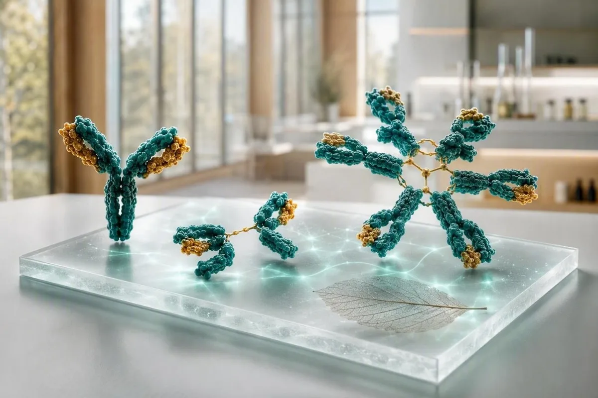
Tes imunoglobulin paling berguna bila IgG, IgA, dan IgM dibaca sebagai pola, bukan tiga penanda yang terisolasi. Nilai tinggi dapat mencerminkan aktivasi kekebalan yang luas atau satu protein abnormal; nilai rendah dapat menunjukkan obat-obatan, kehilangan protein, atau gangguan produksi antibodi.


- IgG biasanya 700-1.600 mg/dL pada orang dewasa; peningkatan yang persisten pada beberapa kelas sering mencerminkan peradangan kronis atau penyakit hati.
- IgA biasanya 70-400 mg/dL pada orang dewasa; defisiensi selektif dapat membuat tes penyakit seliak berbasis IgA yang negatif menjadi tidak dapat diandalkan.
- antibodi IgM biasanya 40-230 mg/dL pada orang dewasa; peningkatan yang terisolasi dan persisten memerlukan elektroforesis protein serum untuk menyingkirkan protein monoklonal.
- Ketiga rendah lebih mengkhawatirkan daripada satu hasil rendah dan memerlukan peninjauan infeksi, obat-obatan, kehilangan protein, dan respons antibodi vaksin.
- Peningkatan poliklonal berarti beberapa kelas imunoglobulin tinggi; ini umum menyertai penyakit autoimun, penyakit hati kronis, atau infeksi yang berkepanjangan.
- Pola monoklonal berarti satu kelas meningkat secara tidak proporsional atau pita sempit muncul pada elektroforesis; ini memerlukan imunofiksasi konfirmasi dan pengujian rantai ringan bebas (free-light-chain).
- Tinjauan segera masuk akal untuk demam dengan infeksi serius berulang, kebingungan baru, sesak napas, penyakit kuning, penurunan berat badan, nyeri tulang, atau peningkatan protein total yang cepat.
- Tren itu penting karena perbedaan 10-20% dapat timbul dari hidrasi, variasi uji, penyakit baru-baru ini, atau metode laboratorium yang berbeda.
Cara membaca tes imunoglobulin sebagai satu pola
Hasil IgG, IgA, dan IgM memiliki arti yang berbeda jika digabungkan dibandingkan jika berdiri sendiri. Panel yang meningkat secara luas biasanya menandakan stimulasi kekebalan poliklonal, sementara satu kelas yang dominan—terutama dengan protein total yang tinggi—menimbulkan pertanyaan terpisah tentang protein monoklonal. Per 5 September 2026, perbedaan tersebut tetap merupakan langkah pertama yang praktis sebelum mengaitkan diagnosis dengan tes darah imunoglobulin.
Interval referensi dewasa bervariasi berdasarkan metode, tetapi banyak laboratorium menggunakan IgG 700-1.600 mg/dL, IgA 70-400 mg/dL, dan IgM 40-230 mg/dL. Nilai 5% di luar kisaran tidak secara otomatis merupakan penyakit; Saya pertama-tama memeriksa interval laboratorium, usia, status kehamilan, infeksi baru-baru ini, kehilangan ginjal, dan apakah sampel diambil setelah imunoglobulin intravena.
Kantesti adalah seorang Analisa tes darah AI yang membaca IgG, IgA, dan IgM di samping protein total, albumin, penanda hati, kreatinin, dan CBC daripada memperlakukan tanda bahaya sebagai diagnosis. Pemeriksaan silang itu berguna karena albumin rendah dengan globulin yang meningkat menceritakan kisah yang sangat berbeda dari dehidrasi dengan kedua nilai yang terkonsentrasi; kami biomarker menjelaskan pengukuran terkait.
Dalam pekerjaan klinis saya, panel yang paling meyakinkan seringkali tidak rata-rata sempurna tetapi stabil: misalnya, IgG 1.690 mg/dL pada tiga pengambilan sampel selama 18 bulan dengan elektroforesis normal dan tanpa gejala. Dr. Thomas Klein masih akan bertanya tentang mata kering, pembengkakan sendi, infeksi berulang, dan obat-obatan, tetapi variasi ringan yang stabil jarang memiliki bobot yang sama dengan kenaikan 600 mg/dL dalam enam bulan.
Tiga imunoglobulin dalam bahasa awam
antibodi IgM adalah respons awal yang luas, IgA melindungi permukaan mukosa di saluran udara dan usus, dan IgG memberikan aktivitas antibodi bersirkulasi yang paling tahan lama. Waktu paruh biologis mereka sangat berbeda: IgG bertahan sekitar 21 hari, IgA sekitar 6 hari dalam serum, dan IgM sekitar 5 hari, sehingga perubahan serial tidak bergerak dengan kecepatan yang sama.
Rentang normal dewasa dan mengapa laboratorium Anda mungkin berbeda
Kisaran referensi yang dicetak di samping hasil Anda sendiri lebih diutamakan daripada kisaran online mana pun. Nephelometry, turbidimetry, dan populasi laboratorium yang dikalibrasi berdasarkan usia menghasilkan batas yang sedikit berbeda, terutama untuk IgA dan IgM.
Interval dewasa umum IgG 700-1.600 mg/dL, IgA 70-400 mg/dL, dan IgM 40-230 mg/dL setara dengan kira-kira 7-16 g/L, 0,7-4,0 g/L, dan 0,4-2,3 g/L. Anak-anak memiliki nilai yang sangat bergantung pada usia, dan kisaran dewasa tidak boleh digunakan untuk menilai bayi atau remaja.
Hasil yang mendekati ambang batas sangat rentan terhadap variasi biologis biasa. Jika IgM adalah 238 mg/dL terhadap batas atas 230 mg/dL setelah penyakit pernapasan, pengulangan dalam 8-12 minggu seringkali lebih mengungkapkan daripada memesan baterai besar segera; lihat panduan kami ke perubahan nyata antar pemeriksaan darah.
Puasa biasanya tidak diperlukan untuk imunoglobulin kuantitatif, tetapi dehidrasi berat dapat mengkonsentrasikan protein dan imunoglobulin IV dapat meningkatkan IgG yang terukur selama berminggu-minggu. Tanyakan kepada dokter yang memesan panel apakah Anda menerima penggantian antibodi, rituximab, kortikosteroid, antikonvulsan, atau pengobatan anti-CD20 dalam 6 bulan terakhir.
Ketika ketiga kelas imunoglobulin tinggi
Peningkatan IgG, IgA, dan IgM bersama-sama biasanya menunjukkan aktivasi kekebalan poliklonal daripada satu klon abnormal. Penyakit hati kronis, kondisi autoimun, infeksi persisten, dan beberapa gangguan radang usus adalah latar klinis yang umum.
Peningkatan poliklonal seringkali memperluas area gamma pada elektroforesis protein serum alih-alih menciptakan lonjakan sempit yang tajam. Dalam praktiknya, IgG 2.100 mg/dL, IgA 520 mg/dL, dan IgM 310 mg/dL dengan CRP yang meningkat menunjukkan jalur yang berbeda dari IgG 2.400 mg/dL dengan dua kelas lainnya ditekan.
Penyakit autoimun dapat menghasilkan pola ini, tetapi gejalanya memandu tes berikutnya. Mata dan mulut kering yang persisten, pembengkakan sendi, ruam, diare, atau neuropati dapat membenarkan pengujian antibodi dan komplemen yang ditargetkan; penjelasan kami tentang C3 dan C4 menjelaskan mengapa penurunan komplemen mengubah interpretasi tersebut.
Infeksi kronis juga dapat meningkatkan beberapa kelas selama berbulan-bulan, meskipun imunoglobulin tidak mengidentifikasi organisme atau membuktikan infeksi aktif. Peningkatan ESR dengan gejala konstitusional memerlukan tinjauan klinis, dan penjelasan kami tentang penyebab ESR yang tinggi menunjukkan mengapa penanda inflamasi nonspesifik tidak dapat menentukan penyebabnya.
Apa arti peningkatan IgM yang terisolasi
Peningkatan IgM yang terisolasi dapat terjadi setelah infeksi baru-baru ini, terjadi pada penyakit hati autoimun kolestatik, atau mewakili protein monoklonal IgM. Persistensi, tingkat peningkatan, gejala, dan elektroforesis lebih penting daripada satu angka batas.
IgM adalah antibodi pentamerik yang besar, dan kadar sedikit di atas 230 mg/dL seringkali mereda setelah tantangan kekebalan. Peningkatan IgM berulang sebesar 800 mg/dL atau 1.200 mg/dL, terutama dengan IgG dan IgA normal atau rendah, harus memicu elektroforesis protein serum, imunofiksasi, dan pengujian rantai ringan bebas serum daripada menebak-nebak.
Kolangitis bilier primer secara klasik terkait dengan peningkatan IgM, terutama ketika alkalinitas fosfatase dan gamma-glutamil transferase juga tinggi. Pedoman EASL menyarankan evaluasi penyakit hati kolestatik ketika alkalinitas fosfatase meningkat secara persisten dan antibodi antimitochondrial ada (EASL, 2017); penjelasan kami tentang pola kolestasis mencakup penanda penyerta.
Protein IgM monoklonal berbeda dari makroglobulinemia Waldenström, dan kebanyakan orang dengan gammopati monoklonal kecil tidak memiliki diagnosis kanker segera. Meskipun demikian, gejala seperti penglihatan kabur, sakit kepala, mimisan, neuropati baru, atau kelelahan yang tidak dapat dijelaskan memerlukan penilaian segera karena IgM yang sangat tinggi dapat mengubah viskositas serum; baca lebih lanjut tentang IgM tinggi menyebabkan.
IgA rendah: jebakan tes penyakit seliak dan petunjuk infeksi
Penurunan IgA dapat membuat skrining celiac transglutaminase jaringan IgA menjadi positif palsu. Defisiensi IgA selektif umumnya didefinisikan sebagai IgA serum di bawah 7 mg/dL pada seseorang yang berusia lebih dari 4 tahun dengan IgG dan IgM normal, meskipun definisi lokal dan pengujian ulang penting.
Jika total IgA rendah, dokter biasanya menggunakan peptida gliadin terdeamidasi berbasis IgG atau tes transglutaminase jaringan IgG bersama riwayat klinis. Jangan memulai diet bebas gluten sebelum pengujian yang tepat kecuali dianjurkan, karena kadar antibodi dapat menurun dan mengaburkan hasil; panduan tantangan gluten menjelaskan masalah penentuan waktu.
Banyak orang dengan defisiensi IgA parsial tidak memiliki masalah kesehatan yang signifikan, sementara yang lain melaporkan infeksi sinus, dada, atau saluran pencernaan yang berulang. Frekuensi, keparahan, dan penyebab bakteri yang terdokumentasi lebih penting daripada sekadar terkena dua kali pilek biasa dalam satu musim dingin; riwayat vaksinasi dan respons antibodi pneumokokus bisa lebih informatif daripada total IgA saja.
Reaksi produk darah jarang terjadi tetapi relevan: orang dengan defisiensi IgA berat dan antibodi anti-IgA mungkin memerlukan komponen yang dipilih secara khusus jika transfusi diperlukan. Ini adalah alasan untuk memberi tahu tim rumah sakit tentang defisiensi berat yang dikonfirmasi, bukan alasan untuk menghindari perawatan yang diperlukan secara medis; lihat diskusi kami yang lebih lengkap tentang hasil IgA rendah.
IgG rendah dan infeksi berulang: kapan hasilnya penting
IgG rendah menjadi signifikan secara klinis ketika menyertai infeksi yang berulang, tidak biasa parah, atau sulit sembuh. IgG dewasa di bawah 400 mg/dL umumnya lebih mengkhawatirkan daripada nilai 650 mg/dL, tetapi respons antibodi vaksin fungsional dapat mengesampingkan asumsi yang hanya didasarkan pada kuantitas.
Infeksi yang membuat ahli imunologi waspada adalah pneumonia berulang, bronkiektasis, infeksi sinus bakteri berulang yang memerlukan beberapa antibiotik, penyakit bakteri invasif, atau giardiasis kronis. Empat kali pilek virus ringan pada pekerja penitipan anak tidak setara dengan tiga pneumonia yang dikonfirmasi secara radiologis dalam 24 bulan.
Penyebab sekunder umum terjadi: rituximab dan terapi lain yang menargetkan sel B, kortikosteroid oral berkepanjangan, beberapa antikonvulsan, kehilangan protein dalam kisaran nefrotik, enteropati kehilangan protein, dan keganasan limfoid dapat menurunkan IgG. Protein urin, albumin, hitung limfosit, dan riwayat pengobatan sering kali memberikan lebih banyak informasi daripada panel antibodi acak lainnya; panduan protein dalam urin menjelaskan jalur kehilangan utama.
Bonilla dkk. (2015) merekomendasikan penilaian imunoglobulin kuantitatif bersama dengan respons antibodi spesifik ketika defisiensi imun primer dicurigai. Menurut pengalaman saya, respons pasca-vaksinasi yang normal bisa meyakinkan bahkan ketika IgG sedikit rendah, sedangkan respons yang buruk dengan IgG di bawah 500 mg/dL memerlukan masukan spesialis.
Ketika IgG, IgA, dan IgM semuanya rendah
Rendahnya IgG, IgA, dan IgM bersama-sama disebut hipogammaglobulinemia dan memerlukan pemeriksaan terstruktur, terutama jika disertai infeksi. Pola ini dapat mencerminkan gangguan produksi antibodi, efek pengobatan, kehilangan protein, penyakit limfoid stadium lanjut, atau, lebih jarang, perubahan sementara pasca-penyakit.
Panel IgG 360 mg/dL, IgA 28 mg/dL, dan IgM 18 mg/dL bukanlah hasil yang bisa ditunggu jika orang tersebut mengalami demam, infeksi dada berulang, atau penurunan berat badan. Seorang klinisi dapat mengulang panel, memeriksa elektroforesis, CBC dengan diferensial, tes ginjal dan hati, protein urin, dan antibodi spesifik vaksin, kemudian merujuk ke imunologi klinis atau hematologi.
Imunodefisiensi variabel umum sering muncul pada masa dewasa, tetapi diagnosisnya memerlukan lebih dari sekadar angka rendah: gejala, penyebab sekunder yang dikecualikan, dan penurunan produksi antibodi fungsional adalah pusatnya. Kantesti AI adalah layanan interpretasi tes lab AI yang dapat mengatur temuan-temuan di sekitarnya menjadi ringkasan kunjungan dokter, tetapi tidak dapat mendiagnosis defisiensi imun atau memutuskan penggantian imunoglobulin.
Penggantian imunoglobulin diresepkan untuk mencegah infeksi serius pada pasien yang terpilih, bukan sekadar untuk menormalkan hasil laboratorium. Regimen penggantian yang khas bersifat individual dan biasanya dimulai sekitar 400-600 mg/kg setiap bulan melalui rute intravena atau subkutan, dengan penyesuaian dosis untuk pengendalian infeksi dan kadar IgG terendah daripada target universal; panduan kami untuk pemeriksaan darah sistem imun menambah konteks yang berguna.
Bagaimana tes imunoglobulin dapat menunjukkan protein monoklonal
Protein monoklonal disarankan ketika satu kelas imunoglobulin sangat tinggi, kelas lain ditekan, atau elektroforesis menunjukkan pita sempit. Imunoglobulin kuantitatif saja tidak dapat membuktikan klonalitas; elektroforesis protein serum dan imunofiksasi adalah alat konfirmasi.
Pertimbangkan IgG 2.900 mg/dL dengan IgA 38 mg/dL dan IgM 21 mg/dL: pola itu lebih mencurigakan daripada jika ketiganya meningkat sedang. Total protein tinggi, rasio albumin-ke-globulin rendah, anemia, penurunan eGFR, kalsium tinggi, nyeri tulang, atau infeksi berulang menambah urgensi klinis; tinjau panduan kami untuk total protein tinggi.
International Myeloma Working Group membedakan MGUS dari mieloma aktif menggunakan jumlah protein, temuan sumsum tulang, dan efek organ—bukan hanya hasil imunoglobulin saja (Rajkumar et al., 2014). M-protein di bawah 3 g/dL masih dapat memerlukan pemantauan, sedangkan nilai yang lebih rendah dengan anemia atau cedera ginjal mungkin memerlukan penilaian hematologi yang lebih cepat.
Jangan terkecoh oleh protein total yang normal. Kelainan rantai ringan saja mungkin tidak menghasilkan peningkatan protein yang jelas, itulah sebabnya mengapa dokter menggabungkan rantai ringan bebas serum, imunofiksasi, dan pemeriksaan urin dalam presentasi yang sesuai; kami jalur pemeriksaan kanker darah menguraikan apa yang terjadi selanjutnya.
Pola penyakit hati: petunjuk IgA, IgG, dan IgM
Penyakit hati dapat meningkatkan imunoglobulin, tetapi kelas yang dominan hanya memberikan petunjuk, bukan diagnosis. IgA sering meningkat dengan cedera hati terkait alkohol, IgG dengan hepatitis autoimun, dan IgM dengan kolangitis bilier primer; tumpang tindih adalah hal biasa.
Peningkatan IgG di atas 1,1 kali batas atas normal mendukung hepatitis autoimun ketika transaminase dan autoantibodi cocok, tetapi tidak cukup sensitif atau spesifik untuk mendiagnosisnya sendiri. Penyakit virus akut, cedera hati kronis, dan penyakit autoimun sistemik semuanya dapat menciptakan sinyal yang serupa, sehingga dokter sering menggunakan pencitraan dan pengujian yang diarahkan oleh spesialis.
Peningkatan IgA bersama dengan peningkatan GGT, AST, dan makrositosis dapat sesuai dengan cedera hati terkait alkohol, namun penggunaan alkohol tidak boleh diasumsikan hanya dari sebuah panel. Efek obat, penyakit hati berlemak metabolik, dan penyebab lain sering terjadi; penjelasan kami tentang hasil ALT membantu menempatkan enzim di samping imunoglobulin.
Pesan keselamatan yang berguna adalah bahwa penyakit kuning, urin gelap, tinja pucat, kebingungan, pembengkakan perut, atau muntah darah memerlukan perawatan medis segera terlepas dari nilai imunoglobulin. Peningkatan IgA terisolasi yang stabil sebesar 430 mg/dL tidak menyebabkan gejala-gejala tersebut dan seharusnya tidak mengalihkan perhatian dari evaluasi segera.
Memisahkan infeksi, peradangan, dan aktivitas autoimun
Kadar imunoglobulin meningkat terlalu lambat dan tidak spesifik untuk mendiagnosis infeksi akut dengan sendirinya. CRP, hitung darah lengkap, kultur, atau tes patogen yang ditargetkan menjawab pertanyaan infeksi segera dengan lebih baik, sementara imunoglobulin menggambarkan latar belakang kekebalan yang lebih luas.
IgM sering digambarkan sebagai antibodi infeksi dini, tetapi IgM serum total berbeda dengan tes IgM spesifik patogen. IgM total sebesar 290 mg/dL tidak dapat mengonfirmasi influenza, virus Epstein-Barr, atau infeksi saluran kemih; gejala dan pengujian langsung menentukan apakah pengobatan diperlukan.
Peradangan autoimun dapat meningkatkan IgG selama bertahun-tahun sebelum diagnosis jelas, terutama dengan hipergammaglobulinemia dan pola elektroforesis yang luas. ANA, faktor rematoid, dan panel ENA harus dipesan untuk menjawab pertanyaan klinis, bukan sebagai ekspedisi memancing; misalnya, kami panduan anti-dsDNA menjelaskan mengapa hasil positif memerlukan konteks ginjal dan komplemen.
Dr. Thomas Klein telah melihat pasien yang ketakutan oleh IgG tinggi setelah penyakit virus yang normal pada pengulangan 3 bulan. Sebaliknya, IgG yang persisten di atas 2.000 mg/dL ditambah C3 rendah, proteinuria, dan gejala sendi bukanlah sesuatu yang akan saya tunda selama setahun—temuan gabungan tersebut membenarkan penilaian klinis yang tepat waktu.
Obat-obatan, kehilangan protein, dan efek waktu yang mengubah hasil
Obat-obatan dan kehilangan protein dapat menurunkan imunoglobulin tanpa kelainan kekebalan primer. Obat anti-CD20 dapat mengurangi IgM terlebih dahulu dan IgG kemudian, sementara kehilangan protein ginjal atau usus dapat menurunkan beberapa kelas bersama dengan albumin.
Hipogammaglobulinemia terkait Rituximab dapat muncul berbulan-bulan setelah infus terakhir karena pemulihan sel B dan pemulihan antibodi tidak identik. Bawa daftar obat lengkap—termasuk biologis, dosis steroid, obat antikejang, dan kemoterapi—ke janji temu; kami panduan tren keamanan obat menjelaskan mengapa tanggal itu penting.
Keadaan yang kehilangan protein seringkali meninggalkan jejak tambahan: albumin rendah di bawah sekitar 3,5 g/dL, edema, diare, rasio albumin-kreatinin urin yang tinggi atau protein total yang rendah. Sebaliknya, dehidrasi dapat membuat imunoglobulin tampak tinggi sementara albumin dan hematokrit meningkat sejajar.
Pengulangan tes biasanya paling baik dilakukan saat Anda stabil secara klinis, menggunakan laboratorium yang sama jika memungkinkan. [Kantesti] dapat membandingkan PDF atau foto bertanggal dengan hasil sebelumnya dan menandai pergeseran besar, tetapi pemeriksaan unggahan yang cermat masuk akal karena unit dan rentang referensi dapat ditangkap secara tidak benar.
Tes lanjutan yang mengklarifikasi panel abnormal
Tes tindak lanjut yang tepat tergantung pada polanya: elektroforesis untuk peningkatan yang tidak proporsional, antibodi vaksin untuk dugaan defisiensi, dan tes urin atau hati untuk kehilangan protein atau petunjuk hati. Mengulang tiga angka yang sama tanpa pertanyaan seringkali tidak menambah banyak.
Untuk hasil yang tinggi atau tidak merata, dokter umum meminta elektroforesis protein serum, imunofiksasi, rantai ringan bebas serum, hitung darah lengkap, kalsium, kreatinin, albumin, dan enzim hati. Pita sempit pada elektroforesis harus diketik dengan imunofiksasi karena kelas—IgG, IgA, atau IgM—mengubah rencana diferensial dan tindak lanjut.
Untuk hasil rendah dengan infeksi, pengujian dapat mencakup subkelas IgG dan titer antibodi terhadap tetanus, Haemophilus influenzae tipe b, atau serotipe pneumokokus sebelum dan sesudah vaksinasi. Tidak ada ambang batas pneumokokus protektif tunggal yang cocok untuk setiap laboratorium dan kelompok usia, yang merupakan salah satu area di mana interpretasi spesialis lebih penting daripada batas daring.
Kantesti adalah seorang Alat analisis pemeriksaan darah berbasis AI yang dapat merakit hasil ini ke dalam garis waktu dan mengidentifikasi konteks yang hilang, seperti albumin atau elektroforesis yang tidak ada, untuk dipertimbangkan oleh dokter Anda. [Our] panduan teknologi menjelaskan bagaimana bendera kontekstual dihasilkan tanpa menggantikan pengambilan keputusan medis.
Gejala yang tidak boleh menunggu pengulangan rutin
Hasil imunoglobulin saja jarang menimbulkan keadaan darurat, tetapi gejala tertentu demikian. Cari penilaian mendesak pada hari yang sama untuk demam tinggi dengan penyakit parah, sesak napas, kebingungan, kelemahan baru, penyakit kuning, urin yang sangat berkurang, sakit kepala parah dengan perubahan visual, atau perdarahan yang tidak terkontrol.
Kadar imunoglobulin rendah ditambah demam 38,0°C atau lebih tinggi lebih memprihatinkan pada seseorang yang menjalani kemoterapi, steroid dosis tinggi, terapi yang ditargetkan pada sel B, atau dengan riwayat infeksi invasif. Jangan menunggu pesan portal jika sulit bernapas, kadar oksigen rendah, atau orang tersebut tampak bingung atau mengantuk secara tidak biasa.
Protein monoklonal yang mungkin memerlukan tinjauan lebih cepat ketika terjadi dengan nyeri tulang baru, anemia yang tidak dapat dijelaskan, peningkatan kreatinin, kalsium di atas rentang atas lokal, neuropati, atau penurunan berat badan yang tidak disengaja. Ini bukan bukti gangguan sel plasma, tetapi ini adalah fitur yang digunakan dokter untuk memprioritaskan pengujian; [our] second-opinion dapat membantu mempersiapkan pertanyaan.
Untuk kelainan ringan yang stabil tanpa tanda bahaya, janji temu yang direncanakan dalam beberapa minggu biasanya masuk akal. Bawa hasil sebelumnya, tanggal infeksi, perubahan pengobatan, dan riwayat keluarga; grafik 2 tahun seringkali lebih berguna secara klinis daripada satu angka yang disorot.
Menggunakan interpretasi AI dengan aman untuk hasil imunoglobulin
AI dapat mengatur pola tes imunoglobulin, tetapi tidak dapat menentukan apakah seseorang memiliki defisiensi kekebalan, penyakit hati, atau gangguan monoklonal. Interpretasi yang aman menjaga rentang laboratorium, gejala, obat-obatan, tren, dan tes konfirmasi tetap terlihat.
[Kantesti] AI meninjau IgG, IgA, dan IgM bersama dengan protein dan penanda organ yang mengubah artinya, kemudian menyajikan pertanyaan tindak lanjut daripada menyatakan diagnosis. Dalam alur kerja analisis kami, IgA rendah yang terisolasi memicu peringatan pengujian coeliac, sedangkan ketiga nilai rendah ditambah albumin rendah memicu pertimbangan kehilangan, efek obat, dan tinjauan imunologi klinis.
Privasi penting dengan hasil terkait kekebalan dan kanker. [Kantesti Ltd] menggunakan penanganan yang selaras dengan GDPR, dan [our] halaman validasi medis menjelaskan pengawasan klinis dan batasan di balik interpretasi lab otomatis.
Laporan yang baik harus membantu Anda bertanya, “Apakah ini aktivasi kekebalan yang luas, kehilangan protein, produksi yang berkurang, atau satu protein tunggal?” Itulah tingkat kepastian yang tepat sebelum elektroforesis atau evaluasi spesialis memberikan jawaban.
Pertanyaan yang harus diajukan setelah panel imunoglobulin abnormal
Pertanyaan lanjutan terbaik biasanya adalah “pola apa ini?” daripada “bagaimana cara menurunkan atau menaikkan angka ini?” Tanyakan apakah hasilnya persisten, poliklonal, atau monoklonal, apakah infeksi menunjukkan gangguan fungsi, dan penyebab sekunder mana yang telah diperiksa.
Pertanyaan yang berguna meliputi: Apakah elektroforesis protein serum dilakukan? Apakah albumin, protein urin, dan enzim hati konsisten dengan kehilangan atau penyakit hati? Haruskah saya melakukan tes antibodi vaksin? Apakah pengujian ulang diperlukan dalam 8-12 minggu? Pertanyaan-pertanyaan tersebut lebih produktif daripada membeli suplemen, yang tidak dapat secara andal memperbaiki defisiensi antibodi yang bermakna secara klinis.
Jika rujukan spesialis direncanakan, catat jumlah infeksi yang diobati dengan antibiotik dalam 12 bulan terakhir, kunjungan rumah sakit, pneumonia yang dikonfirmasi pencitraan, dan paparan obat. Catatan yang tepat—“tiga kali pengobatan antibiotik dan satu pneumonia”—lebih membantu daripada “saya sering sakit”; pelacak hasil lab menawarkan format catatan yang praktis.
Kita Dewan Penasehat Medis mendukung standar tinjauan klinis Kantesti, tetapi dokter Anda sendiri tetap menjadi orang yang dapat memeriksa Anda, memesan pengujian konfirmasi, dan bertindak berdasarkan temuan mendesak. Kebanyakan pasien menemukan bahwa percakapan yang tenang berdasarkan pola mengubah penanda yang mengkhawatirkan menjadi langkah selanjutnya yang dapat dikelola.
Pertanyaan yang Sering Diajukan
Berapa rentang normal tes imunoglobulin untuk orang dewasa?
Rentang imunoglobulin dewasa tipikal adalah IgG 700-1.600 mg/dL, IgA 70-400 mg/dL, dan IgM 40-230 mg/dL, meskipun interval yang tercetak di setiap laboratorium harus digunakan untuk interpretasi. Hasil juga dilaporkan sebagai g/L di banyak negara: 7-16 g/L untuk IgG, 0,7-4,0 g/L untuk IgA, dan 0,4-2,3 g/L untuk IgM. Hasil yang sedikit di luar rentang dapat mencerminkan penyakit baru-baru ini, hidrasi, atau variasi uji. Kelainan dan gejala yang menetap menentukan apakah tindak lanjut diperlukan.
Apa arti tingginya IgG, IgA, dan IgM secara bersamaan?
Peningkatan IgG, IgA, dan IgM yang tinggi secara bersamaan paling sering menunjukkan aktivasi imun poliklonal akibat inflamasi kronis, penyakit autoimun, infeksi persisten, atau penyakit hati. Peningkatan luas di ketiga kelas berbeda dari protein monoklonal, yang biasanya menciptakan satu kelas yang sangat tinggi secara tidak proporsional atau pita elektroforesis yang sempit. Nilai seperti IgG 2.000 mg/dL, IgA 500 mg/dL, dan IgM 300 mg/dL harus diinterpretasikan bersama dengan CRP, tes fungsi hati, albumin, dan elektroforesis protein serum. Pola saja tidak mendiagnosis penyakit autoimun atau infeksi.
Dapatkah kadar imunoglobulin yang rendah menyebabkan infeksi yang sering?
Kadar imunoglobulin yang rendah dapat meningkatkan risiko infeksi bakteri saluran pernapasan atau saluran pencernaan berulang, terutama ketika IgG di bawah 400-500 mg/dL atau respons antibodi vaksin buruk. Pola yang paling mengkhawatirkan adalah pneumonia berulang, bronkiektasis, infeksi bakteri invasif, atau beberapa infeksi sinus yang diobati dengan antibiotik, daripada pilek virus sesekali. Penurunan IgG, IgA, dan IgM bersama-sama harus mendorong peninjauan terhadap obat-obatan, kehilangan protein ginjal atau usus, kondisi limfoid, dan defisiensi imun primer. Pengujian antibodi fungsional sering kali mengklarifikasi risiko lebih baik daripada satu nilai total IgG.
Apakah IgA rendah memengaruhi pengujian penyakit seliak?
Kadar IgA yang rendah dapat menyebabkan hasil tes penyakit seliak IgA transglutaminase jaringan yang negatif palsu karena tes tersebut bergantung pada produksi antibodi IgA. Defisiensi IgA selektif sering didefinisikan sebagai kadar IgA di bawah 7 mg/dL setelah usia 4 tahun dengan IgG dan IgM yang normal. Ketika kadar IgA total rendah, klinisi umumnya menggunakan tes peptida gliadin terdeaminasi IgG atau transglutaminase jaringan IgG sebagai gantinya. Terus konsumsi gluten hingga rencana pengujian disepakati, karena menghindari gluten dapat menurunkan kadar antibodi dan mengurangi sensitivitas tes.
Tes apa yang mengonfirmasi adanya protein imunoglobulin monoklonal?
Elektroforesis protein serum mendeteksi pola protein abnormal, dan imunofiksasi mengonfirmasi apakah imunoglobulin monoklonal ada dan mengidentifikasi kelasnya. Pengujian rantai ringan bebas serum biasanya ditambahkan karena beberapa kelainan sel plasma terutama menghasilkan rantai ringan dan mungkin tidak secara signifikan meningkatkan total protein. Hasil monoklonal memerlukan penilaian konsentrasi protein M, hitung darah, kalsium, fungsi ginjal, dan gejala; protein M saja tidak secara otomatis berarti kanker. Studi protein urin mungkin juga diperlukan dalam kasus-kasus tertentu.
Dapatkah stres menyebabkan IgG, IgA atau IgM tinggi?
Stres psikologis sehari-hari biasanya tidak menyebabkan peningkatan terisolasi yang besar pada IgG, IgA, atau IgM dalam tes darah imunoglobulin. Stres dapat memengaruhi tidur, kerentanan terhadap infeksi, dan pensinyalan inflamasi, tetapi nilai yang persisten di atas batas atas laboratorium tidak boleh diabaikan sebagai stres tanpa meninjau penanda hati, CRP, obat-obatan, dan elektroforesis protein jika sesuai. Perubahan sedang sebesar 10-20% dapat terjadi antar pengambilan sampel karena variasi biologis dan analitik normal. Mengulang panel setelah 8-12 minggu ketika pasien sehat secara klinis seringkali merupakan langkah awal yang masuk akal untuk kenaikan ringan yang tidak dapat dijelaskan.
Dapatkan Analisis Tes Darah Berbasis AI Hari Ini
Bergabunglah dengan lebih dari 2 juta pengguna di seluruh dunia yang mempercayai Kantesti untuk analisis instan dan akurat terhadap tes lab. Unggah hasil tes darah Anda dan terima interpretasi komprehensif biomarker 15,000+ dalam hitungan detik.
📚 Publikasi Riset yang Dirujuk
Klein, T., Mitchell, S., & Weber, H. (2026). Kantesti LTD. (2026). AI Blood Test Analyzer: 2,5M Tes Dianalisis | Global Health Report 2026. Zenodo.. Kantesti Penelitian Medis AI.
Klein, T., Mitchell, S., & Weber, H. (2026). Kantesti LTD. (2026). Tes Darah RDW: Panduan Lengkap untuk RDW-CV, MCV & MCHC. Zenodo.. Kantesti Penelitian Medis AI.
📖 Referensi Medis Eksternal
⚕️ Penafian Medis
Artikel ini hanya untuk tujuan edukasi dan tidak merupakan nasihat medis. Selalu konsultasikan dengan penyedia layanan kesehatan yang berkualifikasi untuk keputusan diagnosis dan perawatan.
Sinyal Kepercayaan E-E-A-T
Pengalaman
Tinjauan klinis yang dipimpin dokter terhadap alur kerja interpretasi hasil lab.
Keahlian
Fokus pada kedokteran laboratorium tentang bagaimana biomarker berperilaku dalam konteks klinis.
Kewenangan
Ditulis oleh Dr. Thomas Klein dengan peninjauan oleh Dr. Sarah Mitchell dan Prof. Dr. Hans Weber.
Kepercayaan
Interpretasi berbasis bukti dengan jalur tindak lanjut yang jelas untuk mengurangi kepanikan.