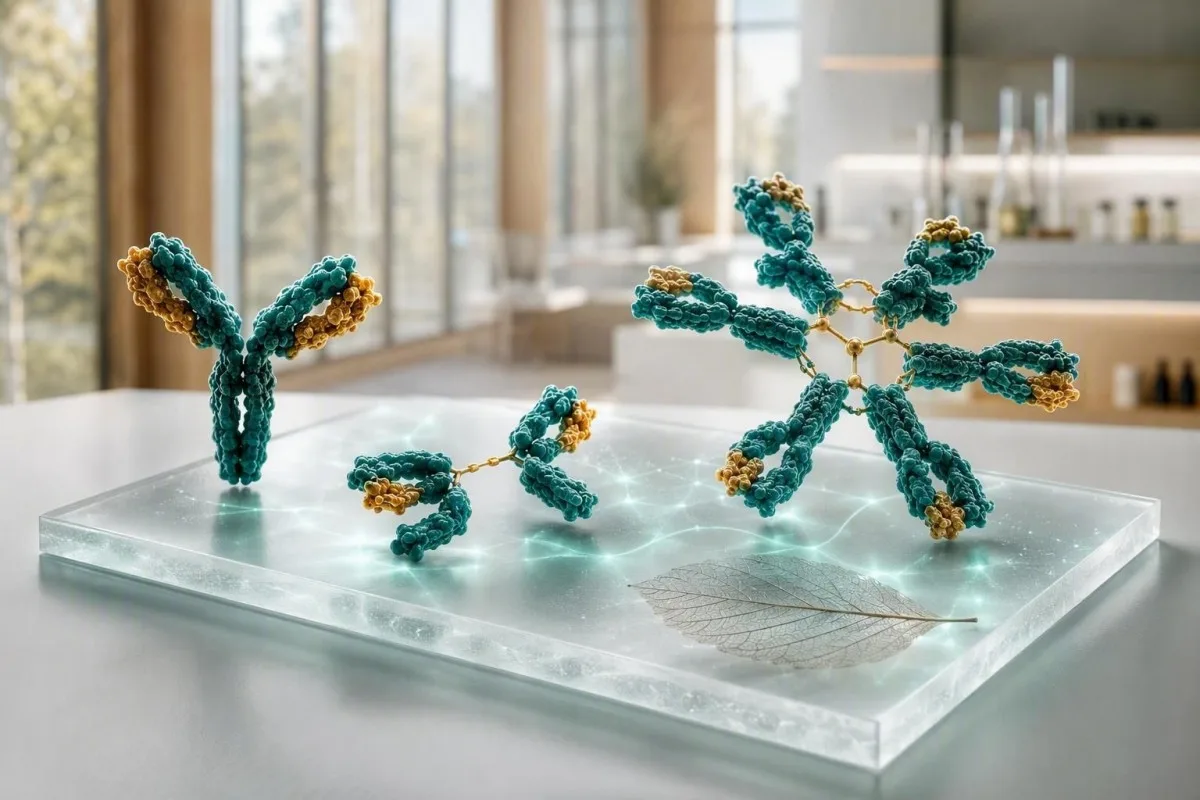
Imunoglobulīnu tests ir visnoderīgākais, ja IgG, IgA un IgM tiek interpretēti kā kopējs rezultāts, nevis trīs atsevišķi rādītāji. Augstas vērtības var atspoguļot plašu imūnsistēmas aktivāciju vai vienu patoloģisku proteīnu; zemas vērtības var norādīt uz medikamentiem, proteīnu zudumu vai traucētu antivielu veidošanos.


- IgG parasti ir 700–1600 mg/dL pieaugušajiem; pastāvīgs vairāku klašu pieaugums bieži atspoguļo hronisku iekaisumu vai aknu slimību.
- IgA parasti ir 70–400 mg/dL pieaugušajiem; selektīvs deficīts var padarīt neuzticamu negatīvu IgA testu celiakijai.
- IgM parasti ir 40–230 mg/dL pieaugušajiem; izolētam, pastāvīgam pieaugumam nepieciešama seruma proteīnu elektroforēze, lai izslēgtu monoklonālu proteīnu.
- Visi trīs zemi ir vairāk satraucoši nekā viens zems rezultāts un prasa infekciju, medikamentu, proteīnu zuduma un vakcīnu antivielu atbilžu pārskatīšanu.
- Poliklonāla paaugstināšanās nozīmē, ka ir paaugstinātas vairākas imunoglobulīnu klases; tas bieži pavada autoimūnas slimības, hroniskas aknu slimības vai ilgstošas infekcijas.
- Monoklonāls paraugs nozīmē, ka viena klase ir nesamērīgi paaugstināta vai uz elektroforēzes parādās šauras joslas; nepieciešama apstiprinoša imunofiksācija un brīvo vieglo ķēžu testēšana.
- steidzama izvērtēšana ir saprātīgs drudzis ar atkārtotām nopietnām infekcijām, jaunu apjukumu, elpas trūkumu, dzeltenumu, svara zudumu, kaulu sāpēm vai strauji augošu kopējo proteīnu.
- Svarīga ir tendence jo 10% atšķirība var rasties no hidratācijas, testa variācijām, nesenas slimības vai atšķirīgas laboratorijas metodes.
Kā interpretēt imunoglobulīnu testu kā kopēju rezultātu
IgG, IgA un IgM rezultāti kopā nozīmē ko citu nekā atsevišķi. Plaši paaugstināts panelis parasti signalizē par policlonālu imūno stimulāciju, savukārt viena dominējošā klase — īpaši ar augstu kopējo proteīnu — rada atsevišķu jautājumu par monoklonālu proteīnu. Sākot ar 2026. gada 5. septembri, šī atšķirība joprojām ir praktisks pirmais solis pirms diagnozes noteikšanas imūnglobulīnu asins analīzei.
Pieaugušo atsauces intervāli atšķiras atkarībā no metodes, bet daudzas laboratorijas izmanto IgG 700–1600 mg/dL, IgA 70–400 mg/dL un IgM 40–230 mg/dL. Vērtība ārpus diapazona nav automātiska slimība; vispirms pārbaudiet laboratorijas intervālu, vecumu, grūtniecības statusu, nesenu infekciju, nieru zudumu un to, vai paraugs tika ņemts pēc intravenoza imūnglobulīna.
Kantesti ir AI asins analīžu analizators kas nolasa IgG, IgA un IgM blakus kopējam proteīnam, albumīnam, aknu marķieriem, kreatinīnam un CBC, nevis uzskata sarkano karogu par diagnozi. Šī krusteniskā pārbaude ir noderīga, jo zems albumīna līmenis ar paaugstinātu globulīnu līmeni stāsta pavisam citu stāstu nekā dehidrācija ar abām koncentrētajām vērtībām; mūsu biomarķieru rokasgrāmata izskaidro saistītos mērījumus.
Manā klīniskajā darbā visatbilstošākais panelis bieži nav perfekti vidējs, bet stabils: piemēram, IgG 1690 mg/dL trīs reizēs 18 mēnešu laikā ar normālu elektroforēzi un bez simptomiem. Dr. Thomas Klein joprojām jautātu par sausām acīm, locītavu pietūkumu, atkārtotām infekcijām un medikamentiem, taču stabila neliela svārstība reti ir tikpat nozīmīga kā 600 mg/dL pieaugums sešos mēnešos.
Trīs imūnglobulīni vienkāršā valodā
IgM ir agrīnais plašais atbildētājs, IgA aizsargā gļotādas virsmas elpceļos un zarnās, un IgG nodrošina visizturīgāko cirkulējošo antivielu aktivitāti. To bioloģiskais pusiznīcināšanās laiks ir ievērojami atšķirīgs: IgG ilgst apmēram 21 dienu, IgA apmēram 6 dienas serumā un IgM apmēram 5 dienas, tāpēc sērijas izmaiņas nenotiek vienātempā.
Tipiskās pieaugušo normas un iemesli, kāpēc jūsu laboratorija var atšķirties
Jūsu rezultātam blakus drukātā atsauces vērtību tabula ir prioritāra pār jebkuru tiešsaistes vērtību tabulu. Nephelometrija, turbidimetrija un vecumam kalibrētas laboratoriju populācijas rada nedaudz atšķirīgas robežas, īpaši IgA un IgM gadījumā.
Kopējie pieaugušo intervāli IgG 700–1600 mg/dL, IgA 70–400 mg/dL un IgM 40–230 mg/dL pārvēršas aptuveni par 7–16 g/L, 0,7–4,0 g/L un 0,4–2,3 g/L. Bērniem ir ievērojami no vecuma atkarīgas vērtības, un pieaugušo vērtību tabulu nekad nedrīkst izmantot, lai novērtētu zīdaini vai pusaudzi.
Rezultāts, kas atrodas netālu no robežvērtības, ir īpaši pakļauts parastajām bioloģiskajām svārstībām. Ja IgM ir 238 mg/dL pret augšējo robežu 230 mg/dL pēc elpceļu slimības, atkārtota analīze pēc 8–12 nedēļām bieži ir informatīvāka nekā tūlītējas plašas analīzes veikšana; skatiet mūsu ceļvedi uz reālām izmaiņām starp asins analīzēm.
Parasti nav nepieciešams gavēnis kvantitatīviem imūnglobulīniem, taču smaga dehidrācija var koncentrēt proteīnus, un IV imūnglobulīns var paaugstināt izmērīto IgG vairākas nedēļas. Jautājiet ārstam, kurš nozīmējis paneli, vai pēdējo 6 mēnešu laikā esat saņēmis antivielu aizstājterapiju, rituksimabu, kortikosteroīdus, pretepilepsijas līdzekļus vai anti-CD20 terapiju.
Kad visas trīs imunoglobulīnu klases ir augstas
Augsts IgG, IgA un IgM kopā parasti norāda uz poliklonālu imūno aktivāciju, nevis vienu nenormālu klonu. Hroniskas aknu slimības, autoimūnas slimības, pastāvīgas infekcijas un daži iekaisuma zarnu traucējumi ir bieži sastopami klīniskie apstākļi.
Poliklonāls paaugstinājums bieži vien paplašina gamma reģionu uz seruma proteīnu elektroforēzes, nevis rada asu šauru smaili. Praktiski, IgG 2,100 mg/dL, IgA 520 mg/dL un IgM 310 mg/dL ar paaugstinātu CRP norāda uz citu ceļu nekā IgG 2,400 mg/dL ar divām citām nomāktām klasēm.
Autoimūna slimība var izraisīt šo modeli, taču simptomi nosaka nākamo testu. Pastāvīgs sausums acīs un mutē, pietūkušas locītavas, izsitumi, caureja vai neiropātija var attaisnot mērķtiecīgu auto antivielu un komplementa testēšanu; mūsu C3 un C4 ceļvedis izskaidro, kāpēc zems komplements maina šo interpretāciju.
Hroniska infekcija arī var paaugstināt vairākas klases mēnešiem, lai gan imūnglobulīni neidentificē organismu vai neapstiprina aktīvu infekciju. Augsts ESR ar vispārējiem simptomiem prasa klīnisku pārskatīšanu, un mūsu izskaidrojums par augstiem ESR cēloņiem parāda, kāpēc nesterils iekaisuma marķieris nevar noteikt cēloni.
Ko var nozīmēt izolēti augsts IgM
Izolēts augsts IgM rezultāts var sekot pēc nesenas infekcijas, rasties ar holestātisku autoimūnu aknu slimību vai attēlot IgM monoklonālu proteīnu. Pastāvība, paaugstinājuma pakāpe, simptomi un elektroforēze ir svarīgāki nekā viens robežzīmes skaitlis.
IgM ir liels pentamērisks antivielu veids, un līmenis nedaudz virs 230 mg/dL bieži vien normalizējas pēc imūnu izaicinājuma. Atkārtots IgM 800 mg/dL vai 1,200 mg/dL, īpaši ar normālu vai zemu IgG un IgA, jāizsauc seruma proteīnu elektroforēze, imunofiksācija un seruma brīvo vieglo ķēžu testēšana, nevis minējumi.
Primārā biliārā holangīts klasiski asociējas ar paaugstinātu IgM, īpaši, ja ir arī paaugstināts sārmainās fosfatāzes un gamma-glutamiltransferāzes līmenis. EASL vadlīnijas iesaka izvērtēt holestātisku aknu slimību, ja sārmainā fosfatāze ir pastāvīgi paaugstināta un ir klātesošs antimito-hondriālu antivielu (EASL, 2017); mūsu holestāzes modeļa ceļvedis aptver pavadošos marķierus.
Monoklonāls IgM proteīns nav tas pats, kas Valdenstrēma makroglobulinēmija, un lielākajai daļai cilvēku ar nelielu monoklonālu gammapātiju nav tūlītējas vēža diagnozes. Tomēr simptomi, piemēram, redzes miglainība, galvassāpes, deguna asiņošana, jauna neiropātija vai neizskaidrojams nogurums, prasa tūlītēju novērtēšanu, jo ļoti augsts IgM var mainīt seruma viskozitāti; lasiet vairāk par augstu IgM cēloņiem.
Zems IgA: kļūda celiakijas testēšanā un infekcijas pazīmes
Zems IgA var padarīt IgA audu transglutamināzes celiakijas skrīningu kļūdaini negatīvu. Selektīvs IgA deficīts parasti tiek definēts kā seruma IgA līmenis zem 7 mg/dL personām vecumā virs 4 gadiem ar normālu IgG un IgM, lai gan vietējās definīcijas un atkārtota testēšana ir svarīga.
Ja kopējais IgA ir zems, klīnicisti parasti izmanto IgG-bāzētu deamidētu gliadīna peptīdu vai IgG audu transglutamināzes testu klīniskās anamnēzes. Nesāciet diētu bez glutēna pirms atbilstošas testēšanas, ja vien nav ieteikts, jo antivielu līmenis var pazemināties un aizsegt rezultātu; mūsu ceļvedis par glutēna izaicinājumu apraksta laika problēmu.
Daudziem cilvēkiem ar daļēju IgA deficītu nav nopietnu veselības problēmu, savukārt citi ziņo par atkārtotām deguna blakusdobumu, krūškurvja vai gastrointestinālām infekcijām. Biežums, smagums un dokumentētie bakteriālie cēloņi ir svarīgāki nekā divas parastas saaukstēšanās ziemā; vakcinācijas vēsture un pneimokoku antivielu reakcija var būt informatīvāka nekā tikai kopējais IgA.
Asins produktu reakcijas ir retas, bet nozīmīgas: cilvēkiem ar dziļu IgA deficītu un anti-IgA antivielām var būt nepieciešamas īpaši izvēlētas sastāvdaļas, ja nepieciešama transfūzija. Tas ir iemesls, kāpēc jāinformē slimnīcas personāls par apstiprinātu smagu deficītu, nevis iemesls, lai izvairītos no medicīniski nepieciešamas aprūpes; skatiet mūsu pilnīgāku apspriedi par zemu IgA rezultātu.
Zems IgG un atkārtotas infekcijas: kad rezultāts ir svarīgs
Zems IgG kļūst klīniski nozīmīgs, ja tas pavada atkārtotas, neparasti smagas vai slikti izzūdošas infekcijas. Pieaugušā IgG zem 400 mg/dL parasti rada lielākas bažas nekā vērtība 650 mg/dL, taču funkcionālās vakcīnu antivielu atbildes var mainīt pieņēmumus, kas balstīti tikai uz daudzumu.
Infekcijas, kas liek imunologiem apstāties, ir atkārtota pneimonija, bronhektāzes, atkārtotas bakteriālas deguna blakusdobumu infekcijas, kas prasa vairākus antibiotiku kursus, invazīvas bakteriālas slimības vai hroniska lamblioze. Četras vieglas vīrusu saaukstēšanās bērnu aprūpes darbiniekam nav ekvivalents trim radioloģiski apstiprinātām pneimonijām 24 mēnešu laikā.
Sekundārie cēloņi ir bieži: rituksimabs un citas B šūnu vērstas terapijas, ilgstoši kortikosteroīdi, daži pretepilepsijas līdzekļi, nieru darbības traucējumi ar proteīnu izdalīšanos urīnā, proteīnu zudums zarnās un limfoīdā audzējs var pazemināt IgG. Urīna proteīns, albumīns, limfocītu skaits un medikamentu vēsture bieži sniedz vairāk nekā cits nejaušs antivielu panelis; mūsu proteīns urīnā ceļvedis paskaidro galveno zuduma ceļu.
Bonilla et al. (2015) iesaka kvantitatīvos imunglobulīnus kopā ar specifiskām antivielu atbildēm, ja tiek aizdomas par primāru imūndeficītu. Manā pieredzē normāla atbilde pēc vakcinācijas var būt nomierinoša pat tad, ja IgG ir nedaudz zems, savukārt vājas atbildes ar IgG zem 500 mg/dL prasa speciālista iesaistīšanos.
Kad IgG, IgA un IgM visi ir zemi
Zems IgG, IgA un IgM kopā tiek saukts par hipogamaglobulinēmiju un prasa strukturālu izmeklēšanu, īpaši ar infekcijām. Shēma var atspoguļot traucētu antivielu veidošanos, medikamentu ietekmi, proteīnu zudumu, progresējošu limfoīdo slimību vai, retāk, pagaidu izmaiņas pēc slimības.
IgG 360 mg/dL, IgA 28 mg/dL un IgM 18 mg/dL panelis nav rezultāts, ko gaidīt un novērot, ja personai ir drudzis, atkārtotas krūškurvja infekcijas vai svara zudums. Klīniskais ārsts var atkārtot paneli, pārbaudīt elektroforēzi, CBC ar diferenciāciju, nieru un aknu testus, urīna proteīnu un vakcīnspecifiskās antivielas, pēc tam nosūtīt uz klīnisko imunoloģiju vai hematoloģiju.
Kopēja mainīga imūndeficīta bieži parādās pieaugušā vecumā, taču tās diagnostika prasa vairāk nekā zemu skaitļu: simptomi, izslēgti sekundārie cēloņi un samazināta funkcionālā antivielu veidošanās ir centrālas. Kantesti AI ir AI laboratorijas testa interpretācijas pakalpojumā kas var sakārtot šos apkārtējos atradumus ārsta apmeklējuma kopsavilkumā, taču tas nevar diagnosticēt imūndeficītu vai noteikt imunglobulīnu aizstāšanu.
Imunglobulīnu aizstāšana tiek izrakstīta, lai novērstu nopietnas infekcijas izvēlētiem pacientiem, nevis tikai, lai normalizētu laboratorijas rezultātu. Tipiski aizstāšanas režīmi ir individuāli un parasti sākas apmēram 400-600 mg/kg katru mēnesi intravenozi vai subkutāni, dozējumu pielāgojot infekciju kontrolei un minimālajam IgG, nevis universālajam mērķim; mūsu ceļvedis par imūnsistēmas asins analīzēm sniedz noderīgu kontekstu.
Kā imunoglobulīnu tests var norādīt uz monoklonālu proteīnu
Monoklonālā proteīna klātbūtne tiek pieņemta, ja viena imunglobulīna klase ir nesamērīgi augsta, citas klases ir nomāktas, vai elektroforēze parāda šauru joslu. Kvantitatīvi imunglobulīni vieni paši nevar pierādīt klonalitāti; seruma proteīnu elektroforēze un imunofiksācija ir apstiprinošie rīki.
Apsveriet IgG 2,900 mg/dL ar IgA 38 mg/dL un IgM 21 mg/dL: šī shēma ir aizdomīgāka nekā visas trīs būtu nedaudz paaugstinātas. Augsts kopējais proteīns, zems albumīna-globulīna attiecība, anēmija, samazināts eGFR, augsts kalcija līmenis, kaulu sāpes vai atkārtotas infekcijas pievieno klīnisku steidzamību; pārskatiet mūsu ceļvedi par augstu kopējo proteīnu.
Starptautiskā mielomas darba grupa atšķir MGUS no aktīvas mielomas, izmantojot olbaltumvielu daudzumu, kaulu smadzeņu atradi un orgānu bojājumu, nevis tikai imūnglobulīnu rezultātu (Rajkumar et al., 2014). M-proteīna līmenim zem 3 g/dL joprojām var būt nepieciešama novērošana, savukārt zemāks līmenis kopā ar anēmiju vai nieru bojājumu var prasīt ātrāku hematologa izvērtējumu.
Neļaujiet sevi maldināt ar normālu kopējo olbaltumvielu līmeni. Vieglo ķēžu slimības var neizraisīt acīmredzamu olbaltumvielu līmeņa paaugstināšanos, tāpēc atbilstošā klīniskā situācijā ārsti apvieno brīvo vieglo ķēžu noteikšanu serumā, imūnfiksāciju un urīna izmeklējumus; mūsu asins vēža testēšanas ceļvedis izklāstā paskaidrots, kas notiek tālāk.
Aknu slimību pazīmes: IgA, IgG un IgM norādes
Aknu slimības var paaugstināt imūnglobulīnu līmeni, taču dominējošā klase sniedz tikai norādi, nevis diagnozi. IgA bieži paaugstinās ar alkoholu saistīta aknu bojājuma gadījumā, IgG — autoimūna hepatīta gadījumā, bet IgM — primāra biliarā holangīta gadījumā; pārklāšanās ir bieža.
IgG līmenis, kas pārsniedz 1,1 reizi normas augšējo robežu, atbalsta autoimūna hepatīta diagnozi, ja transamināžu un autoantivielu rādītāji tam atbilst, taču ar to vien nepietiek, jo šis rādītājs nav pietiekami jutīgs vai specifisks. Līdzīgu ainu var radīt akūta vīrusu infekcija, hronisks aknu bojājums un sistēmiska autoimūna slimība, tāpēc ārsti bieži izmanto attēldiagnostiku un speciālista nozīmētus izmeklējumus.
Paaugstināts IgA līmenis kopā ar paaugstinātu GGT, AST un makrocitozi var atbilst ar alkoholu saistītam aknu bojājumam, tomēr alkohola lietošanu nekad nedrīkst pieņemt tikai pēc analīžu paneļa. Bieži cēloņi ir arī medikamentu ietekme, metaboliski saistīta taukainā aknu slimība un citi faktori; mūsu skaidrojums par ALT rezultātiem palīdz interpretēt enzīmu rādītājus līdztekus imūnglobulīniem.
Būtiskais drošības vēstījums ir tāds, ka dzelte, tumšs urīns, gaišas fēces, apjukums, vēdera pietūkums vai asiņu vemšana neatkarīgi no imūnglobulīnu līmeņa prasa steidzamu medicīnisku palīdzību. Stabils, izolēts IgA līmeņa paaugstinājums līdz 430 mg/dL šādus simptomus neizraisa, un tam nevajadzētu novērst uzmanību no tūlītējas izvērtēšanas.
Infekcijas, iekaisuma un autoimūnas aktivitātes atšķiršana
Imūnglobulīnu līmenis paaugstinās pārāk lēni un nespecifiski, lai ar to vien diagnosticētu akūtu infekciju. CRP, pilna asins aina, uzsējumi vai mērķētas patogēnu analīzes labāk sniedz atbildes uz tūlītējiem jautājumiem par infekciju, savukārt imūnglobulīni raksturo plašāku imūnās sistēmas fonu.
IgM bieži raksturo kā agrīnu infekcijas antivielu, taču kopējais IgM līmenis serumā nav tas pats, kas patogēnam specifiska IgM analīze. Kopējais IgM līmenis 290 mg/dL nevar apstiprināt gripu, Epšteina–Barra vīrusa infekciju vai urīnceļu infekciju; nepieciešamību pēc ārstēšanas nosaka simptomi un tieša izmeklēšana.
Autoimūns iekaisums var paaugstināt IgG līmeni gadiem ilgi, pirms diagnoze kļūst skaidra, īpaši hipergammaglobulinēmijas un plaša elektroforēzes profila gadījumā. ANA, reimatoīdais faktors un ENA paneļi jānosaka, lai atbildētu uz konkrētu klīnisku jautājumu, nevis nejaušas pārbaudes nolūkā; piemēram, mūsu anti-dsDNS ceļvedī izskaidrots, kāpēc pozitīvs rezultāts jāvērtē saistībā ar nieru funkciju un komplementa līmeni.
Dr. Thomas Klein ir redzējis pacientus, kurus pēc vīrusu infekcijas bija satraucis paaugstināts IgG līmenis, kas trīs mēnešus vēlāk atkārtotā analīzē bija normalizējies. Turpretī pastāvīgs IgG līmenis virs 2 000 mg/dL kopā ar zemu C3 līmeni, proteinūriju un locītavu simptomiem nav situācija, kuras izvērtēšanu es atliktu uz gadu — šādu pazīmju kombinācija pamato savlaicīgu klīnisku izvērtēšanu.
Medikamenti, proteīnu zudums un laika efekti, kas maina rezultātus
Medikamenti un olbaltumvielu zudums var pazemināt imūnglobulīnu līmeni arī tad, ja nav primāru imūnsistēmas traucējumu. Anti-CD20 zāles vispirms var samazināt IgM līmeni un vēlāk IgG līmeni, savukārt olbaltumvielu zudums caur nierēm vai zarnām var pazemināt vairāku klašu imūnglobulīnu līmeni vienlaikus ar albumīna līmeni.
Ar rituksimabu saistīta hipogammaglobulinēmija var attīstīties vairākus mēnešus pēc pēdējās infūzijas, jo B šūnu atjaunošanās un antivielu līmeņa atjaunošanās nav identiski procesi. Ņemiet līdzi uz vizīti pilnu lietoto medikamentu sarakstu, tostarp bioloģiskos medikamentus, steroīdu devu, pretkrampju līdzekļus un ķīmijterapijas zāles; mūsu medikamentu drošuma tendenču ceļvedī izskaidrots, kāpēc svarīgi ir lietošanas datumi.
Stāvokļi, kad tiek zaudēti proteīni, bieži atstāj arī citas pazīmes: zems albumīna līmenis — aptuveni zem 3,5 g/dL, tūska, caureja, paaugstināta urīna albumīna un kreatinīna attiecība vai samazināts kopējā proteīna līmenis. Turpretī dehidratācija var radīt iespaidu, ka imūnglobulīnu līmenis ir paaugstināts, kamēr albumīns un hematokrīts palielinās paralēli.
Parasti vislabāk ir atkārtot analīzes, kad klīniskais stāvoklis ir stabils, ja iespējams, izmantojot to pašu laboratoriju. Kantesti var salīdzināt datētu PDF failu vai fotoattēlu ar iepriekšējiem rezultātiem un atzīmēt lielas izmaiņas, taču rūpīga augšupielādes pārbaude ir saprātīga, jo mērvienības un references diapazoni var būt nolasīti nepareizi.
Papildu testi, kas precizē patoloģisku rezultātu kopumu
Atbilstošais turpmākais tests ir atkarīgs no konstatētā modeļa: elektroforēze nesamērīgi paaugstinātu rādītāju gadījumā, vakcīnu antivielu noteikšana, ja ir aizdomas par deficītu, un urīna vai aknu testi proteīnu zuduma vai aknu patoloģijas pazīmju gadījumā. Vienu un to pašu trīs rādītāju atkārtošana bez konkrēta jautājuma bieži sniedz maz papildu informācijas.
Paaugstinātu vai nevienmērīgu rezultātu gadījumā ārsti parasti nozīmē seruma proteīnu elektroforēzi, imūnfiksāciju, brīvās vieglās ķēdes serumā, pilnu asins ainu, kalcija, kreatinīna un albumīna līmeņa noteikšanu, kā arī aknu enzīmu analīzes. Šaura josla elektroforēzes rezultātā jātipizē ar imūnfiksāciju, jo klase — IgG, IgA vai IgM — ietekmē diferenciāldiagnozi un turpmākās novērošanas plānu.
Ja rādītāji ir zemi un ir infekcijas, izmeklēšana var ietvert IgG apakšklases un antivielu titrus pret stingumkrampjiem, Haemophilus influenzae b tipu vai pneimokoku serotipiem pirms un pēc vakcinācijas. Nav viena aizsargājoša pneimokoku antivielu līmeņa sliekšņa, kas būtu piemērots visām laboratorijām un vecuma grupām; tādēļ šajā jomā speciālista interpretācija ir svarīgāka par tiešsaistē norādītu robežvērtību.
Kantesti ir ar AI balstīts asins analīzes rīks kas var apkopot šos rezultātus laika skalā un noteikt trūkstošu kontekstu, piemēram, neesošus albumīna vai elektroforēzes rezultātus, kurus ārsts varētu ņemt vērā. Mūsu tehnoloģiju ceļvedis izskaidro, kā tiek ģenerēti kontekstuālie brīdinājumi, neaizstājot medicīnisko lēmumu pieņemšanu.
Simptomi, kuru dēļ nevajadzētu gaidīt rutīnas atkārtotu analīzi
Imūnglobulīnu rezultāts pats par sevi reti rada neatliekamu situāciju, taču noteikti simptomi to var radīt. Steidzami, tajā pašā dienā, meklējiet medicīnisku palīdzību, ja ir augsta temperatūra ar smagu saslimšanu, elpas trūkums, apjukums, jauns vājums, dzelte, izteikti samazināts urīna daudzums, stipras galvassāpes ar redzes izmaiņām vai nekontrolējama asiņošana.
Zems imūnglobulīnu līmenis kopā ar 38,0 °C vai augstāku temperatūru rada lielākas bažas cilvēkam, kurš saņem ķīmijterapiju, lielas kortikosteroīdu devas vai B šūnu mērķterapiju, vai kuram anamnēzē ir invazīvas infekcijas. Negaidiet ziņu pacientu portālā, ja ir apgrūtināta elpošana, zems skābekļa līmenis vai cilvēks šķiet apjucis vai neparasti miegains.
Iespējamam monoklonālam proteīnam nepieciešama ātrāka izvērtēšana, ja tas konstatēts kopā ar jaunām kaulu sāpēm, neizskaidrojamu anēmiju, kreatinīna līmeņa paaugstināšanos, kalcija līmeni virs vietējās references augšējās robežas, neiropātiju vai neparedzētu ķermeņa masas zudumu. Tas nav pierādījums plazmas šūnu slimībai, taču šīs ir pazīmes, ko ārsti izmanto, lai noteiktu izmeklējumu prioritāti; mūsu otrā viedokļa ceļvedis var palīdzēt sagatavot jautājumus.
Vieglu, stabilu noviržu gadījumā bez brīdinājuma pazīmēm parasti ir saprātīgi ieplānot vizīti dažu nedēļu laikā. Ņemiet līdzi iepriekšējos rezultātus, infekciju datumus, medikamentu izmaiņas un ģimenes anamnēzi; divu gadu grafiks bieži ir klīniski noderīgāks par vienu izceltu rādītāju.
Mākslīgā intelekta interpretācijas droša izmantošana imunoglobulīnu rezultātiem
Mākslīgais intelekts var sakārtot imūnglobulīnu analīžu rezultātu kopumu, taču tas nevar noteikt, vai cilvēkam ir imūndeficīts, aknu slimība vai monoklonāls traucējums. Droša interpretācija ņem vērā laboratorijas references diapazonu, simptomus, medikamentus, tendences un apstiprinošos testus.
Kantesti AI pārskata IgG, IgA un IgM kopā ar proteīniem un orgānu rādītājiem, kas maina to nozīmi, un pēc tam diagnozes noteikšanas vietā piedāvā jautājumus turpmākai izvērtēšanai. Mūsu analīzes procesā izolēti zems IgA līmenis liek ievērot piesardzību, interpretējot celiakijas testus, savukārt visu trīs rādītāju samazināšanās kopā ar zemu albumīna līmeni liek apsvērt zudumu, medikamentu ietekmi un klīniskās imunoloģijas speciālista izvērtējumu.
Privātums ir svarīgs saistībā ar imūnsistēmas un onkoloģijas rezultātiem. Kantesti Ltd izmanto ar GDPR saskaņotu datu apstrādi, un mūsu medicīniskās validācijas lapa apraksta klīnisko uzraudzību un ierobežojumus, kas ir automatizētas laboratorisko analīžu interpretācijas pamatā.
Labam ziņojumam vajadzētu palīdzēt jums jautāt: “Vai tā ir plaša imūnsistēmas aktivācija, proteīnu zudums, samazināta veidošanās vai viens atsevišķs proteīns?” Tas ir atbilstošais noteiktības līmenis, pirms elektroforēze vai speciālista izvērtējums sniedz atbildi.
Jautājumi, kas jāuzdod pēc patoloģiska imunoglobulīnu rezultātu kopuma
Labākais nākamais jautājums parasti ir “kāds ir šis raksturs?”, nevis “kā man šo skaitli pazemināt vai paaugstināt?” Noskaidrojiet, vai rezultāts ir pastāvīgs, poliklonāls vai monoklonāls, vai infekcijas liecina par traucētu funkciju un kuri sekundārie cēloņi ir pārbaudīti.
Noderīgi jautājumi ir šādi: Vai tika veikta seruma olbaltumvielu elektroforēze? Vai albumīna, urīna olbaltumvielu un aknu enzīmu rādītāji atbilst olbaltumvielu zudumam vai aknu slimībai? Vai man būtu jāveic vakcīnu antivielu pārbaude? Vai atkārtota pārbaude nepieciešama pēc 8–12 nedēļām? Šie jautājumi ir produktīvāki nekā uztura bagātinātāju iegāde, jo tie ticami neizlabo klīniski nozīmīgu antivielu deficītu.
Ja ir plānota nosūtīšana pie speciālista, dokumentējiet ar antibiotikām ārstēto infekciju skaitu pēdējo 12 mēnešu laikā, slimnīcas apmeklējumus, ar attēldiagnostiku apstiprinātu pneimoniju un zāļu lietošanu. Precīzs pieraksts — “trīs antibiotiku kursi un viena pneimonija” — palīdz vairāk nekā “es bieži slimoju”; mūsu laboratorijas rezultātu izsekotājs piedāvā praktisku pierakstu formātu.
Mūsu Medicīnas konsultatīvā padome atbalsta Kantesti klīniskās pārskatīšanas standartus, taču jūsu ārsts joprojām ir persona, kas var jūs izmeklēt, nozīmēt apstiprinošas pārbaudes un rīkoties neatliekamu atradņu gadījumā. Lielākā daļa pacientu konstatē, ka mierīga, uz raksturu balstīta saruna satraucošu signālu pārvērš pārvaldāmā nākamajā solī.
Bieži uzdotie jautājumi
Kāds ir normālais imūnglobulīnu (antivielu) testu rādītāju diapazons pieaugušajiem?
Tipiskie pieaugušo imūnglobulīnu līmeņi ir IgG 700–1600 mg/dL, IgA 70–400 mg/dL un IgM 40–230 mg/dL, lai gan interpretācijai jāizmanto katras laboratorijas drukātie intervāli. Daudzās valstīs rezultāti tiek ziņoti arī kā g/L: 7–16 g/L IgG, 0,7–4,0 g/L IgA un 0,4–2,3 g/L IgM. Rezultāts, kas nedaudz pārsniedz diapazonu, var atspoguļot nesenu slimību, hidratācijas vai testa variācijas. Pastāvīgas novirzes un simptomi nosaka, vai nepieciešama tālāka izmeklēšana.
Ko nozīmē paaugstināts IgG, IgA un IgM līmenis kopā?
Augsts IgG, IgA un IgM līmenis kopā visbiežāk norāda uz policlonālu imūno aktivāciju hroniska iekaisuma, autoimūnu slimību, pastāvīgas infekcijas vai aknu slimības dēļ. Visu trīs klašu plašs pieaugums atšķiras no monoklonālās olbaltumvielas, kas parasti rada vienu disproporcionāli augstu klasi vai šauru elektroforēzes joslu. Tādas vērtības kā IgG 2 000 mg/dL, IgA 500 mg/dL un IgM 300 mg/dL jāinterpretē kopā ar CRP, aknu testiem, albumīnu un seruma olbaltumvielu elektroforēzi. Pats par sevi šis modelis neuzstāda autoimūnu slimību vai infekcijas diagnozi.
Vai zems imūnglobulīnu līmenis var izraisīt biežas infekcijas?
Zems imūnglobulīnu līmenis var palielināt atkārtotu bakteriālu elpceļu vai gastrointestinālo infekciju risku, īpaši, ja IgG līmenis ir zem 400-500 mg/dL vai vakcīnu antivielu atbildes ir vājas. Visvairāk satraucošais modelis ir atkārtotas pneimonijas, bronhektāzes, invazīvas bakteriālas infekcijas vai vairākas ar antibiotikām ārstētas deguna blakusdobumu infekcijas, nevis gadījuma rakstura vīrusu saaukstēšanās. Zems IgG, IgA un IgM līmenis kopā rosina izvērtēt medikamentus, nieru vai zarnu proteīnu zudumu, limfoīdās slimības un primāro imūndeficītu. Funkcionālie antivielu testi bieži vien labāk precizē risku nekā viena kopējā IgG vērtība.
Vai zems IgA līmenis ietekmē celiakijas testēšanu?
Zems IgA līmenis var izraisīt viltus negatīvu IgA audu transglutamināzes celiakijas testu, jo tests ir atkarīgs no IgA antivielu ražošanas. Selektīvu IgA deficītu bieži definē kā IgA līmeni zem 7 mg/dL pēc 4 gadu vecuma ar normālu IgG un IgM. Ja kopējais IgA līmenis ir zems, klīnicisti bieži izmanto IgG deamidēto gliadīna peptīdu vai IgG audu transglutamināzes testēšanu. Turpiniet lietot glutēnu uzturā līdz vienošanās panākšanai par testēšanas plānu, jo glutēna izvairīšanās var pazemināt antivielu līmeni un samazināt testa jutīgumu.
Kāda analīze apstiprina monoklonālu imūnglobulīna proteīnu?
Seruma proteīnu elektroforēze atklāj anormālu proteīnu uzņēmumu, un imūnfiksācija apstiprina, vai ir klātesošs monoklonāls imunoglobulīns un identificē tā klasi. Parasti pievieno brīvo vieglo ķēžu testēšanu serumā, jo daži plazmas šūnu traucējumi galvenokārt ražo vieglās ķēdes un var būtiski nepaaugstināt kopējo proteīnu. Monoklonāls rezultāts prasa M-proteīna koncentrācijas, asins skaita, kalcija, nieru darbības un simptomu novērtēšanu; M-proteīns viens pats nenozīmē automātiski vēzi. Atsevišķos gadījumos var būt nepieciešami arī urīna proteīnu izmeklējumi.
Vai stress var paaugstināt IgG, IgA vai IgM līmeni?
Ikdienas psiholoģiskais stress parasti neizraisa ievērojamu izolētu IgG, IgA vai IgM pieaugumu imūnglobulīnu asins analīzēs. Stress var ietekmēt miegu, uzņēmību pret infekcijām un iekaisuma signalizāciju, taču pastāvīgas vērtības, kas pārsniedz laboratorijas augšējo robežu, nedrīkst noraidīt kā stresu, nepārskatot aknu rādītājus, CRP, medikamentus un proteīnu elektroforēzi, ja tas ir lietderīgi. Mērens 10–20% izmaiņas starp analīžu ņemšanu var rasties normālas bioloģiskās un analītiskās variācijas dēļ. Panela atkārtošana pēc 8–12 nedēļām, kad pacients ir klīniski vesels, bieži ir saprātīgs pirmais solis nelielam, neizskaidrojamam pieaugumam.
Iegūstiet AI vadītu asins analīžu analīzi jau šodien
Pievienojieties vairāk nekā 2 miljoniem lietotāju visā pasaulē, kuri uzticas Kantesti tūlītējai, precīzai laboratorijas analīžu interpretācijai. Augšupielādējiet savas asins analīzes rezultātus un dažu sekunžu laikā saņemiet visaptverošu 15,000+ biomarķieru interpretāciju.
📚 Atsauces pētniecības publikācijas
Kleins, T., Mičels, S., un Vēbers, H. (2026). Kantesti LTD. (2026). Mākslīgā intelekta asins analīžu analizators: analizēti 2,5 miljoni testu | Globālais veselības ziņojums 2026. Zenodo.. Kantesti AI Medicīnas pētījumi.
Kleins, T., Mičels, S., un Vēbers, H. (2026). Kantesti LTD. (2026). RDW asins analīze: pilnīgs ceļvedis RDW-CV, MCV un MCHC. Zenodo.. Kantesti AI Medicīnas pētījumi.
📖 Ārējās medicīniskās atsauces
⚕️ Medicīniskā atruna
Šis raksts ir paredzēts tikai izglītojošiem nolūkiem un nav medicīnisks padoms. Vienmēr konsultējieties ar kvalificētu veselības aprūpes speciālistu, lai pieņemtu lēmumus par diagnostiku un ārstēšanu.
E-E-A-T uzticēšanās signāli
Pieredze
Ārstu vadīta klīniskā laboratorijas interpretācijas darbplūsmu pārskatīšana.
Ekspertīze
Laboratorijas medicīnas fokuss uz to, kā biomarķieri uzvedas klīniskā kontekstā.
Autoritāte
Sagatavojis Dr. Thomas Klein, pārskatījusi Dr. Sarah Mitchell un prof. Dr. Hans Weber.
Uzticamība
Uz pierādījumiem balstīta interpretācija ar skaidriem turpmākās rīcības ceļiem, lai mazinātu trauksmi.