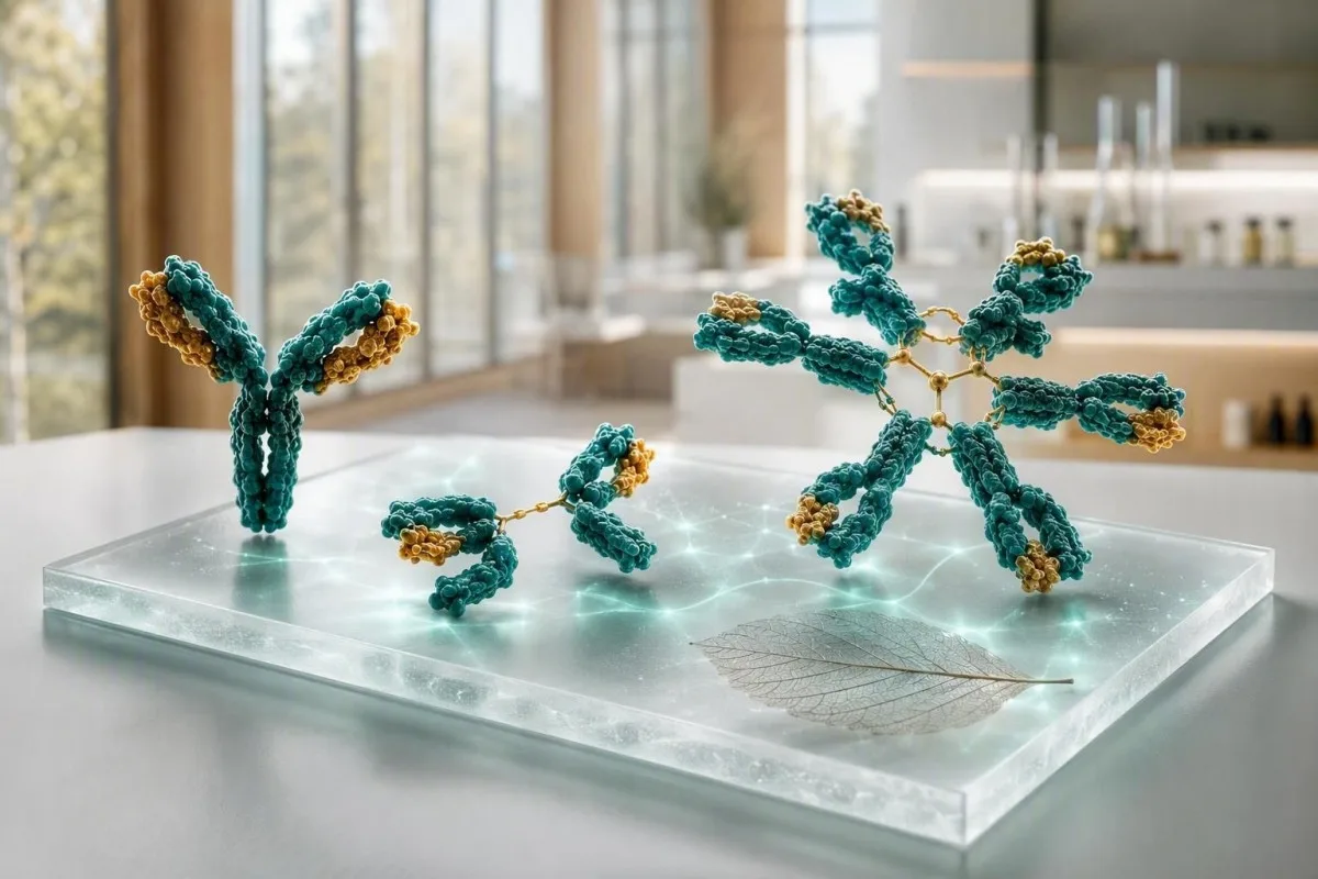
پشکنینی ئیمونۆگلوبولین باشترین سوودی هەیە کاتێک IgG، IgA و IgM وەک شێوازێک بخوێنرێنەوە، نەک سێ نیشانەی جیاواز. بەها بەرزەکان دەتوانن ئاماژە بە چالاکی بەرفراوانی بەرگری یان پرۆتینێکی ناجێگیری تاک بکەن؛ بەها نزمەکان دەتوانن ئاماژە بە دەرمان، لەدەستدانی پرۆتین، یان کەمبونی بەرھەمھێنانی دژەتەن بکەن.


- IgG بە شێوەیەکی گشتی ٧٠٠-١٦٠٠ ملگم/دیسلیترە لە گەورەکاندا؛ بەرزبوونەوەیەکی بەردەوام لە چەندین پۆلدا زۆرجار ئاماژەیە بۆ ھەوکردنی درێژخایەن یان نەخۆشی جگەر.
- IgA بە شێوەیەکی گشتی ٧٠-٤٠٠ ملگم/دیسلیترە لە گەورەکاندا؛ کەمیی ھەڵبژاردە دەتوانێت پشکنینی سیلیاکی نەرێنی لەسەر بنەمای IgA بێ متمانە بکات.
- تاقیکردنەوەی سەرەتایی Lyme ـی نادرست-نەرێنی (false-negative) ڕوودەدات چونکە بە شێوەیەکی گشتی ٤٠-٢٣٠ ملگم/دیسلیترە لە گەورەکاندا؛ بەرزبوونەوەیەکی تاک و بەردەوام پێویستی بە ئەلیکترۆفۆریزیس پرۆتینی سێرمە بۆ دەرکردنی پرۆتینێکی تاک.
- ھەموو سێ نزم نیگەرانی زیاترە لە یەک ئەنجامی نزم و پێویستی بە وردبینی تووشبوون، دەرمان، لەدەستدانی پرۆتین و وەڵامەکانی دژەتەنی ڤاکسینە.
- بەرزبوونەوەی پۆلی کلۆنال مانای ئەوەیە چەندین پۆلی ئیمونۆگلوبولین بەرزن؛ بە شێوەیەکی باو لەگەڵ نەخۆشی خۆبەخۆ، نەخۆشی درێژخایەنی جگەر یان تووشبوونی درێژخایەندا دەبێت.
- شێوازی تاک کلۆنال مانای ئەوەیە یەک پۆل بە شێوەیەکی نادروست بەرزبووەتەوە یان بەندی تەنکێک لەسەر ئەلیکترۆفۆریزیس دەردەکەوێت؛ پێویستی بە پشتڕاستکردنەوەی ئیمونۆفیکسیشن و پشکنینی زنجیرە-سووکەڵەی ئازادە.
- پشکنینی بەهێز/هەڵسەنگاندنی فورس گونجاوە بۆ تا (سەرما) لەگەڵ هەوکردنی جیدی دووبارە بووەوە، تێکچوونی مێشک، بێهەناسەیی، زەردی، دابەزینی کێش، ئازاری ئێسک یان زۆر بەرزبوونەوەی پرۆتینی گشتی.
- ڕێس و ڕەوت گرنگە چونکە جیاوازی 10-20% لەوانەیە لە ئاوهەڵگوشتن، گۆڕانی تاقیکردنەوە، نەخۆشییەکی دوایی یان میتۆدێکی جیاوازی تاقیگە بێت.
چۆن پشکنینی ئیمونۆگلوبولین وەک یەک شێواز بخوێنرێتەوە
ئەنجامەکانی IgG، IgA و IgM پێکەوە واتای جیاواز لە تاک تاک دەگەیەنن. پانێڵێکی بەرفراوان و بەرزکراوە بە گشتی ئاماژەیە بۆ ورووژانی پۆلی کلۆنیال، لە کاتێکدا یەک چینە باڵادەست—بە تایبەت لەگەڵ پرۆتینی گشتی بەرز—پرسیاری جیاواز لە بارەی پرۆتینی مۆنۆکلۆنیالەوە دروست دەکات. لە 5ی ئەیلولی 2026ەوە، ئەو جیاوازییە یەکەم هەنگاوی کردارەکییە پێش ئەوەی ناونیشانی نەخۆشی بدرێتە پشکنینی خوێن بۆ ئیمونوگلوبلین.
سنوورەکانی وەرگرتنەوەی گەورەکان بەپێی میتۆد دەگۆڕێن، بەڵام زۆرێک لە تاقیگەکان بەکاریدەهێنن IgG 700-1,600 mg/dL, IgA 70-400 mg/dL, و IgM 40-230 mg/dL. بەهایەک کە 5% لە سنوورێک دەرکەوێت بە شێوەیەکی خۆکار نەخۆشی نییە؛ من سەرەتا سنووری تاقیگە، تەمەن، بارودۆخی دووگیانی، هەوکردنی دوایی، لەدەستدانی گورچیلە، و ئەوەی ئایا نموونەکە دوای وەرگرتنی ئیمونوگلوبلینی ناوڤینایی وەرگیرابێت، دەستنیشان دەکەم.
Kantestî yek e Analyzerê testa xwînê ya AI کە IgG، IgA و IgM لەگەڵ پرۆتینی گشتی، ئەلبومین، نیشانەکانی جگەر، کرێئاتینین و CBC دەخوێنێتەوە لەبری ئەوەی نیشانەیەکی سوور وەک نەخۆشی مامەڵە بکات. ئەو پشکنینە هاوبەشە بەسوودە چونکە ئەلبومینی کەم لەگەڵ گلوبولینی بەرزبووەوەدا، چیرۆکێکی زۆر جیاواز لە وشکبوونەوە لەگەڵ کۆکردنەوەی هەردوو بەهایەکە دەگێڕێتەوە؛ ئێمە ڕێنمای بیومارکر (biomarker guide) ئەنجامە پەیوەندیدارەکان ڕوون دەکاتەوە.
لە کاری کلینیکیدا، زۆرترین پانێڵی دڵنیایی بە شێوەیەکی تەواو ئاسایی نییە بەڵکو جێگیرە: بۆ نموونە، IgG 1,690 mg/dL لە سێ جار کێشانەوە لە ماوەی 18 مانگدا لەگەڵ ئەلیکترۆفۆڕێسیس ئاسایی و بێ نیشانە. د. تۆماس کلاین هێشتا پرسیار لەبارەی چاوی وشک، ئاوسانی جومگەکان، هەوکردنی دووبارە بووەوە، و دەرمانەکان دەکات، بەڵام گۆڕانی سووکی جێگیر کەمتر بارودۆخی هەمان باری 600 mg/dL بەرزبوونەوە لە شەش مانگدا هەڵدەگرێت.
سێ ئیمونوگلوبلینەکە بە زمانی سادە
تاقیکردنەوەی سەرەتایی Lyme ـی نادرست-نەرێنی (false-negative) ڕوودەدات چونکە وەڵامدەری سەرەتایی و بەرفراوانە،, IgA پارێزگاری لە ڕووەکانی دەموچاو لە ڕێڕەوی هەناسە و گەدە دەکات، و IgG زۆرترین چالاکی دژەتەنی دەمێنێتەوە لە خوێندا. نیوچەژینی بایۆلۆجییان بە ڕوونی جیاوازن: IgG نزیکەی 21 ڕۆژ، IgA نزیکەی 6 ڕۆژ لە خوێن، و IgM نزیکەی 5 ڕۆژ دەمێنێتەوە، بۆیە گۆڕانکارییە یەک لەدوای یەکەکان بە یەک خێرایی جوڵە ناکەن.
ڕێژە ئاساییەکانی گەورەکان و بۆچی تاقیگەکەی تۆ جیاواز دەبێت
سنووری وەرگرتنەوەی چاپکراو لە تەنیشت ئەنجامەکەی خۆتتەوە، لە هەر سنوورێکی ئۆنلایندا پێشینەی هەیە. نیفیلۆمیتری، توربیدیمیتری، و دانیشتووانەکانی تاقیگەی تەمەن-ڕێکخراو سنوورێکی جیاوازی مامناوەندی دروست دەکەن، بە تایبەت بۆ IgA و IgM.
سنوورە باوەکانی گەورەکان IgG 700-1,600 mg/dL, IgA 70-400 mg/dL و IgM 40-230 mg/dL بە نزیکەی 7-16 g/L, 0.7-4.0 g/L و 0.4-2.3 g/L. منداڵان بە شێوەیەکی بەرچاو بەندە بە تەمەن، و سنووری گەورەکان نابێت بەکاربهێنرێت بۆ هەڵسەنگاندنی شیرەخۆرە یان ھەرزەکار.
ئەنجامێک لە نزیک سنوورێک بە تایبەت هەستیارە بەرامبەر گۆڕانی ئاسایی بایۆلۆجی. ئەگەر IgM 238 mg/dL بێت لە بەرامبەر سنووری سەرەوەی 230 mg/dL دوای نەخۆشییەکی هەناسە، دووبارەکردنەوە لە ماوەی 8-12 هەفتەدا زۆر جار زیاتر ئاشکراکەرە لە داواکردنی کۆمەڵێک تاقیکردنەوەی گەورە؛ سەردانی ڕێنماییەکەمان بکە بۆ گۆڕانکارییە ڕاستەقینە لە نێوان تاقیکردنەوەی خوێن.
بەڕۆژوو بوون بە شێوەیەکی ئاسایی بۆ ئیمونوگلوبلینی چەندایەتی پێویست نییە، بەڵام وشکبوونەوەی توند دەتوانێت پرۆتینەکان کۆ بکاتەوە و ئیمونوگلوبلینی ناوڤین بۆ ماوەی هەفتەیەک دەتوانێت IgG ی پێوانەکراو بەرز بکاتەوە. لە کلینیکەکە بپرسە کە پانێڵەکەی داوا کردووە ئایا تۆ لە ماوەی 6 مانگی پێشوودا وەرگرتنی جێگرەوەی دژەتەن، ڕیتوکسیماب، کۆرتیكۆستیرۆید، دژە سکته، یان چارەسەری دژە CD20ت وەرگرتووە.
کاتێک ھەموو سێ پۆلەکانی ئیمونۆگلوبولین بەرزن
IgG, IgA و IgM بەرز لەگەڵ یەکدا، زۆربەی جار ئاماژەیە بۆ چالاکبوونی پۆلیکلۆناڵی بەرگری، نەک یەک کلۆنی نائاسایی. نەخۆشی درێژخایەنی جگەر، حاڵەتە ئۆتۆمینەکان، هەوکردنی بەردەوام و هەندێک نەخۆشی هەوکردنی ڕیخۆڵە، بارودۆخی کلینیکی باوە.
بەرزبوونەوەیەکی پۆلیکلۆناڵ زۆرجار ناوچەی گاما لە ئەلکترۆفۆڕێزی پرۆتینی سرمدا فراوانتر دەکات لەبری دروستکردنی لوتکەیەکی تیژ و تەنک. لە ڕووی کردارییەوە، IgG 2,100 mg/dL، IgA 520 mg/dL و IgM 310 mg/dL لەگەڵ CRP بەرزبوو، ئاماژە بە ڕێگەیەکی جیاواز دەدات لە IgG 2,400 mg/dL لەگەڵ دوو کلاسی دیکەی داپڵۆسراو.
نەخۆشی ئۆتۆمینە دەتوانێت ئەم شێوازە دروست بکات، بەڵام نیشانەکان ڕێنمایی تاقیکردنەوەی داهاتوو دەکەن. وشکبوونی بەردەوامی چاو و دەم، ئاوسانی جومگەکان، پەڵە، سکچوون یان نەخۆشی دەمار، لەوانەیە جێگای دژە-ئۆتۆباڵینی دیاریکراو و تاقیکردنەوەی تەواوکاری بگرێتەبەر؛ ئێمە C3 و C4 ڕووندەکاتەوە بۆچی کەمبوونی تەواوکاری ئەو لێکدانەوەیە دەگۆڕێت.
هەوکردنی درێژخایەنیش دەتوانێت چەندین کڵاس بۆ ماوەی چەند مانگێک بەرز بکاتەوە، هەرچەندە ئیمونوگلوبولینەکان ڕێگر نین لە ناسینەوەی ئۆرگانیزمەکە یان سەلماندنی هەوکردنی چالاک. ESR بەرز لەگەڵ نیشانە بنەڕەتییەکان شایستەی لێدوانێکی کلینیکییە، و ڕوونکردنەوەی ئێمە لەسەر هۆکارە هەڵسوڕانییە بەرزبوونی ESR نیشان دەدات بۆچی نیشانەی هەوکردنی نا-دیاریکراو ناتوانێت هۆکارەکە یەکلایی بکاتەوە.
مانای چی دەبێت IgM ی تاکە بەرز بێت
ئەنجامێکی تاک-ئای جی ئێم (IgM) بەرز دەتوانێت دوای هەوکردنی دوایی بێت، لەگەڵ نەخۆشی هەوکردنی جگەری ئۆتۆمینەی کۆلستاتیک ڕووبدات، یان پرۆتینی مۆنۆکلۆناڵی IgM بێت. بەردەوامی، ئاستی بەرزبوونەوە، نیشانەکان و ئەلکترۆفۆڕێز، گرنگترن لە یەک ژمارەی سنووردار.
IgM دژەتەنی گەورەی پینتامێرە، و ئاستی کەمێک لەسەر 230 mg/dL زۆرجار دوای چاڵاکی بەرگری دادەبێت. دووبارە کردنەوەی IgM ی 800 mg/dL یان 1,200 mg/dL، بە تایبەتی لەگەڵ IgG و IgA ئاسایی یان کەم، دەبێت ببێتە هۆی ئەلکترۆفۆڕێزی پرۆتینی سرم، ئیمونۆفیکسیشن و تاقیکردنەوەی زنجیرەی سووکی ئازادی سرم لەبری گومانی لێکردن.
کۆلەنژیتسی سەرەتایی کلاسی دەبێت بە IgM بەرز، بە تایبەتی کاتێک ئەلکالاین فۆسفاتاز و گاما-گلوتامیل تڕانسفێراز (gamma-glutamyl transferase) یش بەرزن. ڕێنمایی EASL ڕێنمایی دەکات بۆ هەڵسەنگاندنی نەخۆشی جگەری کۆلستاتیک کاتێک ئەلکالاین فۆسفاتاز بەردەوام بەرزە و دژە-مایتی کۆندریا (antimitochondrial antibody) بوونی هەیە (EASL, 2017)؛ ئێمە pattern ی cholestasis نیشانە هاوڕێکان دەگرێتەوە.
پرۆتینی مۆنۆکلۆناڵی IgM یەک شت نییە لەگەڵ ماکرۆگلوبولینیمی Waldenström، و زۆربەی خەڵک لەگەڵ گامۆپاتی مۆنۆکلۆناڵی بچووک، وادەی نزیک بۆ شێرپەنجە نییە. لەگەڵ ئەوەشدا، نیشانە وەک تەمومژی بینین، سەرئێشە، خوێن بەربوونی لووت، نەخۆشی دەماری نوێ یان ماندووبوونی ناڕوون، پێویستی بە هەڵسەنگاندنی خێرا هەیە چونکە IgM ی زۆر بەرز دەتوانێت چڕی سرم بگۆڕێت؛ زیاتر بخوێنەوە لەسەر بەرزبوونی IgM.
نزمبوونی IgA: ھەڵەی پشکنینی سیلیاک و ئاماژەکانی تووشبوون
کەمبوونی IgA دەتوانێت شاشەی سێلیاکی ترانسگلوتامینازی IgA (IgA tissue-transglutaminase) ی درۆینە نەرێنی بکات. کەمبوونی هەڵبژێردراوی IgA زۆرجار بە IgA ی سرم لە خوار 7 mg/dL لە کەسێک کە تەمەنی لە 4 ساڵ زیاترە لەگەڵ IgG و IgM ئاسایی، پێناسە دەکرێت، هەرچەندە پێناسە ناوخۆییەکان و دووبارە کردنەوەی تاقیکردنەوە گرنگن.
ئەگەر کۆی IgA کەم بێت، پزیشکان زۆرجار بەکاردەهێنن پشکنینی پپتیدی گلیادینی دەستکاریکراو بەسەر بنەمای IgG یان پشکنینی IgG tissue-transglutaminase لەگەڵ مێژووی کلینیکی. پێش ئەنجامدانی پشکنینی گونجاو، خۆراکێکی بێ گلوتن مەخۆ، مەگەر ڕێنماییت لێ کرابێت، چونکە ئاستی دژەتەنەکان لەوانەیە داببەزێت و ئەنجامەکە بشارێتەوە؛ ڕێبەری تاقیکردنەوەی گلوتنی ئێمە کێشەی کاتەکە باس دەکات.
زۆرێک لە خەڵکانی کەمئەندامی IgA ی تەواو کێشەی تەندروستی گەورەیان نییە، لەکاتێکدا هەندێکی تر باس لە هەوکردنی دووبارەی دەم و لووت، سنگ یان کۆئەندامی هەرس دەکەن. ڕێژە، توندی و هۆکارە بەکتریاییە پشتڕاستکراوەکان زیاتر گرنگن لەوەی دوو سەرماخواردنی ئاسایی لە زستانێکدا؛ مێژووی کوتان و وەڵامی دژەتەنیی پۆلی ساکارید دەتوانێت زانیاری زیاتری هەبێت لە تەنها IgA ی گشتی.
کاردانەوەی خوێن دەگمەنە بەڵام پەیوەندیدارە: خەڵکانی کەمئەندامی قووڵی IgA و دژەتەنیی دژ بە IgA لەوانەیە پێویستیان بە پێکهاتەی تایبەت هەبێت ئەگەر پێویست بێت گواستنەوەی خوێن. ئەمە هۆکارێکە بۆ ئەوەی تیمەکانی نەخۆشخانە ئاگادار بکەیتەوە سەبارەت بە کەمئەندامییەکی سەختی پشتڕاستکراو، نەک هۆکارێک بۆ خۆلادان لە چاودێری پزیشکی پێویست؛ سەیری باسە فراوانترمان بکە سەبارەت بە ئەنجامە نزمەکانی IgA.
نزمبوونی IgG و تووشبوونی دووبارە: کاتێک ئەنجامەکە گرنگە
نزمبوونی IgG کاتێک گرنگی کلینیکی پەیدا دەکات کە لەگەڵ هەوکردنی دووبارە، نا-سەیر، یان خراپ چاکبووەوە بێت. IgG ی گەورەساڵێک لە خوار 400 mg/dL بە گشتی جێگەی نیگەرانی زیاترە لە بەهای 650 mg/dL، بەڵام وەڵامی دژەتەنیی بازدانی کارا دەتوانێت ئەو پێشبینیانە بگۆڕێت کە تەنها لەسەر بنەمای بڕەکەی دامەزراون.
هەوکردنەکان کە هاندەری چاودێریی دەبێت بریتین لە: هەوکردنی سییەکان، برۆنکیت، هەوکردنی دووبارەی دەم و لووت کە پێویستی بە چەندین ئەنتی بایۆتیک هەیە، نەخۆشیی بەکتریایی وەرگێڕدراو، یان گێاردیازی درێژخایەن. چوار سەرماخواردنی بچووکی منداڵێک لە کارمەندی چاودێری منداڵان هاوتا نییە لەگەڵ سێ هەوکردنی سییەکان کە بە ڕادیۆلۆجی پشتڕاست کرابنەوە لە ماوەی 24 مانگدا.
هۆکارە لاوەکییەکان زۆرن: ڕیتوکسیماپ و چارەسەرەکانی تر کە ئاراستەی خانەکانی B دەکرێن، کۆرتیكۆستیرۆیدی ئۆڕاڵی درێژخایەن، هەندێک دژە-کۆئەکولانت، لەدەستدانی پرۆتینی لە ئاستی نەفروتیدا، ئینتێرۆپاتی پرۆتین-لەدەردەچێت، و شێرپەنجەی لیمفاوی دەتوانێت IgG کەم بکاتەوە. پرۆتینی میز، ئەڵبوومین، ژمارەی خانەکانی لیمفۆسایت و مێژووی دەرمان زۆرجار زیاتر زانیاری دەدەن لە پشکنینی دژەتەنیی ڕێکەوتنی تر؛ ڕێنمایی پڕۆتێن لە ڕێژەدا ڕێگای سەرەکی لەدەستدانەکەمان ڕوون دەکاتەوە.
بونێلا و هاوکارانی (2015) ڕێنمایی دەدەن کە پێویستە ئیمونوگلوبولینە cuantitative لەگەڵ وەڵامی دژەتەنی تایبەتدا هەڵبسەنگێندرێت کاتێک گومانی کەمئەندامیی سەرەتایی هەیە. لە ئەزموونی مندا، وەڵامێکی ئاسایی دوای کوتان دەتوانێت دڵنیایی بدات تەنانەت کاتێک IgG بە کەمێک نزمە، لەکاتێکدا وەڵامی خراپ لەگەڵ IgG ی خوار 500 mg/dL پێویستی بە تێڕوانینی پسپۆڕانە هەیە.
کاتێک IgG، IgA و IgM ھەموویان نزم دەبن
نزمبوونی IgG، IgA و IgM پێکەوە پێی دەوترێت هایپۆگاماگلوبولینیمیا و پێویستی بە لێکۆڵینەوەیەکی ڕێکخراو هەیە، بەتایبەتی لەگەڵ هەوکردندا. شێوازەکە لەوانەیە ڕەنگدانەوەی کەمبوونەوەی بەرهەمهێنانی دژەتەن، کاریگەریی دەرمان، لەدەستدانی پرۆتین، نەخۆشییەکی پێشکەوتووی لیمفاوی، یان کەمتر، گۆڕانکارییەکی کاتی دوای نەخۆشی بێت.
کۆمەڵێک لە IgG 360 mg/dL، IgA 28 mg/dL و IgM 18 mg/dL ئەنجامێکی چاوەڕوانینینینین نییە ئەگەر کەسەکە تا یان، هەوکردنی دووبارەی سنگ، یان دابەزینی کێش. پزیشکێک لەوانەیە کۆمەڵەکە دووبارە بکاتەوە، ئەلیکترۆفۆڕێز، CBC لەگەڵ جیاکاری، پشکنینی گورچیلە و جگەر، پرۆتینی میز و دژەتەنییە تایبەتەکانی کوتان، پاشان ڕەوانەی immunology کلینیکی یان hematology بکات.
کەمئەندامیی ڤاریانتی هاوبەش زۆرجار لە گەورەساییدا دەردەکەوێت، بەڵام دیاریکردنی پێویستی بە زیاتر لە ژمارەی کەمە: نیشانەکان، هۆکارە لاوەکییە دوورخراوەکان و بەرهەمهێنانی دژەتەنیی کارای کەمبوو، ناوەندییەکانن. خزمەتگوزاری تێکست/تێگەیشتنی تاقیکردنی لابراتۆریی AI کە دەتوانێت ئەم دۆزینە دەوروبەرە ڕێک بخاتە ناو پوختەی سەردانی پزیشک، بەڵام ناتوانێت کەمئەندامیی بەرگری دیاری بکات یان بڕیاری گۆڕینەوەی ئیمونوگلوبولین بدات.
گۆڕینەوەی ئیمونوگلوبولین بۆ ڕێگرتن لە هەوکردنی قورس لە نەخۆشی هەڵبژێردراو دەردەکرێت، نەک تەنها بۆ ئاساییکردنەوەی ئەنجامێکی تاقیگەیی. ڕێژەکانی گۆڕینەوەی ئاسایی بە تاک تێپەڕ دەکرێن و زۆرجار دەستپێدەکەن لە دەوروبەری 400-600 mg/kg لە مانگێکدا بە ڕێگای وێناویی یان ژێرپێست، لەگەڵ دۆزین کە بۆ کۆنترۆڵی هەوکردن و نزمترین ئاستی IgG ڕێکدەخرێت نەک ئامانجێکی جیهانی؛ ڕێبەری ئێمە بۆ تاقیکردنەوەی خوێنی سیستەمی ئیمون ـە بەکارهێنانی زۆر بەجێ دەکات بۆ پێدانی کۆنتێکستی سودبەخش.
چۆن پشکنینی ئیمونۆگلوبولین دەتوانێت ئاماژە بە پرۆتینێکی تاک بکات
پرۆتینێکی مۆنۆکلۆنال پێشنیار دەکرێت کاتێک پۆلێکی ئیمونوگلوبولین بە شێوەیەکی ناڕێکوپڕ بەرزە، پۆلەکانی تر سەرکوت کراون، یان ئەلیکترۆفۆڕێز نوارێکی تەنگ پیشان دەدات. ئیمونوگلوبولینە cuantitative تەنها ناتوانێت کلۆنیالیزم بسەلمێنێت؛ ئەلیکترۆفۆڕێزی پرۆتینی سیروم و ئیمونۆفیکسەشن ئامرازە پشتڕاستکەرەوەکانن.
IgG 2,900 mg/dL لەگەڵ IgA 38 mg/dL و IgM 21 mg/dL لەبەرچاو بگرە: ئەو شێوازە زیاتر گوماناوییە لەوەی هەر سێیان بە کەمێک بەرزبنەوە. پرۆتینی گشتی بەرز، ڕێژەی ئەڵبوومین بۆ گلۆبوولینی نزم، کەمخوێنی، کەمبوونی eGFR، کالسیۆمی بەرز، ئازاری ئێسک یان هەوکردنی دووبارە، بەپەلەیی کلینیکی زیاد دەکەن؛ ڕێبەری ئێمە بۆ پروتین تام بالا.
گروپی کاری میلومای نێودەوڵەتی (International Myeloma Working Group) جیاوازی لە نێوان MGUS و میلومای چالاک دەکات بە بەکارهێنانی بڕی پروتین، دۆزینەوەکانی مەغزی ئێسک و کاریگەرییەکانی لەسەر ئەندامەکان—نەک تەنها ئەنجامی ئیمێنوگڵۆبولین (Rajkumar et al., 2014). پروتینی M لەخوار 3 g/dL هێشتا دەتوانێت پێویستی بە چاودێری هەبێت، لە کاتێکدا بەهایەکی کەمتر لەگەڵ کەمخونی یان زیانی گورچیلەکان دەکرێت پێویستی بە هەڵسەنگاندنی خێراتری پزیشکییەکی پسپۆڕی خوێن هەبێت.
بە پروتینی تامی ئاسایی بە هەڵە مەچۆ. نەخۆشییە تەنها-زنجیرە-سووکەکان لەوانەیە بەرزبوونەوەیەکی ئاشکرای پروتین دروست نەکەن؛ بۆیە لە کەیسێکی گونجاودا پزیشکان زنجیرە سووکە ئازادەکانی سێرەم، ئیمینۆفیکسیشن و پشکنینەکانی میز پێکەوە بەکاردەهێنن؛ ڕوونکردنەوەکەمان ڕێگای تاقیکردنەوەی نەخۆشی سەربەخۆری خوێن ئەوەی دواتر ڕوودەدات ڕوون دەکاتەوە.
شێوازەکانی نەخۆشی جگەر: ئاماژەکانی IgA، IgG و IgM
نەخۆشییەکانی جگەر دەتوانن ئیمێنوگڵۆبولینەکان بەرز بکەن، بەڵام پۆلی زاڵ تەنها ئاماژەیەکە، نەک تشخیص. IgA زۆرجار لەگەڵ زیانی جگەری پەیوەست بە خواردنەوەی ئەلکۆل زیاد دەبێت، IgG لەگەڵ هەپاتیتی ئۆتۆئیمین، و IgM لەگەڵ کۆڵانژیتی صفراوی سەرەتایی؛ بەڵام تێکەڵبوونەکان باون.
بەرزبوونەوەی IgG بۆ سەرووی 1.1 جارەی سنووری سەرەوەی ئاسایی، لە کاتێکدا ترانسئامینازەکان و ئەنتیبۆدییە ئۆتۆئیمینەکان لەگەڵ یەکتر بگونجێن، پشتگیری لە هەپاتیتی ئۆتۆئیمین دەکات؛ بەڵام بە تەنها هەستیاری و تایبەتمەندیی پێویستی نییە بۆ تشخیصکردنی. نەخۆشییە ڤایرۆسییە توندەکان، زیانی درێژخایەنی جگەر و نەخۆشییە ئۆتۆئیمینە سیستەمییەکان هەموویان دەتوانن هەمان نیشانە دروست بکەن، بۆیە پزیشکان زۆرجار وێنەبرداری و پشکنینی ئاراستەکراوی پسپۆڕ بەکاردەهێنن.
بەرزبوونەوەی IgA لەگەڵ بەرزبوونەوەی GGT، AST و ماکروسایتۆز دەتوانێت لەگەڵ زیانی جگەری پەیوەست بە خواردنەوەی ئەلکۆل بگونجێت، بەڵام نابێت هیچکات لەسەر بنەمای پەنێڵێکی پشکنینەوە بەکارهێنانی ئەلکۆل گریمانە بکرێت. کاریگەریی دەرمانەکان، نەخۆشیی جگەری چەورەی میتابۆلیک و هۆکارەکانی دیکە زۆرن؛ ڕوونکردنەوەکەمان لەبارەی ئەنجامەکانی ALT یارمەتیدەرە بۆ ئەوەی ئەنجامەکانی ئەنزیمەکان لە تەنیشت ئیمێنوگڵۆبولینەکان دابنرێن.
پەیامی گرنگی پاراستن ئەوەیە کە زەردبوونەوە، میزەی تاریک، پیسایی ڕەنگپەڕیو، سەرلێشێواوی، ئاوسانی شکم یان ڕشانەوەی خوێن پێویستیان بە چاودێریی پزیشکیی فۆری هەیە، بەبێ گوێدانە بڕی ئیمێنوگڵۆبولینەکان. بەرزبوونەوەی جیاکراوە و جێگیری IgA بە بڕی 430 mg/dL ئەو نیشانانە دروست ناکات و نابێت سەرنج لە هەڵسەنگاندنی فۆری لاببات.
جیاکردنەوەی تووشبوون، ھەوکردن و چالاکی خۆبەخۆ
ئاستی ئیمێنوگڵۆبولینەکان بە هێواشی و بە شێوەیەکی ناتایب بەرز دەبن، بۆیە بە تەنها ناتوانن تووشبوونی توند بە نەخۆشییەکی تووشبەری تشخیص بکەن. CRP، ژمێری تەواوی خوێن، کەلتوورەکان یان پشکنینە ئاراستەکراوەکانی پاتۆجینەکان پرسیارەکانی تووشبوونی فۆری بە باشتر وەڵام دەدەن، لە کاتێکدا ئیمێنوگڵۆبولینەکان پاشبنەمای گشتیی بەرگریی جەستە باس دەکەن.
زۆرجار IgM وەک ئەنتیبۆدییەکی سەرەتاییی تووشبوون باس دەکرێت، بەڵام IgM ی تامی سێرەم هەمان IgM ی تایبەت بە پاتۆجین نییە. IgM ی تام بە بڕی 290 mg/dL ناتوانێت هەڵگریکردنی ئەنفلوانزا، ڤایرۆسی ئێپشتاین-بار یان تووشبوونی ڕێڕەوی میز پشتڕاست بکاتەوە؛ نیشانەکان و پشکنینی ڕاستەوخۆ دیاری دەکەن کە چارەسەر پێویستە یان نا.
ئاوسانی ئۆتۆئیمین دەتوانێت بۆ ساڵان IgG بەرز بکات پێش ئەوەی تشخیصەکە ڕوون بێت، بە تایبەتی لەگەڵ هایپەرگامماگڵۆبولینیمیا و نەخشەیەکی فراوانی ئەلیکترۆفۆرێسیس. پەنێڵەکانی ANA، فاکتەری ڕۆماتۆید و ENA دەبێت بۆ وەڵامدانەوەی پرسیارێکی کلینیکی داوا بکرێن، نەک بۆ گەڕان بەدوای هەر شتێکدا؛ بۆ نموونە، ڕێبەری anti-dsDNA ـمان ڕوون دەکاتەوە بۆچی ئەنجامێکی ئەرێنی پێویستی بە زانیارییەکانی گورچیلە و کۆمپلەمێنت هەیە.
د. Thomas Klein نەخۆشانی بینیوە کە دوای نەخۆشییەکی ڤایرۆسی بەهۆی بەرزبوونەوەی IgG ترساون، بەڵام لە دووبارە پشکنینەوە دوای 3 مانگ ئاستەکە ئاسایی بووە. بە پێچەوانەوە، بەردەوامبوونی IgG لەسەر 2,000 mg/dL لەگەڵ C3 ی نزم، پڕۆتینوریا و نیشانەکانی جومگە شتێک نییە کە من بۆ ساڵێک دواخی بخەم—ئەم دۆزینەوانەی پێکەوە هەڵسەنگاندنی کلینیکی لە کاتی خۆیدا پشتڕاست دەکەن.
کاریگەرییەکانی دەرمان، لەدەستدانی پرۆتین و کات کە ئەنجامەکان دەگۆڕن
دەرمانەکان و لەدەستدانی پروتین دەتوانن ئیمێنوگڵۆبولینەکان کەم بکەن، بەبێ ئەوەی کێشەیەکی سەرەتاییی سیستەمی بەرگری هەبێت. دەرمانەکانی دژە-CD20 دەتوانن سەرەتا IgM و دواتر IgG کەم بکەن، لە کاتێکدا لەدەستدانی پروتین لە گورچیلە یان ڕوودەکان دەتوانێت چەندین پۆل لەگەڵ ئەلبومین کەم بکات.
هایپۆگامماگڵۆبولینیمیای پەیوەست بە Rituximab دەتوانێت مانگەکان دوای دواهەمین دۆزەکە دەربکەوێت، چونکە گەڕانەوەی خانەکانی B و گەڕانەوەی ئەنتیبۆدی یەکسان نین. لیستێکی تەواوی دەرمانەکانت—لەوانە دەرمانە بیۆلۆژیکییەکان، دۆزی ستێرۆید، دەرمانەکانی دژە-تشنج و کیمیاویدەرمانی—لەگەڵ خۆت بۆ وادەکە بهێنە؛ ڕێبەری ڕەوتەکانی سەلامەتیی دەرمانەکانمان ڕوون دەکاتەوە بۆچی بەروارەکان گرنگن.
دۆخەکانی لەدەستدانی پرۆتین زۆرجار نیشانەی زیادە بەجێدەهێڵن: ئەلبومینی نزم، کەمتر لە نزیکەی 3.5 g/dL، وەرەم، ئیسهال، بەرزبوونەوەی ڕێژەی ئەلبومینی پیشاب بە کرێئاتینین، یان کەمبوونەوەی کۆی پرۆتین. بە پێچەوانەوە، وشکبوونەوەی جەستە دەتوانێت وا بکات ئیمێنۆگڵۆبۆلینەکان بەرز دیاربن، لە کاتێکدا ئەلبومین و هێماتۆکریت بە شێوەیەکی هاوتا زیاد دەبن.
زۆرجار باشترە تاقیکردنەوەکە دووبارە بکرێتەوە کاتێک لە ڕووی کلینیکییەوە جێگیر بیت، و ئەگەر بکرێت هەمان تاقیگە بەکاربهێنیت. Kantesti دەتوانێت فایلێکی PDF یان وێنەیەکی بەروارنووسراو لەگەڵ ئەنجامەکانی پێشوو بەراورد بکات و گۆڕانێکی گەورە دیاری بکات، بەڵام پشکنینی وردی بارکردن کارێکی عاقڵانەیە، چونکە یەکەکان و مەوداکانی ئاماژە لەوانەیە بە هەڵە تۆمار کرابن.
پشکنینی دوایین کە ڕووناکی دەخەنە سەر پەڕەیەکی ناجێگیر
تاقیکردنەوەی شوێنکەوتنی گونجاو بە پێی ئەو شێوەیەیە کە دەردەکەوێت: ئەلیکترۆفۆرێزی بۆ بەرزبوونەوەی ناتەبا، ئەنتیبۆدی واکسین بۆ گومانی کەمبوونی بەرگری، و تاقیکردنەوەی پیشاب یان جگەر بۆ لەدەستدانی پرۆتین یان نیشانەکانی نەخۆشیی جگەر. دووبارەکردنەوەی هەمان سێ ژمارە بەبێ پرسیارێکی دیاریکراو زۆرجار زانیارییەکی زۆر زیاد ناکات.
بۆ ئەنجامە بەرز یان ناتەبا، پزیشکان بە شێوەی باو داوای ئەلیکترۆفۆرێزی پرۆتینی سیرەم، ئیمێنۆفیکسەیشن، زنجیرە لاوازە ئازادەکانی سیرەم، ژمارەی تەواوی خانەکانی خوێن، کەلسیۆم، کرێئاتینین، ئەلبومین و ئەنزیمەکانی جگەر دەکەن. باندێکی تەنگ لە ئەلیکترۆفۆرێزیدا دەبێت بە ئیمێنۆفیکسەیشن جۆربەندی بکرێت، چونکە جۆرەکە—IgG، IgA یان IgM—جیاوازییەکەی نەخۆشییە گومانلێکراوەکان و پلانی شوێنکەوتن دەگۆڕێت.
بۆ ئەنجامە نزمەکان لەگەڵ تووشبوون بە نەخۆشییەکان، تاقیکردنەوەکان لەوانەیە پۆلە لاوەکییەکانی IgG و تایتەری ئەنتیبۆدییەکان بۆ تیتانۆس، Haemophilus influenzae جۆری b یان سیرۆتایپەکانی پەنیومۆکۆک لە پێش و دوای واکسینەکردن لەخۆ بگرن. هیچ سنوورێکی تاکی پاراستنی پەنیومۆکۆکی نییە کە بۆ هەموو تاقیگە و گرووپە تەمەنەکان بگونجێت؛ ئەمە یەکێکە لەو بوارانەی لێکدانەوەی پسپۆڕ لە کۆتاییپێهێنانی ئۆنلاین گرنگترە.
Kantestî yek e ئامێری توێژینەوەی تاقیکردنەوەی خوێن بە پشتبەستن بە AI کە دەتوانێت ئەم ئەنجامانە لە شێوەی هێڵی کاتدا کۆبکاتەوە و زانیارییە نەبووەکان، وەک نەبوونی ئەلبومین یان ئەلیکترۆفۆرێزی، دیاری بکات بۆ ئەوەی پزیشکەکەت لەبەرچاوی بگرێت. هی ئێمە ڕێنمایی تەکنەلۆژی ڕوون دەکاتەوە چۆن ئاگادارکردنەوە کۆنتێکستییەکان بەبێ جێگرتنەوەی بڕیاردانی پزیشکی دروست دەکرێن.
نیشانەکانی کە نابێت چاوەڕێی دووبارەکردنەوەی ئاسایی بکەن
ئەنجامی ئیمێنۆگڵۆبۆلین بە تەنیا بە دەگمەن دۆخێکی فۆری دروست دەکات، بەڵام هەندێک نیشانە وای دەکەن. بۆ هەڵسەنگاندنی فۆری هەمان ڕۆژ داوا بکە ئەگەر تبی بەرز لەگەڵ نەخۆشییەکی توند، کورتەهەناسە، سەرلێشێواوی، لاوازییەکی تازە، زەردبوون، کەمبوونەوەی زۆری پیشاب، سەرئێشەی توند لەگەڵ گۆڕانی بینین، یان خوێنبەربوونی کۆنترۆڵنەکراو هەبێت.
ئاستی نزمەکانی ئیمێنۆگڵۆبۆلین لەگەڵ تبی 38.0°C یان زیاتر لە کەسێکدا نیگەرانیی زیاتر دروست دەکات کە کیمۆتێراپی، ستێرۆیدی دۆزی بەرز، چارەسەری ئاراستەکراوی خانەکانی B وەردەگرێت، یان مێژووی تووشبوونی هەیە بە نەخۆشییە هێرشبەرەکان. ئەگەر هەناسەدان قورسە، ئاستی ئۆکسجین نزمە، یان کەسەکە سەرلێشێواو یان بە شێوەیەکی نائاسایی خەواڵوو دیارە، چاوەڕێی پەیامی پۆرتاڵ مەکە.
پرۆتینێکی مۆنۆکلۆنال پێویستی بە پشکنینی خێراتر هەیە کاتێک لەگەڵ ئازاری نوێی ئێسک، کەمخونییەکی بێڕوونکردنەوە، بەرزبوونەوەی کرێئاتینین، کەلسیۆمی سەرووی سنووری سەرەوەی ناوخۆیی، نێوروپاتی یان کەمبوونەوەی نەخوازراوی کێشدا ڕوودەدات. ئەمانە بەڵگەی سەلمێنەری نەخۆشیی خانە پلازماییەکان نین، بەڵام ئەو تایبەتمەندییانەن کە پزیشکان بۆ پێشخستنی تاقیکردنەوە بەکاریان دەهێنن؛ هی ئێمە دووەم ڕاڤە/سەردانی ڕەخنە دەتوانێت یارمەتیت بدات پرسیارەکانت ئامادە بکەیت.
بۆ نائاسایییە سووک و جێگیرەکان بەبێ نیشانەی مەترسیدار، دانانی وادەیەکی پلاندانراو لە ماوەی چەند هەفتەیەکدا زۆرجار گونجاوە. ئەنجامەکانی پێشوو، بەروارەکانی تووشبوون، گۆڕانکارییەکانی دەرمان و مێژووی خێزانی لەگەڵ خۆت بهێنە؛ گرافی دوو ساڵە زۆرجار لە یەک ژمارەی نیشانەکراو بۆ کلینیک بەسوودترە.
بەکارھێنانی لێکدانەوەی AI بە سەلامەتی بۆ ئەنجامەکانی ئیمونۆگلوبولین
AI دەتوانێت شێوەی ئەنجامەکانی تاقیکردنەوەی ئیمێنۆگڵۆبۆلین ڕێکبخات، بەڵام ناتوانێت دیاری بکات کە کەسێک کەمبوونی بەرگری، نەخۆشیی جگەر یان نەخۆشییەکی مۆنۆکلۆنال هەیە. لێکدانەوەی سەلامەت مەودای تاقیگە، نیشانەکان، دەرمانەکان، ڕەوتی گۆڕانکارییەکان و تاقیکردنەوە پشتڕاستکەرەکان بە ڕوونی دەهێڵێتەوە.
Kantesti AI پێکەوە IgG، IgA و IgM لەگەڵ ئەو پرۆتین و نیشانە ئەندامییەکاندا پێداچوونەوە دەکات کە ماناکانیان دەگۆڕن، پاشان پرسیارەکانی شوێنکەوتن پێشکەش دەکات، نەک ئەوەی دەستنیشانی نەخۆشی بکات. لە ڕەوتی شیکاریی ئێمەدا، نزمبوونی تەنهای IgA ئاگادارییەک بۆ تاقیکردنەوەی سێلیاک دروست دەکات، لە کاتێکدا نزمبوونی هەرسێ بەهاکە لەگەڵ ئەلبومینی نزم، بیرکردنەوە لە لەدەستدان، کاریگەریی دەرمانەکان و پێداچوونەوەی ئیمێنۆلۆژیی کلینیکی پێشنیار دەکات.
پاراستنی نهێنی لە ئەنجامە پەیوەندیدارەکان بە بەرگری و شێرپەنجە گرنگە. Kantesti Ltd مامەڵەکردنێکی هاوتەریب لەگەڵ GDPR بەکاردەهێنێت، و هی ئێمە لە لاپەڕەی تاییدکردنی پزیشکی چاودێریی کلینیکی و سنوورەکانی پشت لە لێکدانەوەی ئۆتۆماتیکیی تاقیکردنەوەکانی تاقیگە ڕوون دەکاتەوە.
ڕاپۆرتێکی باش دەبێت یارمەتیت بدات بپرسیت: “ئایا ئەمە چالاکبوونەوەی بەرینی بەرگرییە، لەدەستدانی پرۆتینە، کەمبوونەوەی بەرهەمهێنانە، یان تەنها یەک پرۆتینە؟” ئەمە ئاستی گونجاوی دڵنیاییە پێش ئەوەی ئەلیکترۆفۆرێسیس یان هەڵسەنگاندنی پسپۆڕ وەڵامێک بدات.
پرسیارەکان کە دەبێت دوای پەڕەیەکی ناجێگیری ئیمونۆگلوبولین بکرێن
باشترین پرسیاری دواتر زۆرجار ئەوەیە “ئەمە چ شێوەیەکە؟” نەک “چۆن ئەم ژمارەیە نزم یان بەرز بکەمەوە؟” بپرسە ئایا ئەنجامەکە بەردەوامە، پۆلیکڵۆنالە یان مۆنۆکڵۆنالە، ئایا تووشبوونەکانی بە نەخۆشییەکانی توانا لاوازییەکانی کارکردن نیشان دەدەن، و کام هۆکارە لاوەکییەکان پشکنراون.
پرسیارە بەسوودەکان ئەمانەن: ئایا ئەلیکترۆفۆرێسیسی پرۆتینی سێرەم کراوە؟ ئایا ئەلبومین، پرۆتینی میز و ئەنزیمەکانی جگەر لەگەڵ لەدەستدانی پرۆتین یان نەخۆشیی جگەر دەگونجێن؟ ئایا پێویستە تاقیکردنەوەی ئەنتیبۆدی ڤاکسین بکەم؟ ئایا دووبارەکردنەوەی تاقیکردنەوەکە لە ٨–١٢ هەفتەدا پێویستە؟ ئەم پرسیارانە بەرهەمدارترن لە کڕینی پاشکۆکان، چونکە بە شێوەیەکی باوەڕپێکراو کەمیی ئەنتیبۆدییەکی گرنگی کلینیکی چاک ناکەن.
ئەگەر پلانی ناردنت بۆ پسپۆڕ هەیە، ژمارەی ئەو تووشبوونانە بە نەخۆشی تۆمار بکە کە لە ١٢ مانگی ڕابردوودا بە ئەنتیبایۆتیک چارەسەر کراون، هەروەها سەردانەکانی نەخۆشخانە، سینهپەنۆمۆنیی پشتڕاستکراوە بە وێنەبرداری، و مەودای بەکارهێنانی دەرمانەکان. تۆمارێکی ورد—“سێ جار کۆرسی ئەنتیبایۆتیک و یەک جار سینهپەنۆمۆنی”—لە “زۆر نەخۆش دەکەوم” زیاتر یارمەتیدەرە؛ ڕێکخستنی تۆمارە پراکتیکییەکەمان تۆمارکەری ئەنجامی لابراتۆری فۆرماتێکی پراکتیکی بۆ تۆمارکردن پێشکەش دەکات.
Yên me Lijneya Şêwirmendiya Bijîşkî ستانداردەکانی پێداچوونەوەی کلینیکی Kantesti پشتگیری دەکات، بەڵام پزیشکی خۆت هەر ئەو کەسەیە کە دەتوانێت تۆ بپشکنێت، تاقیکردنەوەی پشتڕاستکەرەوە داوا بکات و لەگەڵ ئەنجامە پەلەییەکاندا هەنگاو بنێت. زۆربەی نەخۆشان دەبینن کە گفتوگۆیەکی ئارام و بنەما-شێوە، ئاڵایەکی نیگەرانیبەخش دەگۆڕێت بۆ هەنگاوێکی دواتری بەڕێوەبەرایەتیکراو.
Pirsên Pir tên Pirsîn
ئایا ڕێژەی ئاسایی پشکنینی ئیمونوگلوبولین بۆ گەورەکان چەندە؟
ئاستی ئاسایی گشتگیری ئیمونوگلوبولین لە کەسانی پێگەیشتوودا بریتییە لە IgG 700-1,600 mg/dL، IgA 70-400 mg/dL و IgM 40-230 mg/dL، سەرەڕای ئەوەی کە دەبێت ماوەی چاپکراوی هەر تاقیگەیەک بەکاربهێنرێت بۆ لێکدانەوە. ئەنجامەکان لە زۆرێک لە وڵاتاندا بە g/L ڕاپۆرت دەکرێن: 7-16 g/L بۆ IgG، 0.7-4.0 g/L بۆ IgA و 0.4-2.3 g/L بۆ IgM. ئەنجامێک کە کەمێک لە دەرەوەی ئاستەکە بێت ڕەنگە ئاماژە بێت بۆ نەخۆشییەکی دوایی، شیدەبونی جەستە یان گۆڕانی تاقیکردنەوە. نائاسایی بوونی بەردەوام و نیشانەکان دیاری دەکەن کە ئایا پێویستە چاودێری زیاتر بکرێت.
مانای بەرزبوونی IgG و IgA و IgM پێکەوە چییە؟
بەرزبوونی IgG، IgA و IgM پێکەوە زۆر جار ئاماژەیە بۆ چالاکبوونی بەرگری پۆلیکلۆناڵ لەبەر هەوکردنی درێژخایەن، نەخۆشی خۆبەخۆ (autoimmune disease)، هەوکردنی بەردەوام یان نەخۆشی جگەر. زیادبوونی فراوان لە هەرسێ پۆل جیاوازە لە پڕۆتینی مۆنۆکلۆناڵ، کە بە شێوەیەکی گشتی یەک پۆلی زۆر بەرز دروست دەکات یان باندێکی تەسکی ئەلیکترۆفۆڕێسیس. بەهای وەک IgG 2,000 mg/dL، IgA 500 mg/dL و IgM 300 mg/dL دەبێت لەگەڵ CRP، پشکنینی جگەر، ئەلبومین و ئەلیکترۆفۆڕێسیسی پڕۆتینی سیرمدا لێکبدرێتەوە. تەنها ئەم شێوازە نەخۆشی خۆبەخۆ (autoimmune disease) یان هەوکردن دیاری ناکات.
ئایا کەمبوونەوەی ئاستی ئیمونوگلوبولین دەبێتە هۆی هەوکردنی بەردەوام؟
کەمبوونەوەی ئاستی ئیمونوگلوبولین دەکرێت مەترسی تووشبوون بە هەوکردنی بەکتریایی دووبارەبووەوەی ڕێڕەوی هەناسە یان گەدە و ڕیخۆڵە زیاد بکات، بەتایبەتی کاتێک ئاستی IgG لە خوار 400-500 mg/dL بێت یان وەڵامدانەوەی دژەتەنەکان بۆ ڤاکسین لاواز بێت. دیارترین شێوەی نیگەرانکەر بریتییە لە پنومۆنیای دووبارە، برۆنشیئێکتازی، هەوکردنی بەکتریایی داگیرکەر یان چەندین هەوکردنی قوڕگ کە بە دژەزین دەرمان چارەسەر کرابێت، نەک سەرمابوونەوەی ڤایرۆسی. کەمبوونەوەی IgG, IgA و IgM پێکەوە دەبێت هۆکارێک بێت بۆ وردبینی کردن لە دەرمانەکان، لەدەستدانی پرۆتینی گورچیلە یان ڕیخۆڵە، نەخۆشییە لیمفۆییەکان و کەمبوونەوەی سەرەتایی بەرگری. تاقیکردنەوەی functionally antibody زۆرجار مەترسی باشتر ڕوون دەکاتەوە لە یەک نرخی گشتی IgG.
ئایا IgAی کەم کاریگەری لەسەر پشکنینی نەخۆشی سیلیاک هەیە؟
کەمبوونی IgA دەبێتە هۆی ئەنجامی نەرێنیی هەڵەی پشکنینی IgA tissue-transglutaminase بۆ سیلیاک، چونکونکە پشکنینەکە پشت بە بەرهەمهێنانی دژەتەنی IgA دەبەستێت. کەمبوونی هەڵبژێردراوی IgA زۆرجار بە IgA کەمتر لە 7 mg/dL دوای تەمەنی 4 ساڵ بەبێ کەمبوونی IgG و IgM پێناسە دەکرێت. کاتێک IgA گشتی کەمە، پزیشکان بە شێوەیەکی گشتی پشکنینی IgG deamidated gliadin peptide یان IgG tissue-transglutaminase بەکاردەهێنن. بەردەوام بە گلیسیۆم بخۆ تا کاتێک پلانێکی پشکنین ڕێکبکەوێت، چونکە خۆبەدوورگرتن لە گلیۆم دەتوانێت ئاستی دژەتەنی کەم بکاتەوە و هەستیاری پشکنین کەم بکات.
چ پشکنینێک پشتڕاستی بوونی پرۆتینی گლوبولینی مۆنۆکلۆناڵ دەکات؟
سێروم پرۆتین ئەلێکترۆفۆڕێسیس (Serum protein electrophoresis) شێوازێکی نائاسایی پرۆتینی دەستنیشان دەکات، و ئیمونۆفیکساسیۆن (immunofixation) پشتڕاست دەکاتەوە کە ئایا ئیمونوگلوبولینی مۆنۆکلۆنال (monoclonal immunoglobulin) هەیە یان نا و پۆلەکەی دیاری دەکات. پشکنینی زنجیرەی سووکی ئازادی سێروم (serum free-light-chain) بەزۆری زیاد دەکرێت چونکە هەندێک لە تێکچوونەکانی خانەی پلاسما (plasma-cell disorders) بە شێوەیەکی سەرەکی زنجیرە سووکەکان بەرهەم دەهێنن و ڕەنگە پرۆتینی گشتی بە شێوەیەکی بەرچاو بەرز نەکەنەوە. ئەنجامێکی مۆنۆکلۆنال پێویستی بە هەڵسەنگاندنی کۆنتراتراشنی پرۆتینی M (M-protein concentration)، ژمارەی خوێن (blood count)، کالسیۆم (calcium)، فرمانی گورچیلە (kidney function) و نیشانەکان هەیە؛ پرۆتینی M بە تەنها بە شێوەیەکی خۆکار مانای شێرپەنجە نادات. لێکۆڵینەوەکانی پرۆتینی میز (Urine protein studies) ڕەنگە لە هەندێک حاڵەتی دیاریکراودا پێویست بن.
ئایا سترێس دەبێتە هۆی بەرزبوونەوەی IgG, IgA یان IgM؟
سترێسی دەرونی ڕۆژانە بە شێوەیەکی گشتی هۆکاری زیادبونێکی گەورە و جیاوازی IgG، IgA یان IgM نابێت لە پشکنینی خوێندا بۆ ئیمونوگلوبولین. سترێس دەتوانێت کاریگەری لەسەر خەو، بەرگری لە نەخۆشی و ئاماژەکانی هەوکردن هەبێت، بەڵام بەهای بەردەوام کە لە سنووری سەرەوەی تاقیگە تێپەڕ بکات نابێت بە سترێس دانرێت بەبێ پشکنینی نیشانەکانی جگەر، CRP، دەرمانەکان و ئەلکترۆفۆڕێزی پرۆتین کاتێک گونجاو بێت. گۆڕانکارییەکی مامناوەند لە 10-20% دەتوانێت لە نێوان کێشانەکاندا ڕووبدات بەهۆی جیاوازی ئاسایی بایۆلۆجی و شیکاری. دووبارەکردنەوەی پشکنینەکە دوای 8-12 هەفتە کاتێک نەخۆشەکە باشە، زۆرجار هەنگاوی یەکەمی ژیرانەیە بۆ بەرزبوونەوەیەکی کەم و ناڕوون.
ئەمڕۆ AI-پاوەرد لەسەر تاقیکردنەوەی خوێن بەدەست بهێنە
بە یارمەتی زیاتر لە 2 ملیۆن بەکارهێنەر لە هەموو جیهاندا کە Kantesti دەستپێدەکەن بۆ تاقیکردنەوەی لابراتۆری ڕاست و بەهێز لە کاتێکی کەم. ڕەخنەی تاقیکردنەوەی خوێنت بنێرە و تفسیرێکی تەواو لە 15,000+ نیشانەی زیستی (biomarkers) لە ماوەی چرکەکاندا وەرگرە.
📚 توێژینەوە سەرچاوە پەیوەندیدارەکان
کلاین، ت.، میچێڵ، س.، & وێبەر، ه. (2026). Kantesti LTD. (2026). AI Blood Test Analyzer: 2.5M Tests Analyzed | Global Health Report 2026. Zenodo.. Kantesti توێژینەوەی پزیشکی AI.
کلاین، ت.، میچێڵ، س.، & وێبەر، ه. (2026). Kantesti LTD. (2026). تاقیکردنەوەی خوێنی RDW: ڕێنمای تەواو بۆ RDW-CV، MCV و MCHC. Zenodo.. Kantesti توێژینەوەی پزیشکی AI.
📖 سەرچاوەی پزیشکی دەرەکی
⚕️ Daxuyaniya Bijîşkî
ئەم مادەیە تەنها بۆ. I think I must continue but user expects all items.
سەنگەری باوەڕپێکردن E-E-A-T
Tecribe
ڕەوی پشکنینی کلینیکی لەلایەن پزیشکەوە بۆ ڕێکخستنی ڕووداوەکانی لێکدانەوەی ئازمایش.
Pisporî
گرێدانی لابراتۆرییەی پزیشکی بەوەی چۆن بایۆمارکەرەکان لە کۆنتێکستی کلینیکی دەگۆڕن.
Desthilatdarî
نووسراوە لەلایەن د. توماس کلاین بە پشکنینی لەلایەن د. سارا میچێڵ و پڕۆف. د. هانس وێبەر.
Bawerî
لێکدانەوە بە بنەمای دڵنیابوون (Evidence-based) بە ڕێڕەوی دوایینەوەی ڕوون بۆ کەمکردنەوەی هەست بە ترس/هەڵوەشاندن.