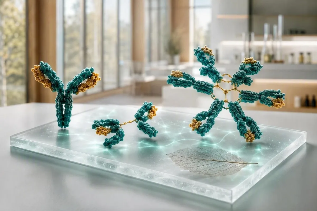
Test na imunoglobuline je najkorisniji kada se IgG, IgA i IgM očitavaju kao obrazac, a ne kao tri izolirana pokazatelja. Visoke vrijednosti mogu odražavati široku imunološku aktivaciju ili jedan nenormalan protein; niske vrijednosti mogu ukazivati na lijekove, gubitak proteina ili oslabljenu proizvodnju antitijela.


- IgG je obično 700-1.600 mg/dL u odraslih; uporno povišenje u nekoliko klasa često odražava kroničnu upalu ili bolest jetre.
- IgA je obično 70-400 mg/dL u odraslih; selektivni nedostatak može učiniti negativni test na celijakiju na bazi IgA nepouzdanim.
- protutijela pojavljuju se sa zakašnjenjem, a ne na dan kada uzročnik uđe u kožu. Ako vam je ta “abecedna juha” naporna, naš je obično 40-230 mg/dL u odraslih; izolirano, uporno povišenje zahtijeva elektroforezu serumskih proteina kako bi se isključio monoklonski protein.
- Sve tri niske je zabrinjavajuće od jednog niskog rezultata i zahtijeva pregled infekcija, lijekova, gubitka proteina i odgovora antitijela na cjepiva.
- Poliklonalna elevacija znači da su povišene višestruke klase imunoglobulina; često prati autoimune bolesti, kronične bolesti jetre ili dugotrajne infekcije.
- Monoklonski obrazac znači da je jedna klasa nesrazmjerno povišena ili se na elektroforezi pojavljuje uski pojas; zahtijeva potvrdu imunofiksacije i testiranje slobodnih lakih lanaca.
- hitna provjera je smisleno kod vrućice s ponavljajućim ozbiljnim infekcijama, nove zbunjenosti, nedostatka daha, žutice, gubitka težine, bolova u kostima ili naglo rastućeg ukupnog proteina.
- Važan je trend jer razlika od 10-20% može proizaći iz hidratacije, varijacije testa, nedavne bolesti ili drugačije laboratorijske metode.
Kako očitati test na imunoglobuline kao jedan obrazac
rezultati IgG, IgA i IgM zajedno znače različite stvari nego sami za sebe. Široko povišen panel obično signalizira poliklonsku imunološku stimulaciju, dok jedna dominantna klasa—posebno s visokim ukupnim proteinom—postavlja odvojeno pitanje monoklonske proteine. Od 5. rujna 2026. ta razlika ostaje praktičan prvi korak prije pridavanja dijagnoze testu krvi na imunoglobuline.
Referentni intervali za odrasle variraju ovisno o metodi, ali mnogi laboratoriji koriste IgG 700-1.600 mg/dL, IgA 70-400 mg/dL i IgM 40-230 mg/dL. Vrijednost izvan raspona 5% nije automatski bolest; prvo provjeravam laboratorijski interval, dob, status trudnoće, nedavnu infekciju, bubrežni gubitak i je li uzorak uzet nakon intravenskog imunoglobulina.
Kantesti je Analizator krvnih testova s umjetnom inteligencijom koji očitava IgG, IgA i IgM uz ukupni protein, albumin, jetrene markere, kreatinin i CBC umjesto da crvenu zastavicu tretira kao dijagnozu. Ta unakrsna provjera je korisna jer niska razina albumina s povišenim globulinima govori vrlo drugačiju priču od dehidracije s obje koncentrirane vrijednosti; naš vodič za biomarkere objašnjava povezane mjere.
U svom kliničkom radu, najutješniji panel često nije savršeno prosječan, ali je stabilan: na primjer, IgG 1.690 mg/dL u tri uzorka tijekom 18 mjeseci s normalnom elektroforezom i bez simptoma. Dr. Thomas Klein bi i dalje pitao o suhim očima, oticanju zglobova, ponavljajućim infekcijama i lijekovima, ali stabilna blaga varijacija rijetko nosi istu težinu kao porast od 600 mg/dL u šest mjeseci.
Tri imunoglobulina jednostavnim jezikom
protutijela pojavljuju se sa zakašnjenjem, a ne na dan kada uzročnik uđe u kožu. Ako vam je ta “abecedna juha” naporna, naš je rani široki odgovor, IgA štiti mukozne površine u dišnim putevima i crijevima, i IgG pruža najtrajniju cirkulirajuću aktivnost antitijela. Njihovi biološki poluživoti se značajno razlikuju: IgG traje oko 21 dan, IgA oko 6 dana u serumu, a IgM oko 5 dana, tako da se serijske promjene ne odvijaju istom brzinom.
Uobičajene referentne vrijednosti za odrasle i zašto se vaš laboratorij može razlikovati
Referentni raspon ispisan uz vlastiti rezultat ima prednost pred bilo kojim online rasponom. Nefelometrija, turbidimetrija i laboratorijske populacije kalibrirane prema dobi proizvode umjereno različita ograničenja, posebno za IgA i IgM.
Uobičajeni intervali za odrasle IgG 700-1.600 mg/dL, IgA 70-400 mg/dL i IgM 40-230 mg/dL pretvaraju se u otprilike 7-16 g/L, 0,7-4,0 g/L i 0,4-2,3 g/L. Djeca imaju znatno vrijednosti ovisne o dobi, a raspon za odrasle nikada se ne smije koristiti za procjenu dojenčeta ili tinejdžera.
Rezultat blizu granične vrijednosti posebno je osjetljiv na uobičajenu biološku varijaciju. Ako je IgM 238 mg/dL naspram gornje granice od 230 mg/dL nakon respiratorne bolesti, ponavljanje za 8-12 tjedana često je otkrivajuće nego odmah naručivanje velike baterije testova; pogledajte naš vodič za stvarne promjene između krvnih pretraga.
Postenje obično nije potrebno za kvantitativne imunoglobuline, ali jaka dehidracija može koncentrirati proteine, a IV imunoglobulin može povećati izmjereni IgG tjednima. Pitajte kliničara koji naručuje panel jeste li primili nadomjestak antitijela, rituksimab, kortikosteroide, antikonvulzive ili anti-CD20 terapiju u prethodnih 6 mjeseci.
Kada su sve tri klase imunoglobulina visoke
Visoki IgG, IgA i IgM zajedno obično ukazuju na poliklonsku imunološku aktivaciju, a ne na jedan abnormalni klon. Kronična bolest jetre, autoimune bolesti, perzistentne infekcije i neka upalna bolest crijeva česta su klinička stanja.
Poliklonsko povišenje često proširuje gama regiju na elektroforezi serumskih proteina umjesto da stvara oštar, uzak šiljak. U praktičnom smislu, IgG 2.100 mg/dL, IgA 520 mg/dL i IgM 310 mg/dL s povišenim CRP ukazuju na drugačiji put od IgG 2.400 mg/dL s potisnutim ostalim dvjema klasama.
Autoimune bolesti mogu proizvesti ovaj obrazac, ali simptomi usmjeravaju sljedeće testiranje. Perzistentna suhoća očiju i usta, otečeni zglobovi, osip, proljev ili neuropatija mogu opravdati ciljana autoantitijela i testiranje komplementa; naše vodič za C3 i C4 objašnjava zašto niska razina komplementa mijenja tu interpretaciju.
Kronična infekcija također može povisiti nekoliko klasa mjesecima, iako imunoglobulini ne identificiraju uzročnika niti dokazuju aktivnu infekciju. Visoka ESR s konstitucijskim simptomima zaslužuje klinički pregled, a naše objašnjenje uzroka visokog ESR-a pokazuje zašto nespecifični upalni marker ne može utvrditi uzrok.
Što izolirano visoki IgM može značiti
Izolirano povišeni IgM može uslijediti nakon nedavne infekcije, javiti se kod kolestatske autoimune bolesti jetre ili predstavljati monoklonski protein IgM. Perzistencija, stupanj povišenja, simptomi i elektroforeza važniji su od jednog graničnog broja.
IgM je veliko pentamerno antitijelo, a razine malo iznad 230 mg/dL često se normaliziraju nakon imunološkog izazova. Ponovljeni IgM od 800 mg/dL ili 1.200 mg/dL, osobito s normalnim ili niskim IgG i IgA, trebao bi pokrenuti elektroforezu serumskih proteina, imunofiksaciju i testiranje serumskih slobodnih lakih lanaca umjesto nagađanja.
Primarni bilijarni kolangitis klasično se povezuje s povišenim IgM-om, osobito kada su alkalna fosfataza i gama-glutamiltransferaza također visoke. Smjernice EASL-a savjetuju procjenu kolestatske bolesti jetre kada je alkalna fosfataza trajno povišena i prisutna su antimitoondrijalna antitijela (EASL, 2017.); naše vodič za obrazac kolestaze pokriva prateće markere.
Monoklonski IgM protein nije isto što i Waldenströmova makroglobulinemija, a većina ljudi s malom monoklonskom gamapatijom nema dijagnozu neposrednog raka. Ipak, simptomi poput zamućenog vida, glavobolja, krvarenja iz nosa, nove neuropatije ili neobjašnjivog umora zahtijevaju hitnu procjenu jer vrlo visoki IgM može promijeniti viskoznost seruma; pročitajte više o visoki IgM uzrokuje.
Niska razina IgA: zamka testa na celijakiju i tragovi infekcije
Niska razina IgA može učiniti koelijakijski skrining na IgA tkivnu transglutaminazu lažno negativnim. Selektivni nedostatak IgA obično se definira kao serumski IgA niži od 7 mg/dL u osobe starije od 4 godine s normalnim IgG i IgM, iako lokalne definicije i ponovljeno testiranje imaju važnost.
Ako je ukupni IgA nizak, kliničari obično koriste IgG-bazirani deamidirani glijadinski peptid ili IgG test tkivne transglutaminaze uz kliničku anamnezu. Nemojte započeti dijetu bez glutena prije odgovarajućeg testiranja osim ako vam se drugačije ne savjetuje, jer razine antitijela mogu pasti i zamutiti rezultat; naš vodič za izazivanje glutena opisuje problem s vremenskim rasporedom.
Mnogi ljudi s djelomičnim nedostatkom IgA nemaju ozbiljnih zdravstvenih problema, dok drugi prijavljuju ponavljajuće sinusne, prsne ili gastrointestinalne infekcije. Učestalost, težina i dokumentirani bakterijski uzroci važniji su od dva obična prehlada zimi; povijest cijepljenja i odgovor antitijela na pneumokoke mogu biti informativniji od ukupnog IgA.
Reakcije na krvne pripravke su rijetke, ali relevantne: osobe s teškim nedostatkom IgA i anti-IgA antitijelima možda će trebati posebno odabrane komponente ako je potrebna transfuzija. Ovo je razlog za obavještavanje bolničkih timova o potvrđenom teškom nedostatku, a ne razlog za izbjegavanje medicinski nužne skrbi; pogledajte našu opširniju raspravu o niskoj razini IgA.
Niska razina IgG i ponavljajuće infekcije: kada rezultat ima važnost
Niska razina IgG postaje klinički značajna kada prati ponavljajuće, neuobičajeno teške ili sporo prolazeće infekcije. IgG u odraslih ispod 400 mg/dL općenito je veća briga nego vrijednost od 650 mg/dL, ali funkcionalni odgovori antitijela na cjepivo mogu preokrenuti pretpostavke temeljene samo na količini.
Infekcije zbog kojih imunolozi zastanu su ponavljajuća upala pluća, bronhiektazije, ponavljajuće bakterijske infekcije sinusa koje zahtijevaju nekoliko antibiotika, invazivna bakterijska bolest ili kronična giardijaza. Četiri blage virusne prehlade kod radnika u dječjem vrtiću nisu ekvivalentne trima radiološki potvrđenim upalama pluća u 24 mjeseca.
Sekundarni uzroci su česti: rituksimab i drugi terapijski lijekovi usmjereni na B-stanice, dugotrajni oralni kortikosteroidi, neki antikonvulzivi, gubitak proteina u nefrotskom rasponu, enteropatija s gubitkom proteina i limfoidna malignost mogu sniziti IgG. Proteini u urinu, albumin, broj limfocita i povijest medikacije često daju više informacija od još jednog slučajnog panelu antitijela; naš vodič za protein u urinu objašnjava ključni put gubitka.
Bonilla i sur. (2015.) preporučuju procjenu kvantitativnih imunoglobulina zajedno s odgovorima specifičnih antitijela kada se sumnja na primarnu imunodeficijenciju. Po mom iskustvu, normalan odgovor nakon cijepljenja može biti umirujući čak i kada je IgG blago nizak, dok siromašni odgovori s IgG ispod 500 mg/dL zaslužuju stručni unos.
Kada su IgG, IgA i IgM svi niski
Niska razina IgG, IgA i IgM zajedno naziva se hipogamaglobulinemija i zaslužuje strukturiranu obradu, posebno kod infekcija. Obrazac može odražavati oštećenu proizvodnju antitijela, učinke lijekova, gubitak proteina, naprednu limfoidnu bolest ili, rjeđe, privremenu promjenu nakon bolesti.
Panel IgG 360 mg/dL, IgA 28 mg/dL i IgM 18 mg/dL nije rezultat koji treba čekati ako osoba ima vrućicu, ponavljajuće infekcije prsnog koša ili gubitak težine. Kliničar može ponoviti panel, provjeriti elektroforezu, CBC s diferencijalnom krvnom slikom, bubrežne i jetrene testove, proteine u urinu i cjepivu specifična antitijela, a zatim uputiti na kliničku imunologiju ili hematologiju.
Varijabilna imunodeficijencija (CVID) često se javlja u odrasloj dobi, ali njezina dijagnoza zahtijeva više od niskih brojeva: simptomi, isključeni sekundarni uzroci i smanjena funkcionalna proizvodnja antitijela su ključni. Kantesti AI je usluzi za tumačenje laboratorijskih testova AI koji može organizirati ove okolne nalaze u sažetak liječničkog posjeta, ali ne može dijagnosticirati imunodeficijenciju niti odlučiti o nadomjesnoj terapiji imunoglobulinima.
Nadomjesna terapija imunoglobulinima propisuje se za sprječavanje ozbiljnih infekcija kod odabranih pacijenata, a ne samo za normalizaciju laboratorijskog rezultata. Tipični režimi nadomjesne terapije su individualizirani i obično započinju oko 400-600 mg/kg mjesečno intravenskim ili supkutanim putem, s doziranjem prilagođenim kontroli infekcije i minimalnom razinom IgG, a ne univerzalnim ciljem; naš vodič za krvnih pretraga imunološkog sustava dodaje koristan kontekst.
Kako test na imunoglobuline može ukazati na monoklonski protein
Monoklonski protein sugerira se kada je jedna klasa imunoglobulina nesrazmjerno visoka, druge klase su potisnute, ili elektroforeza pokazuje usku traku. Kvantitativni imunoglobulini sami po sebi ne mogu dokazati klonalnost; serumsko elektroforezom proteina i imunofiksacijom su potvrdni alati.
Razmotrite IgG 2.900 mg/dL s IgA 38 mg/dL i IgM 21 mg/dL: taj obrazac je sumnjiviji nego da su sve tri umjereno povišene. Visok ukupni protein, nizak omjer albumina i globulina, anemija, smanjeni eGFR, visok kalcij, bol u kostima ili ponavljajuća infekcija dodaju kliničku hitnost; pregledajte naš vodič za povišen ukupni protein.
Međunarodna radna skupina za multipli mijelom razlikuje MGUS od aktivnog mijeloma koristeći količinu proteina, nalaze koštane srži i organske učinke – ne samo rezultat imunoglobulina (Rajkumar et al., 2014). M-protein ispod 3 g/dL i dalje može zahtijevati praćenje, dok niža vrijednost s anemijom ili bubrežnom ozljedom može zahtijevati bržu hematološku procjenu.
Nemojte se zavarati normalnim ukupnim proteinom. Poremećaji samo lakih lanaca možda neće uzrokovati očito povećanje proteina, zato kliničari kombiniraju serumske slobodne lake lance, imunofiksaciju i pretrage urina kod odgovarajuće prezentacije; naš put za testiranje krvnih karcinoma opisuje što se događa dalje.
Obrasci bolesti jetre: tragovi IgA, IgG i IgM
Bolest jetre može povisiti imunoglobuline, ali dominantna klasa pruža samo trag, ne dijagnozu. IgA je često povišen kod bolesti jetre povezane s alkoholom, IgG kod autoimunog hepatitisa, a IgM kod primarnog bilijarnog kolangitisa; preklapanje je uobičajeno.
Povišen IgG iznad 1,1 puta gornje granice normale podupire autoimuni hepatitis kada transaminaze i autoantitijela odgovaraju, ali nije ni dovoljno osjetljiv ni specifičan za samostalnu dijagnozu. Akutna virusna bolest, kronična bolest jetre i sustavna autoimuna bolest mogu stvoriti sličan signal, stoga kliničari često koriste slikovne prikaze i testiranje usmjereno od strane specijalista.
Povišenje IgA uz povišene GGT, AST i makrocitozu može odgovarati bolesti jetre povezane s alkoholom, ali konzumacija alkohola nikada se ne smije pretpostaviti iz panela. Nuspojave lijekova, metabolička bolest masne jetre i drugi uzroci su česti; naše objašnjenje ALT rezultata pomaže smjestiti enzime pored imunoglobulina.
Korisna poruka o sigurnosti je da žutica, tamni urin, blijeda stolica, konfuzija, oticanje trbuha ili povraćanje krvi zahtijevaju hitnu medicinsku skrb bez obzira na vrijednosti imunoglobulina. Stabilno izolirano povišenje IgA od 430 mg/dL ne uzrokuje te simptome i ne smije odvraćati od hitne procjene.
Razlikovanje infekcije, upale i autoimune aktivnosti
Razine imunoglobulina rastu presporo i nespecifično da bi sami po sebi dijagnosticirali akutnu infekciju. CRP, potpuna krvna slika, kulture ili ciljani testovi na patogene bolje odgovaraju na hitna pitanja o infekciji, dok imunoglobulini opisuju širu imunološku pozadinu.
IgM se često opisuje kao protutijelo rane infekcije, ali ukupni serumski IgM nije isto što i specifični IgM test na patogen. Ukupni IgM od 290 mg/dL ne može potvrditi gripu, Epstein-Barr virus ili urinarnu infekciju; simptomi i izravno testiranje utvrđuju treba li liječenje.
Autoimuna upala može povisiti IgG godinama prije nego što je dijagnoza jasna, osobito kod hipergamaglobulinemije i širokog elektroforetskog obrasca. ANA, reumatoidni faktor i ENA paneli trebaju se naručiti kako bi se odgovorilo na kliničko pitanje, a ne kao izvidničko istraživanje; na primjer, naš vodič za anti-dsDNA objašnjava zašto pozitivan rezultat zahtijeva bubrežni i komplementni kontekst.
Dr. Thomas Klein vidio je pacijente preplašene visokim IgG nakon virusne bolesti koja se normalizirala na ponovljenom testiranju nakon 3 mjeseca. Nasuprot tome, uporni IgG iznad 2.000 mg/dL plus niska razina C3, proteinurija i zglobni simptomi nisu nešto što bih odgodio na godinu dana – ti kombinirani nalazi opravdavaju pravovremenu kliničku procjenu.
Lijekovi, gubitak proteina i vremenski učinci koji mijenjaju rezultate
Lijekovi i gubitak proteina mogu sniziti imunoglobuline bez primarnog imunološkog poremećaja. Anti-CD20 lijekovi mogu prvo smanjiti IgM, a zatim IgG, dok gubitak proteina bubrega ili crijeva može sniziti nekoliko klasa uz albumin.
Hipogamaglobulinemija povezana s rituksimabom može se pojaviti mjesecima nakon posljednje infuzije jer oporavak B-stanica i oporavak antitijela nisu identični. Ponesite potpuni popis lijekova – uključujući biološke lijekove, dozu steroida, lijekove protiv napadaja i kemoterapiju – na pregled; naš vodič o trendovima sigurnosti lijekova objašnjava zašto su datumi važni.
Stanja gubitka proteina često ostavljaju dodatne tragove: nizak albumin ispod oko 3,5 g/dL, edem, proljev, povišen omjer albumin-kreatinin u urinu ili smanjeni ukupni proteini. Nasuprot tome, dehidracija može uzrokovati da imunoglobulini izgledaju visoko, dok albumin i hematokrit rastu paralelno.
Ponovljeno testiranje je obično najbolje kada ste klinički stabilni, koristeći istu laboratoriju ako je moguće. Kantesti može usporediti datirani PDF ili fotografiju s prethodnim rezultatima i označiti veliku promjenu, ali pažljiva provjera učitavanja je razumna jer se jedinice i referentni rasponi mogu pogrešno zabilježiti.
Kontrolni testovi koji pojašnjavaju abnormalnu panelu
Pravo naknadno testiranje ovisi o uzorku: elektroforeza za nesrazmjerne povišenja, antitijela na cjepiva za sumnju na nedostatak, te pretrage urina ili jetre za gubitak proteina ili jetrene tragove. Ponavljanje iste tri brojke bez pitanja često malo doprinosi.
Za visoke ili neujednačene rezultate, kliničari obično naručuju elektroforezu proteina seruma, imunofiksaciju, slobodne lake lance seruma, kompletna krvna slika, kalcij, kreatinin, albumin i jetrene enzime. Uski pojas na elektroforezi treba tipizirati imunofiksacijom jer klasa—IgG, IgA ili IgM—mijenja diferencijalnu dijagnozu i plan naknadnog liječenja.
Za niske rezultate s infekcijama, testiranje može uključivati IgG podklase i titar antitijela na tetanus, Haemophilus influenzae tip b ili pneumokokne serotipove prije i poslije cijepljenja. Ne postoji jedinstveni zaštitni pneumokokni prag koji odgovara svakom laboratoriju i dobnoj skupini, što je jedno područje gdje specijalistička interpretacija ima veću važnost od internetskog graničnog određenja.
Kantesti je Alat za analizu krvnih nalaza uz pomoć AI-a koji mogu sastaviti ove rezultate u vremensku crtu i identificirati nedostajući kontekst, poput odsutnog albumina ili elektroforeze, kako bi vaš kliničar mogao razmotriti. Naš vodič za tehnologiju objašnjava kako se generiraju kontekstualne zastavice bez zamjene medicinskog odlučivanja.
Simptomi koji ne bi smjeli čekati rutinsko ponavljanje
Sam rezultat imunoglobulina rijetko stvara hitnu situaciju, ali određeni simptomi stvaraju. Potražite hitnu procjenu istog dana kod visoke temperature sa teškom bolešću, otežanog disanja, zbunjenosti, nove slabosti, žutice, znatno smanjene količine urina, jake glavobolje s promjenama vida ili nekontroliranog krvarenja.
Niske razine imunoglobulina plus temperatura od 38,0°C ili više zabrinjavajuće su kod osoba na kemoterapiji, visokim dozama steroida, terapiji usmjerenoj na B-stanice ili s poviješću invazivnih infekcija. Nemojte čekati poruku portala ako je disanje otežano, razina kisika niska, ili se osoba čini zbunjena ili neuobičajeno pospana.
Mogući monoklonski protein zahtijeva bržu procjenu kada se pojavi uz novu bol u kostima, neobjašnjivu anemiju, porast kreatinina, kalcij iznad lokalne gornje granice, neuropatiju ili nenamjerni gubitak težine. Ovo nije dokaz poremećaja plazma stanica, ali su to značajke koje kliničari koriste za prioritetizaciju testiranja; naš vodič za drugo mišljenje može pomoći u pripremi pitanja.
Za blage stabilne abnormalnosti bez crvenih zastavica, planirani pregled u roku od nekoliko tjedana je obično razuman. Ponesite prethodne rezultate, datume infekcija, promjene lijekova i obiteljsku anamnezu; grafikon od 2 godine često je klinički korisniji od jednog istaknutog broja.
Sigurna primjena AI tumačenja za rezultate imunoglobulina
AI može organizirati uzorak testiranja imunoglobulina, ali ne može utvrditi ima li osoba imunodeficijenciju, bolest jetre ili monoklonski poremećaj. Sigurna interpretacija zadržava laboratorijski raspon, simptome, lijekove, trendove i potvrdne testove vidljivima.
Kantesti AI pregledava IgG, IgA i IgM uz proteine i organske markere koji mijenjaju njihovo značenje, a zatim predstavlja pitanja za praćenje umjesto proglašavanja dijagnoze. U našem tijeku analize, izolirani niski IgA potiče oprez pri testiranju na celijakiju, dok sva tri niska vrijednosti plus niski albumin potiču razmatranje gubitka, učinaka lijekova i kliničke imunološke revizije.
Privatnost je važna kod rezultata povezanih s imunitetom i rakom. Kantesti Ltd koristi rukovanje usklađeno s GDPR-om, a naš stranica medicinske verifikacije opisuje klinički nadzor i granice iza automatizirane laboratorijske interpretacije.
Dobar nalaz trebao bi vam pomoći da se zapitate: “Je li ovo široka aktivacija imunološkog sustava, gubitak proteina, smanjena proizvodnja ili jedan pojedinačni protein?” To je odgovarajuća razina sigurnosti prije nego što elektroforeza ili procjena specijalista daju odgovor.
Pitanja koja treba postaviti nakon abnormalne panelne pretrage imunoglobulina
Najbolje sljedeće pitanje obično je “Koji je ovo obrazac?” umjesto “Kako mogu sniziti ili povisiti ovu vrijednost?” Pitajte je li rezultat trajan, poliklonski ili monoklonski, upućuju li infekcije na oslabljenu funkciju te koji su sekundarni uzroci provjereni.
Korisna pitanja uključuju: Je li učinjena elektroforeza serumskih proteina? Jesu li albumin, proteini u mokraći i jetreni enzimi u skladu s gubitkom proteina ili bolešću jetre? Trebam li testiranje protutijela na cjepiva? Treba li ponoviti testiranje za 8–12 tjedana? Ta su pitanja korisnija od kupnje dodataka prehrani, koji ne ispravljaju pouzdano klinički značajan nedostatak protutijela.
Ako je planirano upućivanje specijalistu, zabilježite broj infekcija liječenih antibioticima u posljednjih 12 mjeseci, posjete bolnici, upalu pluća potvrđenu slikovnim pretragama i izloženost lijekovima. Precizan zapis — “tri kure antibiotika i jedna upala pluća” — korisniji je od izjave “često se razbolim”; naš pratitelj laboratorijskih rezultata nudi praktičan format za vođenje zapisa.
Naše Medicinski savjetodavni odbor podupire kliničke standarde pregleda Kantesti-a, ali vaš liječnik i dalje ostaje osoba koja vas može pregledati, zatražiti potvrdne pretrage i postupiti u slučaju hitnih nalaza. Većina pacijenata smatra da smiren razgovor utemeljen na obrascima zabrinjavajući nalaz pretvara u sljedeći korak kojim se može upravljati.
Često postavljana pitanja
Koja je normalna referentna vrijednost testiranja imunoglobulina za odrasle?
Uobičajeni rasponi imunoglobulina u odraslih su IgG 700-1.600 mg/dL, IgA 70-400 mg/dL i IgM 40-230 mg/dL, iako bi interval ispisan u svakom laboratoriju trebalo koristiti za tumačenje. Rezultati se također izvješćuju kao g/L u mnogim zemljama: 7-16 g/L za IgG, 0,7-4,0 g/L za IgA i 0,4-2,3 g/L za IgM. Rezultat malo izvan raspona može odražavati nedavnu bolest, hidrataciju ili varijaciju analize. Uporne abnormalnosti i simptomi određuju je li potreban daljnji nadzor.
Što zajedno znači visok IgG, IgA i IgM?
Visoki IgG, IgA i IgM zajedno najčešće ukazuju na poliklonsku imunološku aktivaciju uslijed kronične upale, autoimune bolesti, perzistentne infekcije ili bolesti jetre. Široko povećanje sve tri klase razlikuje se od monoklonskog proteina, koji obično stvara jednu nesrazmjerno visoku klasu ili usku elektroforetsku traku. Vrijednosti poput IgG 2.000 mg/dL, IgA 500 mg/dL i IgM 300 mg/dL trebaju se tumačiti zajedno s CRP-om, testovima jetre, albuminom i elektroforezom serumskih proteina. Sam obrazac ne dijagnosticira autoimunu bolest ili infekciju.
Mogu li niske razine imunoglobulina uzrokovati česte infekcije?
Niske razine imunoglobulina mogu povećati rizik od ponavljajućih bakterijskih respiratornih ili gastrointestinalnih infekcija, osobito kada je IgG ispod 400-500 mg/dL ili su cjepne antigenske reakcije slabe. Najzabrinjavajući obrazac su ponavljajuće upale pluća, bronhiektazije, invazivne bakterijske infekcije ili nekoliko liječenih sinusnih infekcija antibioticima, umjesto povremenih virusnih prehlada. Niske razine IgG, IgA i IgM zajedno trebale bi potaknuti provjeru lijekova, gubitka proteina putem bubrega ili crijeva, limfoidnih stanja i primarne imunodeficijencije. Testiranje funkcionalnih antitijela često bolje pojašnjava rizik nego samo jedna vrijednost ukupnog IgG.
Utječe li nizak IgA na testiranje na celijakiju?
Niska razina IgA može uzrokovati lažno negativan test na celijakiju pomoću antitijela IgA protiv tkivne transglutaminaze jer test ovisi o proizvodnji antitijela IgA. Selektivni nedostatak IgA često se definira kao razina IgA ispod 7 mg/dL nakon navršene 4 godine života uz normalne razine IgG i IgM. Kada je ukupna razina IgA niska, kliničari umjesto toga često koriste testiranje na IgG deamidirani glijadinski peptid ili IgG tkivnu transglutaminazu. Nastavite konzumirati gluten dok se ne dogovorite o planu testiranja, jer izbjegavanje glutena može sniziti razine antitijela i smanjiti osjetljivost testa.
Koji test potvrđuje proteinski monoklonski imunoglobulin?
Elektroforeza serumskih proteina otkriva nenormalan proteinski profil, a imunofiksacija potvrđuje prisutnost monoklonskog imunoglobulina i identificira njegov razred. Testiranje slobodnih lakih lanaca u serumu obično se dodaje jer neki poremećaji plazma-stanica proizvode uglavnom lake lance i možda neće značajno povisiti ukupne proteine. Monoklonski nalaz zahtijeva procjenu koncentracije M-proteina, krvne slike, kalcija, bubrežne funkcije i simptoma; sam M-protein ne znači automatski rak. U odabranim slučajevima mogu biti potrebne i pretrage urina na proteine.
Može li stres povisiti IgG, IgA ili IgM?
Svakodnevni psihološki stres obično ne uzrokuje veliko izolirano povećanje IgG, IgA ili IgM na testu imunoglobulina iz krvi. Stres može utjecati na san, podložnost infekcijama i upalnu signalizaciju, ali postojane vrijednosti iznad gornje laboratorijske granice ne treba pripisivati stresu bez provjere jetrenih markera, CRP, lijekova i proteinske elektroforeze kada je to prikladno. Skromna promjena od 10-20% može se pojaviti između uzimanja uzoraka zbog normalnih bioloških i analitičkih varijacija. Ponavljanje testa nakon 8-12 tjedana kada je pacijent klinički dobro često je razuman prvi korak kod blagog neobjašnjivog porasta.
Isprobajte analizu krvne slike uz AI već danas
Pridružite se više od 2 milijuna korisnika diljem svijeta koji vjeruju Kantesti-u za trenutačnu i točnu analizu laboratorijskih nalaza. Prenesite svoje rezultate krvne slike i dobijte sveobuhvatno tumačenje 15,000+ biomarkera u sekundama.
📚 Referirane znanstvene publikacije
Klein, T., Mitchell, S., & Weber, H. (2026). Kantesti LTD. (2026). AI analizator krvne slike: 2,5M testova analizirano | Globalni zdravstveni izvještaj 2026. Zenodo.. Kantesti AI Medical Research.
Klein, T., Mitchell, S., & Weber, H. (2026). Kantesti LTD. (2026). RDW krvni test: potpuni vodič za RDW-CV, MCV i MCHC. Zenodo.. Kantesti AI Medical Research.
📖 Vanjske medicinske reference
⚕️ Medicinska izjava o odricanju odgovornosti
Ovaj je članak samo u obrazovne svrhe i ne predstavlja medicinski savjet. Za odluke o dijagnozi i liječenju uvijek se posavjetujte s kvalificiranim pružateljem zdravstvenih usluga.
E-E-A-T signal(i) povjerenja
Iskustvo
Liječnički vođena klinička revizija radnih tokova tumačenja laboratorijskih nalaza.
Stručnost
Fokus laboratorijske medicine na to kako se biomarkeri ponašaju u kliničkom kontekstu.
Autoritativnost
Napisao dr. Thomas Klein, uz recenziju dr. Sarah Mitchell i prof. dr. Hans Weber.
Pouzdanost
Tumačenje utemeljeno na dokazima, s jasnim putovima praćenja kako bi se smanjila uzbuna.