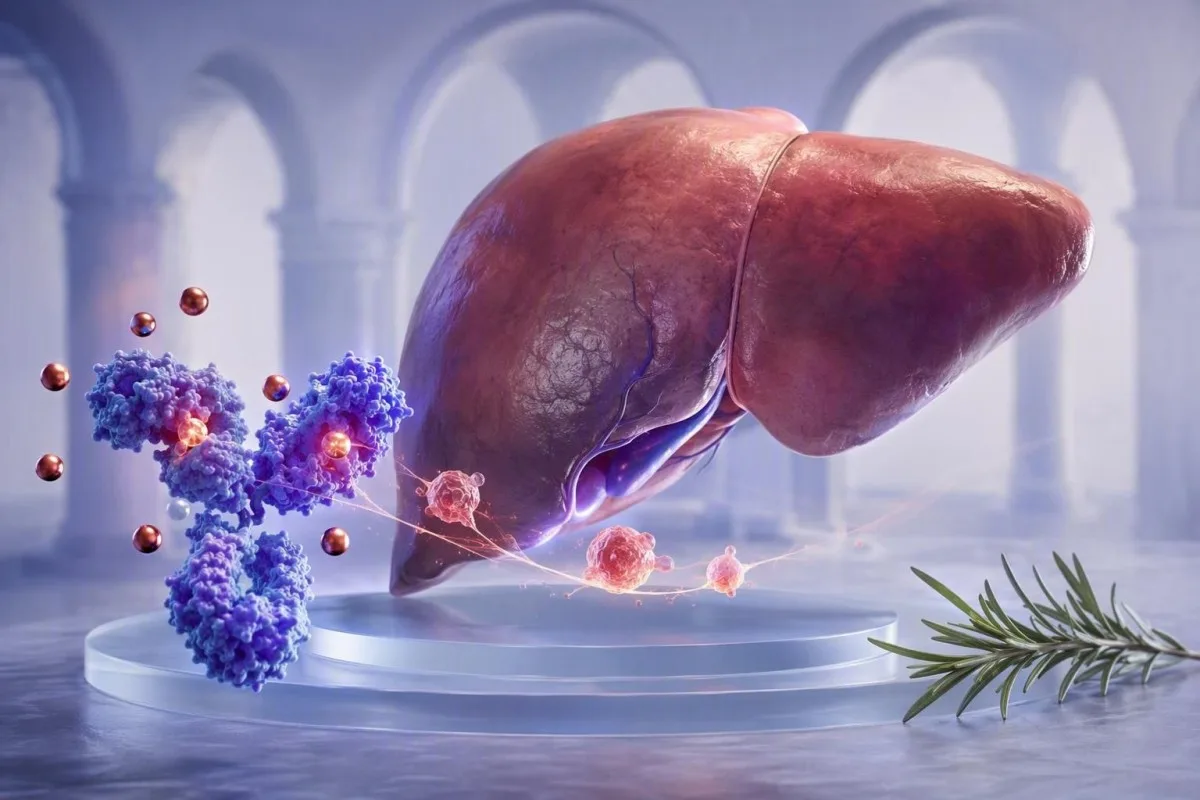
Нізкі вынік трансферрыну можа адлюстроўваць адказ печані на запаленне, а не знясіленыя запасы жалеза. Сумеснае чытанне сыроватачнага жалеза, ферытыну, CRP і трансферрыну прадухіляе распаўсюджаныя памылкі.


- Адмоўны бялок вострай фазы: Трансферрын звычайна падае падчас інфекцый, аутоіммунных захворванняў, хірургічных аперацый і іншых запаленчых станаў, нават калі запасы жалеза ў арганізме дастатковыя.
- Тыповы дыяпазон для дарослых: Большасць лабараторый выкарыстоўваюць рэферэнтны інтэрвал трансферрыну каля 200-360 мг/дл (2,0-3,6 г/л), але мясцовыя дыяпазоны аналізаў маюць прыярытэт.
- Разлік TSAT: Насычэнне трансферрыну роўна сыроватачнаму жалезу, падзеленаму на агульную жалезазлучальную здольнасць сыроваткі, памножанаму на 100; нізкі TIBC можа зрабіць насычэнне менш ненармальным, чым чакалася.
- Заўвага адносна фе Ферытын павялічваецца пры запаленні, таму ферытын ніжэй за 30 нг/мл моцна сведчыць аб дэфіцыце жалеза, у той час як значэнне 100 нг/мл не можа надзейна выключыць яго пры павышаным CRP.
- Карысны спадарожны тэст: Растваральны рэцэптар трансферрыну звычайна менш падвяргаецца ўплыву запалення, чым ферытын або трансферрын, і можа дапамагчы ўдакладніць змешаную анемію.
- Калі паўторна правяраць: Пры неадкладных анамаліях паўторныя даследаванні жалеза прыкладна праз 2-4 тыдні пасля таго, як кароткая хвароба прайшла, часта даюць больш рэпрэзентатыўны базавы ўзровень.
- Не займайцеся самалячэннем наўслеп: Дабаўкі жалеза могуць быць карыснымі пры пацверджаным дэфіцыце, але могуць быць непрыдатнымі пры перагрузцы жалезам, актыўных захворваннях печані ці анеміі, якая ў асноўным выклікана запаленнем.
Чаму тэст на трансферрын падае пры запаленні?
Запаленне зніжае трансферын, паколькі трансферын з'яўляецца негатыўным бялком вострай фазы. Цытакіны, асабліва інтэрлейкін-6, сігналізуюць печані зніжаць выпрацоўку трансферыну, у той час як гепцыдын абмяжоўвае вызваленне жалеза ў плазму; гэта можа прывесці да нізкага ўзроўню трансферыну і нізкага ўзроўню жалеза ў сыроватцы крыві без пацверджання дэплецыі запасаў жалеза.
Кароткая версія: арганізм часова робіць жалеза менш даступным падчас імуннай актывацыі. Гепцыдын зніжае экспарт жалеза з энтэрацытаў і макрафагаў праз ферапорцін, таму жалеза ў сыроватцы крыві часта падае на працягу 24-48 гадзін; печань можа адначасова зніжаць сінтэз трансферыну. Гэтая карціна з'яўляецца рэакцыяй абароны гаспадара, а не прамым вымярэннем спажывання жалеза з ежай.
У сваёй клінічнай практыцы ў чалавека з CRP 68 мг/л пасля бактэрыяльнай пнеўманіі можа быць жалеза ў сыроватцы крыві 22 мкг/дл і трансферын 165 мг/л, але феритин 240 нг/мл. Называць гэтую ізаляваную карціну “перагрузкай жалезам” або ўпэўнена выключаць дэфіцыт жалеза будзе памылкай. Даследаванні жалеза кіруюць больш падрабязна тлумачыць асноўныя разлікі.
Кантэсці — гэта Аналізатар крыві са штучным інтэлектам які чытае трансферын побач з CRP, феритин, паказчыкамі поўнага аналізу крыві, маркерамі печані і папярэднімі вынікамі, замест таго, каб разглядаць адно нізкае значэнне як дыягназ. Практычнае правіла доктара Томаса Кляйна простае: вынік трансферыну, атрыманы падчас ліхаманкі, абвастрэння або аднаўлення, павінен быць пазначаны як кантэкстна-залежны перад прыняццем любога рашэння аб лячэнні.
Напрамак вострай фазы мае значэнне
CRP, феритин, фібрынаген і гаптаксіген звычайна павышаюцца як станоўчыя бялкі вострай фазы, у той час як трансферын і альбумін могуць зніжацца. CRP вышэй за 10 мг/л робіць адзінае значэнне феритина значна больш складаным для інтэрпрэтацыі, хоць ніякі пастаянны парог CRP не можа выправіць вынік кожнага пацыента.
Якія нармальныя дыяпазоны трансферрыну і агульнай жалезазлучальнай здольнасці сыроваткі?
Звычайны дыяпазон трансферыну для дарослых звычайна складае ад 200-360 мг/л, а агульная ёмістасць жалеза злучэння (TIBC) звычайна складае ад 250-450 мкг/л. Гэтыя інтэрвалы адрозніваюцца ў залежнасці ад лабараторнага метаду, полу, цяжарнасці і мясцовых адзінак справаздачнасці, таму дыяпазон, надрукаваны побач з вашым вынікам, з'яўляецца тым, які трэба выкарыстоўваць.
Трансферын звычайна вымяраецца напрамую ў мг/л або г/л, у той час як агульная ёмістасць жалеза злучэння, або ТІБК, ацэньвае, колькі жалеза можа звязаць даступны трансферын. Шматлікія лабараторыі атрымліваюць TIBC з трансферыну, а не вымяраюць яго асобна; прыблізная канверсія: TIBC у мкг/л роўна трансферын у мг/л, памножаны на 1.25. Гэта суадносіны карысна для арыентацыі, а не для адмены ўласнага разліку лабараторыі.
Насычэнне трансферыну, скарочана TSAT, разлічваецца як жалеза сыроваткі, падзеленае на TIBC, памножанае на 100. TSAT каля 20-45% з'яўляецца тыповым для многіх дарослых лабараторый; значэнні ніжэй 20% паказваюць на абмежаванае цыркулюючае жалеза, але яны самі па сабе не адрозніваюць абсалютны дэфіцыт жалеза ад секвестрацыі жалеза, выкліканай запаленнем. Нізкая насычанасць трансферыну ахоплівае гэтае адрозненне.
Некаторыя еўрапейскія лабараторыі паведамляюць трансферын у г/л, дзе 2.0-3.6 г/л прыблізна адпавядае 200-360 мг/л. Вынік можа знаходзіцца крыху ніжэй за норму пасля віруснага захворвання і нармалізавацца пры паўторным аналізе, у той час як пастаянныя значэнні ніжэй за 150 мг/л патрабуюць наўмыснага пошуку праблем з сінтэзам печані, страты бялку, значнага запалення або недаядання.
Як інтэрпрэтаваць даследаванні жалеза як патэ́рн?
Даследаванні жалеза найбольш дакладныя, калі адначасова аналізуюцца сыроватачнае жалеза, трансферын або TIBC, TSAT, ферритин і маркер запалення. Сыроватачнае жалеза сама па сабе значна змяняецца на працягу дня і можа зніжацца больш чым на 30% паміж узяццямі проб у аднаго і таго ж чалавека.
Класічны неўскладнены дэфіцыт жалеза звычайна выклікае нізкі ўзровень сыроватачнага жалеза, высокі трансферын або TIBC, TSAT ніжэй за 16%, і ферритин ніжэй за 30 нг/мл. Высокі TIBC узнікае таму, што печань павялічвае выпрацоўку трансферыну, калі даступнасць жалеза нізкая. Павышэнне шырыні размеркавання эрытрацытаў можа з'явіцца да зніжэння гемаглабіну; гл. наша кіраўніцтва па Змены RDW пасля тэрапіі жалезам для часовай працягласці.
Анемія запалення часцей выклікае нізкі ўзровень сыроватачнага жалеза, нізкі або нармальны трансферын, нізкі TSAT і ферритин, які з'яўляецца нармальным або высокім. Вайс і Гуднаў апісвалі гэта як эрытрапаэз, абмежаваны жалезам, выкліканы парушэннем даступнасці жалеза, а не абавязкова адсутнасцю запасаў жалеза (Weiss & Goodnough, 2005). Дзве станы часта сустракаюцца разам, асабліва пры запаленчых захворваннях кішачніка, рэўматоідным артрыце, хранічнай хваробе нырак і раку.
Адной з менш відавочных пастак з'яўляецца арыфметыка: калі сыроватачнае жалеза складае 30 мкг/дл, а TIBC зніжаны да 180 мкг/дл, TSAT складае 17%. Калі б TIBC быў 360 мкг/дл, тое ж сыроватачнае жалеза дало б 8%. Ніжні знаменальнік можа хаваць, наколькі мала цыркулюе жалеза, таму нізкае сыроватачнае жалеза патрабуе кантэксту, а не рэфлекторнага прызначэння.
Практычны падказка змешанага тыпу
Ферритин паміж 30 і 100 нг/мл з TSAT ніжэй за 16% і CRP вышэй за 10 мг/л часта з'яўляецца "шэрай зонай", а не падставай для супакаення. У такой сітуацыі клініцысты могуць выкарыстоўваць растваральны рэцэптар трансферыну, утрыманне гемаглабіну ў рэтыкулацытах, даныя тэндэнцый альбо тэрапеўтычны план, прыстасаваны да асноўнага захворвання.
Чаму ферытын можа выглядаць нармальным, калі жалеза мала
Ферритин з'яўляецца як бялком, які захоўвае жалеза, так і станоўчым бялком вострай фазы, таму запаленне можа павялічыць яго незалежна ад запасаў жалеза. Ферритин ніжэй за 30 нг/мл моцна падтрымлівае дэфіцыт жалеза ў большасці дарослых, але больш высокае значэнне не можа надзейна выключыць дэфіцыт, калі CRP або ESR павышаны.
Кіраўніцтва Сусветнай арганізацыі аховы здароўя 2020 года па ферритину рэкамендуе вымяраць маркеры запалення разам з ферритинам, калі інфекцыя або запаленне распаўсюджаныя. У дарослых з запаленнем СААЗ мяркуе, што ферритин ніжэй за 70 мкг/л можа сведчыць аб дэфіцыце жалеза; гэта парог, заснаваны на ацэнцы насельніцтва, а не замена індывідуальнай клінічнай ацэнкі (WHO, 2020).
Я часта бачу пацыентаў, якія стурбаваны ферритинам 180 нг/мл пасля запаленчага абвастрэння, мяркуючы, што іх запасы жалеза павінны быць багатымі. Часам гэта так. Аднак ферритин можа павышацца пасля інтэнсіўных фізічных нагрузак, пашкоджання клеткамі печані, метабалічных захворванняў, уздзеяння алкаголю і імуннай актыўнасці, таму гэтае значэнне не з'яўляецца самастойнай інвентарызацыйнай адзнакай. Ферритин і CRP з'яўляецца карысным суправаджальным абмеркаваннем.
Кантэсці — гэта Платформа AI для расшыфроўкі аналізу крыві які пазначае супярэчлівае спалучэнне нізкага трансферыну, нізкага TSAT і павышэння CRP як магчымае абмежаванне жалеза, звязанае з запаленнем. Наш набор дадзеных карыстальнікаў 2M+ не з'яўляецца заменай для дыягнастычных даследаванняў, але ён шматразова пацвярджае стары ўрок клініцыста: ферритин паводзіць сябе па-рознаму, калі пацыент актыўна хворы.
Значэнні ферритина, якія патрабуюць іншай размовы
Ферритин вышэй за 1000 нг/мл патрабуе своечасовага медыцынскага агляду, таму што сур'ёзнае запаленне, значнае пашкоджанне печані, сіндромы перагрузкі жалезам і некалькі іншых станаў могуць выклікаць такі дыяпазон. Неадкладнасць залежыць ад сімптомаў, узроўню ферментаў печані, насычэння трансферыну і хуткасці змены, а не толькі ад значэння ферритина.
Якія тэсты ўдакладняюць дэфіцыт жалеза падчас запалення?
Растваральны трансферрынавы рэцэптар і гемаглабін рэтыкулацытаў могуць дапамагчы вызначыць жалезна-абмежаваную выпрацоўку эрытрацытаў, калі феритин і трансферрин не згаджаюцца. Растваральны трансферрынавы рэцэптар звычайна павышаецца пры патрэбе клетак у жалезе і менш скажаецца вострым запаленнем, чым феритин.
Растваральны трансферрынавы рэцэптар, або sTfR, адлюстроўвае экспрэсію трансферрынавага рэцэптара з эрытроідных прекурсораў. Звычайна ён павышаецца пры абсалютным дэфіцыце жалеза і часта бывае нармальным пры чыстым запаленні; аднак рэферэнсныя інтэрвалы аналізаў не стандартызаваныя, а гемоліз або высокая эрытрапаэтычная актыўнасць могуць яго павышаць. Гэта адна з тых абласцей, дзе дакладны лабараторны метад мае большае значэнне, чым онлайн-парогі.
Змест гемаглабіну ў рэтыкулацытах, пазначаны як CHr або Ret-He ў залежнасці ад аналізатара, адлюстроўвае жалеза, даступнае для новаствораных эрытрацытаў за прыблізна папярэднія 2-4 дні. Значэнні ніжэй за прыблізна 28-30 пг могуць сведчыць аб жалезна-абмежаваным эрытрапаэзе, хоць мясцовыя парогі і пратаколы нырачных захворванняў адрозніваюцца. Тэставанне растваральнага трансферрынавага рэцэптара тлумачыць, дзе ён знаходзіцца.
Афарбоўка жалезам касцявога мозгу застаецца гістарычным эталонным метадам, але рэдка бывае неабходнай выключна для вырашэння руціннага амбулаторнага аналізу жалеза. Доктар Томас Кляйн звычайна аддае перавагу паўторнаму ўзяццю проб пасля аднаўлення, аналізу крывацёку і харчавання, а таксама выкарыстанню sTfR або вымярэнняў рэтыкулацытаў, калі адказ сапраўды зменіць лячэнне; дадатковыя тэсты без прынятага рашэння, як правіла, ствараюць шум.
Індэкс sTfR-феритин
Некаторыя спецыялісты разлічваюць індэкс sTfR/log феритин, які можа палепшыць дыскрымінацыю ў запаленчых умовах. Парогі значна адрозніваюцца ў залежнасці ад аналізу, часта ад прыблізна 1,0 да 3,2, таму вынік павінен інтэрпрэтавацца з улікам валідаваных лабараторыяй метадаў, а не запазычанага інтэрнэт-парога.
Калі варта паўтарыць тэст на трансферрын?
Аналіз на трансферрын лепш паўтарыць пасля таго, як кароткая інфекцыя, ліхаманка або буйнае запаленчае падзея ўстараніліся, звычайна праз 2-4 тыдні, калі сітуацыя не тэрміновая. Ранішні збор і адпаведныя перадтэставыя ўмовы зніжаюць непазбежныя варыяцыі жалеза ў сыроватцы і TSAT.
Жалеза ў сыроватцы мае сутачныя варыяцыі і можа быць ніжэй у пазнейшы час дня, у той час як нядаўняе пероральнае прыём жалеза можа часова павысіць яго. Многія клініцысты прызначаюць ранішнюю пробу пасля начной галадання прыблізна 8-12 гадзін, калі ім патрэбна дакладнае параўнанне, хоць галаданне не з'яўляецца абавязковым для ўсіх даследаванняў жалеза. Выконвайце інструкцыі лекара, які прызначыў аналіз, а не спыняйце прызначаныя лекі самастойна.
Вынік аналізу на трансферрын, узяты праз 48 гадзін пасля марафону, стаматалагічнай працэдуры, вакцынацыі або вострага рэспіраторнага захворвання, можа адлюстроўваць вострую фазу рэакцыі, а не ўстойлівы шаблон жалеза. Фізічныя нагрузкі таксама могуць змяніць КФ, аб'ём плазмы і феритин; спартсмены на цягавітасць могуць знайсці наш кіраўніцтва па аналізе жалеза для бегуноў карысным.
Аналіз тэндэнцый Kantesti параўноўвае даты, а не проста эталонныя знакі, і можа вылучыць падзенне з 310 мг/дл да 180 мг/дл, якое супадае з ростам CRP ад 1 да 52 мг/л. Гэта клінічна больш інфарматыўна, чым любы асобны вынік. Азнаёмцеся з падрыхтоўкай да аналізу TIBC перад планаваннем паўторнага аналізу.
Калі не варта чакаць паўторнага аналізу
Не адкладайце медыцынскі агляд для паўторнага тэставання, калі прысутнічае моцная дыхавіца, боль у грудзях, непрытомнасць, чорны крэсла, моцны бягучы крывацёк, жаўтуха або хуткае пагаршэнне слабасці. Гемаглабін ніжэй за 80 г/л (8 г/дл) часта клінічна значны, але тэрміновасць залежыць ад сімптомаў, хуткасці зніжэння, статусу цяжарнасці і сардэчна-сасудзiстых захворванняў.
Як CRP, ESR і гепцыдын тлумачаць нізкі трансферрын?
CRP і ESR не вымяраюць запасы жалеза, але яны паказваюць, ці можа запаленне скажаць аналіз на трансферрын. CRP павышаецца і зніжаецца на працягу гадзін да дзён, у той час як ESR можа заставацца павышаным на працягу некалькіх тыдняў, таму што на яго ўплываюць фібрынаген, анемія, узрост і імунаглабуліны.
Інтэрлейкін-6 стымулюе выпрацоўку гепцыдыну ў печані, а гепцыдын звязваецца з феропарцінам, што прыводзіць да зніжэння вываду жалеза з макрафагаў і клетак кішэчніка. У аглядзе Ganz і Nemeth гэты шлях апісваецца як цэнтральны для анеміі запалення і эрытрапоэзу з абмежаванай даступнасцю жалеза (Ganz & Nemeth, 2012). У выніку ўзровень жалеза ў сыроватцы можа знізіцца на працягу сутак, тады як трансферын паніжаецца як частка той жа сістэмнай рэакцыі.
CRP ніжэй за 5 мг/л часта лічыцца паказчыкам у межах рэферэнтнага дыяпазону, хоць у розных лабараторыях нормы адрозніваюцца. Узровень CRP 40 мг/л не вызначае прычыну запалення, але павінен прымусіць клініцыста больш асцярожна ставіцца да дыягностыкі дэфіцыту жалеза толькі на падставе ферытыну або трансферыну. Причини високої ШОЕ тлумачыць, чаму ESR павышаецца павольней і з'яўляецца менш спецыфічным паказчыкам.
Kantesti AI інтэрпрэтуе вынікі аналізу на трансферын, параўноўваючы дынаміку CRP, ферытыну, альбуміну, колькасці лейкацытаў і нядаўнія тэндэнцыі. Гэта не сістэма дыягностыкі інфекцыйных або аўтаімунных захворванняў; гэта структураванае пытанне аб тым, ці быў аналіз паказчыкаў абмену жалеза праведзены ў перыяд біялагічнай нестабільнасці.
Чаму гепцыдын звычайна не вымяраюць
Даступнасць, стандартызацыя і час атрымання выніку аналізаў на гепцыдын у звычайнай практыцы па-ранейшаму абмежаваныя. Вызначэнне ўзроўню гепцыдыну можа быць інфарматыўным у спецыялізаваных даследаваннях або пры незвычайных выпадках анеміі, але ферытын у сыроватцы, TSAT, CRP, функцыя нырак і паказчыкі агульнага аналізу крыві застаюцца практычнымі інструментамі першай лініі.
Чым анемія запалення адрозніваецца ад дэфіцыту жалеза?
Абсалютны дэфіцыт жалеза азначае, што агульнай колькасці жалеза ў арганізме недастаткова, тады як анемія запалення азначае, што жалеза прысутнічае, але дрэнна даступнае для выпрацоўкі эрытрацытаў. Абодва станы могуць выклікаць стомленасць, нізкі TSAT і зніжэнне ўзроўню гемаглабіну, а таксама часта сустракаюцца адначасова.
Пры неўскладненым дэфіцыце жалеза ферытын звычайна паніжаны, а трансферын часта павышаецца вышэй за 360 мг/дл па меры павелічэння жалезазвязвальнай здольнасці. Пры анеміі запалення трансферын звычайна зніжаецца ніжэй за 200 мг/дл, ферытын часта перавышае 100 нг/мл, а CRP можа быць павышаны. Ні адзін з гэтых узораў не з'яўляецца абсалютным; пры хранічнай хваробе нырак і хваробах печані асабліва часта назіраецца перакрыцце паказчыкаў.
Гемаглабін і MCV могуць адставаць ад развіцця абмежавання даступнасці жалеза. У чалавека можа быць ферытын 18 нг/мл, TSAT % і нармальны гемаглабін 132 г/л, асабліва на ранняй стадыі дэфіцыту; наадварот, запаленне можа выклікаць анемію пры нармальным MCV 82–100 фл. Што азначае гемаглабін дапамагае разглядаць CBC разам з даследаваннямі абмену жалеза.
Прычына, па якой нас непакоіць спалучэнне нізкага трансферыну з нізкім альбумінам, заключаецца ў тым, што разам яны могуць сведчыць пра больш выражаную запаленчую нагрузку, парушэнне сінтэзу ў печані або страту бялку, тады як ізалявана нізкі трансферын пасля прастуды часта мае часовы характар. Клініцыст павінен ацаніць функцыю нырак, бялок у мачы, пячоначныя пробы, харчаванне, лекавыя прэпараты, анамнез крывацёкаў і дынаміку паказчыкаў як мінімум у двух аналізах.
Лячэнне не з'яўляецца ўзаемазаменным
Пероральнае жалеза часта паляпшае стан пры абсалютным дэфіцыце, але можа мець абмежаваны эфект, пакуль запаленне падтрымлівае высокі ўзровень гепцыдыну. Пры пэўных станах, такіх як хранічная хвароба нырак або актыўнае запаленчае захворванне кішэчніка, клініцысты могуць прымяняць нутравеннае жалеза або спачатку лячыць прычыну запалення; падыход залежыць ад дыягназу, сімптомаў, узроўню гемаглабіну і мер бяспекі.
Чаму нізкі трансферрын можа скажаць насычэнне трансферрыну
Нізкі трансферын можа ствараць уражанне, што насычэнне трансферыну зніжана менш, паколькі TSAT разлічваецца з выкарыстаннем TIBC у якасці назоўніка. Нармальны або нязначна паніжаны TSAT не заўсёды азначае, што дастаўка жалеза дастатковая, калі TIBC прыгнечаны запаленнем або парушэннем функцыі печані.
Разгледзім узровень жалеза ў сыроватцы 36 мкг/дл. Пры TIBC 360 мкг/дл TSAT складае 10%; пры TIBC 180 мкг/дл TSAT складае 20%. Другі вынік матэматычна выглядае менш трывожным, але абедзве пробы ўтрымліваюць аднолькава нізкі ўзровень жалеза ў крывацёку. Таму клініцысты павінны аналізаваць зыходныя значэнні, а не толькі працэнтныя суадносіны.
Адваротная праблема ўзнікае пры выяўленым пашкоджанні печані або вострым пашкоджанні гепатацытаў: выпрацоўка трансферыну можа зніжацца, а ўзровень жалеза ў сыроватцы можа павышацца з-за парушэння яго метабалізму, што прыводзіць да павышэння TSAT. У адпаведным клінічным кантэксце ўстойліва павышаны TSAT вышэй за 45% патрабуе абследавання на перагрузку жалезам, але гэты паказчык не варта выкарыстоўваць для дыягностыкі спадчыннага гемахраматозу падчас вострага эпізоду захворвання печані. Прачытайце нашы прыкметы цырозу ў аналізах крыві для больш шырокага разумення стану печані.
Кантэсці — гэта Інструмент аналізу вынікаў аналізу крыві з падтрымкай AI выкарыстоўваецца ў 127+ краінах, таму перад разлікам насычэння нармалізуе адзінкі і пазначае вынікі, якія могуць быць матэматычна нестабільнымі з-за незвычайна нізкага TIBC. Вынік павінен дапамагаць клініцысту, а не замяняць яго, бо толькі клініцыст ведае, ці ёсць у пацыента ліхаманка, гепатыт, нефратычны сіндром або нядаўняе лячэнне жалезам.
Не выкарыстоўвайце TSAT як маркер гідратацыі
Абязводжванне можа канцэнтраваць некалькі паказчыкаў сыроваткі, але не стварае надзейнай карціны перагрузкі жалезам. Калі альбумін, гематакрыт, мачавіна і натрый паказваюць на памяншэнне аб’ёму плазмы, можа быць разумна паўтарыць панэль пасля нармалізацыі гідратацыі, перш чым надаваць значэнне памежнаму TSAT.
Што яшчэ выклікае нізкі трансферрын, акрамя запалення?
Нізкі трансферын таксама сустракаецца пры зніжаным сінтэзе ў печані, страце бялку праз ныркі або кішэчнік, недастатковым паступленні бялку і энергіі, а таксама, рэдка, пры прыроджаных захворваннях. Запаленне сустракаецца часта, але яно ніколі не павінна станавіцца універсальным тлумачэннем без праверкі астатніх паказчыкаў панэлі.
Печань сінтэзуе трансферын, таму нізкі трансферын разам з павышанымі білірубінам, INR, AST, ALT або нізкім альбумінам можа сведчыць хутчэй пра захворванне печані, чым пра статус жалеза. Цяжкая дысфункцыя печані можа зніжаць трансферын ніжэй за 150 мг/дл. Вынікі пячоначнай панэлі дапамагаюць вызначыць, ці верагоднае такое тлумачэнне.
Страта бялку з мочой у нефратычным дыяпазоне можа выводзіць трансферын разам з альбумінам і іншымі бялкамі. Суадносіны альбумін-крэатынін у мачы звыш 300 мг/г або 30 мг/ммоль азначаюць цяжкую альбумінурыю і патрабуюць ацэнкі з акцэнтам на ныркі; аднаго бялку ў мачы, выяўленага тэст-палоскай, недастаткова для поўнага адказу. Глядзіце наша кіраўніцтва па бялку ў мачы.
Недастатковае харчаванне, мальабсорбцыя або цяжкае катабалічнае захворванне могуць зніжаць трансферын, хоць трансферын занадта адчувальны да запалення, каб выкарыстоўваць яго як самастойны харчовы маркер. Прыроджаная атрансферынэмія сустракаецца выключна рэдка і звычайна выяўляецца значна раней у жыцці цяжкай анеміяй і парадаксальнай сістэмнай перагрузкай жалезам. Такое незвычайнае спалучэнне патрабуе кансультацыі спецыяліста-гематолага.
Уплыў лекаў і гармонаў
Уздзеянне эстрагенаў і цяжарнасць могуць павышаць узровень трансферыну, тады як андрогены могуць умерана яго зніжаць. Гэтыя змены звычайна меншыя за наступствы значнага запалення або захворвання печані, але могуць тлумачыць памежны вынік, калі астатнія паказчыкі панэлі жалеза стабільныя.
Як цяжарнасць і страта менструальнай крыві змяняюць трансферрын?
Цяжарнасць часта павышае ўзровень трансферыну і TIBC, пры гэтым патрэба ў жалезе найбольш рэзка ўзрастае ў другім і трэцім трыместрах. Таму нізкі ўзровень трансферыну падчас цяжарнасці патрабуе асаблівай увагі да запалення, функцыі печані, страты бялку і лабараторнага кантэксту.
Падчас цяжарнасці аб’ём плазмы павялічваецца, а эстраген павышае сінтэз трансферыну, таму TIBC часта перавышае дыяпазон, характэрны для нецяжарных жанчын. Узровень ферытыну таксама звычайна зніжаецца па меры прагрэсавання цяжарнасці; ферытын ніжэй за 30 мкг/л звычайна выкарыстоўваюць для выяўлення вычарпаных запасаў жалеза падчас цяжарнасці, хоць мясцовыя акушэрскія рэкамендацыі могуць адрознівацца. Ферытын па трыместрах дае практычныя дыяпазоны.
Багатыя менструальныя крывацёкі з’яўляюцца частай прычынай абсалютнага дэфіцыту жалеза, асабліва калі ферытын ніжэй за 30 нг/мл, а трансферын павышаны, а не паніжаны. Аднак у чалавека з аўтаімунным захворваннем і багатымі месячнымі могуць адначасова прысутнічаць дэфіцыт з прычыны кровастраты і запаленне; “нармальны” ферытын 75 нг/мл не вырашае пытанне, калі CRP складае 24 мг/л.
Новае зніжэнне трансферыну ў спалучэнні з высокім артэрыяльным ціскам, ацёкамі, протэінурыяй, галаўным болем, болем у правай верхняй частцы жывата або паталагічнымі пячоначнымі паказчыкамі падчас цяжарнасці патрабуе неадкладнай акушэрскай ацэнкі. Гэта не спосаб дыягностыкі преэклампсіі, але агульная карціна змяненняў бялкоў і паказчыкаў печані можа мець значна большае значэнне, чым адзін толькі паказчык жалеза.
Як інтэрпрэтуецца трансферрын пры захворваннях нырак і хранічных захворваннях?
Хранічная хвароба нырак часта выклікае функцыянальны дэфіцыт жалеза, паколькі запаленне і зніжэнне выпрацоўкі эрытрапаэтыну абмяжоўваюць даступнасць жалеза для ўтварэння эрытрацытаў. У гэтай сітуацыі TSAT ніжэй за 20% і ферытын ніжэй за 100 нг/мл часта пацвярджаюць дэфіцыт жалеза да пачатку дыялізу, хоць парогі лячэння адрозніваюцца ў залежнасці ад рэкамендацый і клінічнай сітуацыі.
Рэкамендацыі KDIGO гістарычна выкарыстоўвалі падыход пробнага прызначэння жалеза ў многіх дарослых з ХБП, калі TSAT складае 30% або менш, а ферытын — 500 нг/мл або менш, пры ўмове, што клінічная мэта — павысіць гемаглабін або знізіць патрэбу ў тэрапіі, якая стымулюе эрытрапаэз. Гэта меркаванні адносна лячэння, а не ўніверсальныя вызначэнні нармальных запасаў жалеза, і верхні парог 500 нг/мл часта разумеюць няправільна.
У пацыента з eGFR 28 мл/мін/1,73 м², гемаглабінам 96 г/л, TSAT 16%, ферытыном 220 нг/мл і CRP 12 мг/л можа быць функцыянальны дэфіцыт жалеза, нягледзячы на не нізкі ўзровень ферытыну. Усмоктванне пероральнага жалеза можа быць зніжана, і клініцысты таксама павінны ацаніць B12, фолат, схаваную кровастрату, прымяненне эрытрапаэтыну і дынаміку захворвання нырак. Стадыяванне ХБП прапануе карысны агульны кантэкст.
Kantesti можа змясціць трансферын у кантэкст дынамікі стану нырак, але не можа вызначыць, ці патрэбныя нутравеннае ўвядзенне жалеза, стымулюючая эрытрапаэз тэрапія або накіраванне да спецыяліста. Для гэтага рашэння неабходна ўлічыць сімптомы, артэрыяльны ціск, наяўнасць інфекцыі, агляд лекавых сродкаў і поўную нырачную гісторыю.
Якія дэталі анамнезу робяць вынік трансферрыну больш карысным?
Найбольш карысная інтэрпрэтацыя ўзроўню трансферыну ўключае ўлік сімптомаў, нядаўняга захворвання, менструальнай або страўнікава-кішачнай страты крыві, харчавання, ужывання алкаголю, лекавых сродкаў, фізічных нагрузак і папярэдніх значэнняў. Адзін аналіз — гэта толькі імгненны здымак; два супастаўныя ўзоры часта паказваюць, ці з'яўляецца змяненне фізіялагічным, запаленчым або ўстойлівым.
Удакладніце, ці былі ў 14 дзён перад узяццем крыві ліхаманка, вакцынацыя, траўма, шпіталізацыя, стаматалагічнае лячэнне, інтэнсіўныя трэніроўкі або вядомае абвастрэнне захворвання. Таксама зафіксуйце прыём таблетак жалеза, полівітамінаў, інгібітараў пратоннай помпы, нестэроідных супрацьзапаленчых прэпаратаў, антыкаагулянтаў і нядаўнія змены ў харчаванні. Гэтыя падрабязнасці могуць змяняць альбо ўсмоктванне жалеза, альбо верагоднасць схаванай страты крыві.
Змяненне ўзроўню трансферыну з 290 да 205 мг/дл мае большае значэнне, калі CRP павысіўся з 2 да 35 мг/л, а альбумін знізіўся з 44 да 37 г/л. Наадварот, змяненне на 10 мг/дл пры стабільным CRP і аднолькавым часе ўзяцця ўзораў можа знаходзіцца ў межах біялагічнай і аналітычнай варыябельнасці. Параўнанне аналізаў крыві тлумачыць, чаму невялікія змены не заўсёды з'яўляюцца сапраўднымі зменамі.
Шматмоўныя інструменты Kantesti для вядзення запісаў дазваляюць захоўваць кантэкст побач з вынікам, у тым ліку даты захворванняў і змены прыёму дабавак, на 75+ мовах. Такая гісторыя робіць агляд урачом больш бяспечным; наш кіраўніцтва па доўгатэрміновых аналізах крыві пералічвае практычныя дэталі, якія варта захоўваць.
Сімптомы не з'яўляюцца спецыфічнымі
Стомленасць, зніжэнне пераноснасці фізічных нагрузак, выпадзенне валасоў, сіндром неспакойных ног і пагаршэнне канцэнтрацыі ўвагі могуць назірацца пры дэфіцыце жалеза, запаленні, захворваннях шчытападобнай залозы, парушэннях сну, дэпрэсіі і многіх іншых станах. Сімптомы павінны быць падставай для адпаведнага абследавання, але не могуць паказаць, ці сведчыць нізкі ўзровень трансферыну аб вычарпанні запасаў жалеза.
Калі нізкі вынік трансферрыну патрабуе неадкладнага разгляду?
Нізкі ўзровень трансферыну патрабуе неадкладнай медыцынскай ацэнкі, калі ён спалучаецца са значнай анеміяй, жаўтухай, ацёкамі, моцным крывацёкам, чорным крэслам, невытлумачальнай стратай вагі або прыкметамі парушэння функцыі нырак ці печані. Сам па сабе гэты вынік рэдка з'яўляецца неадкладным станам, але захворванне, якое стаіць за ім, часам можа быць такім.
Ацэнка ў той жа дзень мэтазгодная пры болі ў грудзях, страце прытомнасці, моцнай дыхавіцы, спутанасці свядомасці, чорным дэгцевападобным крэсле, ванітах з крывёю або хутка нарастаючых ацёках. Узровень гемаглабіну ніжэй за 70 г/л (7 г/дл), білірубіну вышэй за 50 мкмоль/л пры жаўтусе або нечакана высокі INR патрабуюць індывідуальнай клінічнай ацэнкі і могуць патрабаваць тэрміновага абследавання. Не спрабуйце выпраўляць такія станы аднымі безрэцэптурнымі прэпаратамі жалеза.
Пры стабільна нізкім, але нязначна зніжаным узроўні трансферыну 185 мг/дл, нармальным гемаглабіне, нармальных паказчыках функцыі печані і CRP 18 мг/л на фоне дакументальна пацверджанай рэспіраторнай інфекцыі часта разумна паўтарыць панэль аналізаў пасля выздараўлення. Калі змяненне захоўваецца больш за 6–8 тыдняў, лекары звычайна пашыраюць абследаванне, уключаючы пячоначную панэль, аналіз бялку ў мачы, ацэнку харчавання, актыўнасці запаленчага захворвання і крыніц крывацёку.
Наш клінічны кантэнт правяраецца пры падтрымцы Медыцынская кансультатыўная рада, а логіка інтэрпрэтацыі Kantesti дакументуецца ў нашым медыцынская праверачная структура. Па стане на 5 верасня 2026 года найбольш бяспечнае паведамленне застаецца нязменным: нізкі ўзровень трансферыну — гэта падказка адносна транспарту жалеза і сістэмнай фізіялогіі, а не самастойны дыягназ.
Часта задаваныя пытанні
Ці можа запаленне выклікаць нізкі трансферын без дэфіцыту жалеза?
Так. Запаленне можа знізіць трансферын, нават калі запасы жалеза дастатковыя, паколькі трансферын з'яўляецца негатыўным бялком вострай фазы, які выпрацоўваецца печанню. Чалавек з CRP вышэй за 10 мг/л можа мець нізкі трансферын, нізкае сыроватачнае жалеза і феррытын вышэй за 100 нг/мл з-за запасу жалеза, звязанага з запаленнем, а не з-за чыстага дэфіцыту жалеза. Дэфіцыт жалеза ўсё яшчэ можа суіснаваць, асабліва калі TSAT ніжэй за 20% або феррытын ніжэй за 30 нг/мл. Клініцыст павінен інтэрпрэтаваць поўную карціну і прычыну запалення.
Ці азначае нізкі трансферын нізкі ўзровень жалеза?
Нізкі трансферын не абавязкова азначае нізкі агульны запас жалеза ў арганізме. Нізкі трансферын часта ўзнікае пры запаленні, захворваннях печані, страце бялку ныркамі і недастатковым спажыванні бялку, у той час як класічны дэфіцыт жалеза часта павышае трансферын прыкладна вышэй за 360 мг/дл. Серумнае жалеза ніжэй за 50 мкг/дл можа назірацца як пры дэфіцыце жалеза, так і пры запаленні, таму неабходныя феритын, CRP, TIBC, TSAT і аналіз крыві. Вынік трансферыну ніжэй за 200 мг/дл трэба інтэрпрэтаваць з улікам рэферэнтнага дыяпазону лабараторыі і клінічнай гісторыі.
Ці можа нізкі трансферын рабіць насычэнне трансферыну нармальным?
Так. Насычэнне трансферыну - гэта сыроватачнае жалеза, падзеленае на АЖЖБ, памножанае на 100, таму нізкі АЖЖБ, выкліканы нізкім трансферынам, стварае меншы знаменальнік. Напрыклад, сыроватачнае жалеза 36 мкг/дл дае TSAT 10% пры АЖЖБ 360 мкг/дл, але 20% пры АЖЖБ 180 мкг/дл. Гэта азначае, што мяжа TSAT можа недаацэньваць абмежаванне жалеза, калі трансферын падаўлены. Клініцысты павінны правяраць як працэнт, так і сырыя значэнні сыроватачнага жалеза і АЖЖБ.
Які ўзровень феSetryну пацвярджае дэфіцыт жалеза падчас запалення?
Ферытын ніжэй за 30 нг/мл або мкг/л з'яўляецца моцным пацвярджэннем дэфіцыту жалеза ў большасці дарослых, у тым ліку ў многіх людзей з лёгкім запаленнем. Кіраўніцтва СААЗ 2020 года мяркуе, што ферытын ніжэй за 70 мкг/л можа сведчыць аб дэфіцыце жалеза ў дарослых з прыкметамі запалення, але гэты паказчык не з'яўляецца абсалютным для кожнага чалавека. Ферытын можа павышацца пры павышэнні CRP, пашкоджанні печані і метабалічных захворваннях, таму значэнні ад 30 да 100 нг/мл часта патрабуюць тэставання TSAT, CRP, а часам і тэсціравання растваральнага рэцэптара трансферыну. Ферытын вышэй за 100 нг/мл не заўсёды выключае дэфіцыт жалеза пры наяўнасці запаленчай актыўнасці.
Як доўга пасля хваробы варта паўтарыць тэст на трансферын?
Для нязначнага самаабмежаванага захворвання часта мэтазгодным з'яўляецца паўторнае правядзенне аналізу на трансферын прыкладна праз 2-4 тыдні пасля таго, як сімптомы і ліхаманка знікнуць, калі няма тэрміновых фактараў. CRP можа нармалізавацца на працягу некалькіх дзён, але ESR, фе C21, альбумін і трансферын могуць даўжэй вяртацца да зыходных паказчыкаў. Выкарыстоўвайце падобныя ўмовы для паўторнага забору крыві, у ідэале ранішні забор і ў той жа лабараторыі, калі гэта магчыма. Не адкладайце агляд, калі гемаглабін падае, крывацёк працягваецца, або паказчыкі функцыі печані і нырак адхіляюцца ад нормы.
Ці варта мне прымаць жалезныя дабаўкі пры нізкім трансферыне?
Нізкі трансферын сам па сабе не з'яўляецца падставай для пачатку прыёму прэпаратаў жалеза. Пероральны прыём жалеза звычайна разглядаецца пры наяўнасці прыкмет абсалютнага дэфіцыту жалеза, такіх як феритин ніжэй за 30 нг/мл, нізкі TSAT, сумяшчальныя сімптомы або відавочная крыніца страты; доза і графік павінны быць індывідуальныя. Пры анеміі запалення жалеза можа дрэнна засвойвацца або быць недаступным з-за высокага ўзроўню гепцидина, і лячэнне асноўнай прычыны можа быць больш важным. Жалеза можа быць шкодным або ўводзіць у зман пры парушэннях перагрузкі жалезам і некаторых захворваннях печані, таму пацвердзіце карціну ў клініцыста.
Атрымаць аналіз крыві з AI сёння
Далучайцеся больш чым да 2 мільёнаў карыстальнікаў па ўсім свеце, якія давяраюць Kantesti для імгненнага, дакладнага аналізу лабараторных тэстаў. Загрузіце вынікі аналізу крыві і атрымайце поўную інтэрпрэтацыю біямаркераў 15,000+ за лічаныя секунды.
📚 Публікацыі даследаванняў са спасылкамі
Кляйн, Т., Мітчэл, С., і Вебер, Г. (2026). Папярэдне зарэгістраваны, рубрыка-аснаваны аўтаматызаваны тэхнічны бенчмарк інтэрпрэтацыйнага рухавіка Kantesti для аналізу крыві на 100 000 сінтэтычных тэставых выпадках. Kantesti AI Medical Research.
Кляйн, Т., Мітчэл, С., і Вебер, Г. (2026). Клінічная валідацыйная структура v2.0 (старонка медыцынскай валідацыі). Kantesti AI Medical Research.
📖 Знешнія медыцынскія спасылкі
Сусветная арганізацыя аховы здароўя (2020). Рэкамендацыі СААЗ па выкарыстанні канцэнтрацый ферытыну для ацэнкі статусу жалеза ў асоб і папуляцыях. Сусветная арганізацыя аховы здароўя.
⚕️ Медыцынская адмова ад адказнасці
Гэты артыкул прызначаны толькі для адукацыйных мэт і не з’яўляецца медычнай парадай. Заўсёды кансультуйцеся з кваліфікаваным спецыялістам аховы здароўя для прыняцця рашэнняў па дыягностыцы і лячэнні.
Сігналы даверу E-E-A-T
Вопыт
Клінічны агляд, які вядзе лекар, працэсаў інтэрпрэтацыі лабараторных паказчыкаў.
Экспертыза
Фокус лабараторнай медыцыны на тым, як біямаркеры паводзяць сябе ў клінічным кантэксце.
Аўтарытэтнасць
Напісана доктарам Томасам Кляйнам, з аглядам доктара Сара Мітчэл і праф. доктара Ганса Вебера.
Надзейнасць
Інтэрпрэтацыя на аснове доказаў з выразнымі шляхамі далейшых дзеянняў, каб паменшыць трывогу.