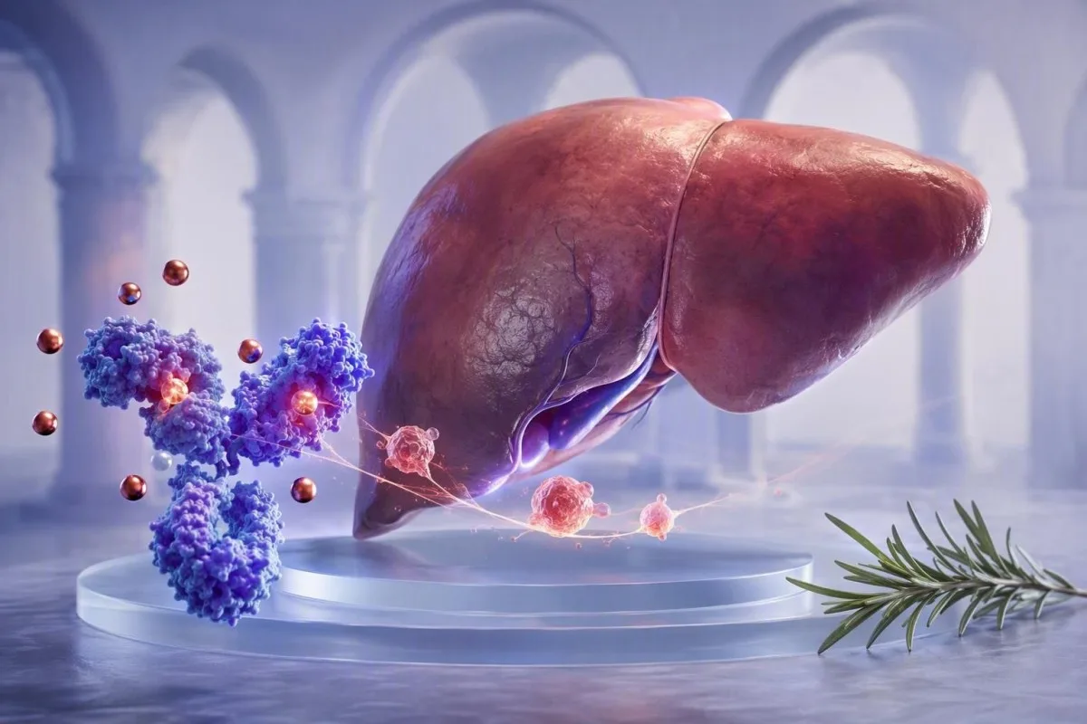
A low transferrin result can reflect the liver’s response to inflammation rather than depleted iron stores. Reading serum iron, ferritin, CRP, and transferrin together prevents common mistakes.


- Negative acute-phase protein: Transferrin commonly falls during infection, autoimmune activity, surgery, and other inflammatory states, even when body iron stores are adequate.
- Typical adult range: Most laboratories use a transferrin reference interval near 200-360 mg/dL (2.0-3.6 g/L), but local assay ranges take priority.
- TSAT calculation: Transferrin saturation equals serum iron divided by TIBC multiplied by 100; a low TIBC can make saturation look less abnormal than expected.
- Ferritin caveat: Ferritin rises with inflammation, so a ferritin below 30 ng/mL strongly supports iron deficiency, while a value of 100 ng/mL does not reliably exclude it when CRP is elevated.
- Useful companion test: Soluble transferrin receptor is usually less affected by inflammation than ferritin or transferrin and can help clarify mixed anemia.
- Retest timing: For non-urgent abnormalities, repeating iron studies about 2-4 weeks after a short illness has resolved often gives a more representative baseline.
- Do not self-treat blindly: Iron supplements can be useful in proven deficiency but may be inappropriate in iron overload, active liver disease, or anemia driven mainly by inflammation.
Why does a transferrin test fall during inflammation?
Inflammation lowers transferrin because transferrin is a negative acute-phase protein. Cytokines, especially interleukin-6, signal the liver to reduce transferrin production while hepcidin restricts iron release into plasma; this can produce low transferrin and low serum iron without proving iron stores are depleted.
The short version is that the body temporarily makes iron less available during immune activation. Hepcidin reduces ferroportin-mediated iron export from enterocytes and macrophages, so serum iron often drops within 24-48 hours; the liver can simultaneously reduce transferrin synthesis. This pattern is a host-defense response, not a direct measurement of dietary iron intake.
In my clinical work, a person with a CRP of 68 mg/L after bacterial pneumonia may have serum iron of 22 µg/dL and transferrin of 165 mg/dL, yet ferritin of 240 ng/mL. Calling that isolated pattern “iron overload” or confidently excluding iron deficiency would both be errors. Iron studies guide explains the underlying calculations in more depth.
Kantesti is an AI blood test analyzer that reads transferrin beside CRP, ferritin, full blood count indices, liver markers, and prior results rather than treating one low value as a diagnosis. Dr. Thomas Klein’s practical rule is simple: a transferrin result obtained during fever, flare, or recovery should be labelled as context-sensitive before any treatment decision is made.
The acute-phase direction matters
CRP, ferritin, fibrinogen, and haptoglobin generally rise as positive acute-phase proteins, whereas transferrin and albumin may fall. A CRP above 10 mg/L makes a single ferritin value materially harder to interpret, although no universal CRP threshold can correct every patient’s result.
What are normal transferrin and TIBC ranges?
Adult transferrin commonly ranges from about 200-360 mg/dL, and TIBC commonly ranges from about 250-450 µg/dL. These intervals differ by laboratory method, sex, pregnancy status, and local reporting units, so the range printed beside your own result is the one to use.
Transferrin is usually measured directly in mg/dL or g/L, while total iron-binding capacity, or TIBC, estimates how much iron the available transferrin can bind. Many laboratories derive TIBC from transferrin rather than measuring it separately; a rough conversion is TIBC in µg/dL equals transferrin in mg/dL multiplied by 1.25. That relationship is useful for orientation, not for overriding the laboratory’s own calculation.
Transferrin saturation, abbreviated TSAT, is calculated as serum iron divided by TIBC times 100. A TSAT near 20-45% is typical in many adult laboratories; values below 20% indicate restricted circulating iron, but they do not by themselves distinguish absolute iron deficiency from inflammation-driven iron sequestration. Low transferrin saturation covers that distinction.
Some European laboratories report transferrin in g/L, where 2.0-3.6 g/L broadly corresponds to 200-360 mg/dL. A result can sit just below range after a viral illness and normalize on repeat, whereas persistent values below 150 mg/dL deserve a deliberate search for liver synthesis problems, protein loss, significant inflammation, or undernutrition.
How should iron studies be interpreted as a pattern?
Iron studies are safest when serum iron, transferrin or TIBC, TSAT, ferritin, and an inflammatory marker are interpreted together. Serum iron alone changes markedly across the day and may fall by more than 30% between samples in the same person.
Classic uncomplicated iron deficiency usually produces low serum iron, high transferrin or TIBC, TSAT below 20%, and ferritin below 30 ng/mL. The high TIBC occurs because the liver increases transferrin production when iron availability is low. A rising red-cell distribution width can appear before hemoglobin falls; see our guide to RDW changes after iron therapy for the time course.
Anemia of inflammation more often produces low serum iron, low or normal transferrin, low TSAT, and ferritin that is normal or high. Weiss and Goodnough described this as iron-restricted erythropoiesis caused by impaired iron availability rather than necessarily absent stored iron (Weiss & Goodnough, 2005). The two states commonly coexist, particularly in inflammatory bowel disease, rheumatoid arthritis, chronic kidney disease, and cancer.
One less obvious trap is arithmetic: if serum iron is 30 µg/dL and TIBC is suppressed to 180 µg/dL, TSAT is 17%. If TIBC were 360 µg/dL, the same serum iron would yield 8%. The lower denominator can conceal how little iron is circulating, which is why low serum iron needs context rather than a reflex prescription.
A practical mixed-pattern clue
Ferritin between 30 and 100 ng/mL with TSAT below 20% and CRP above 10 mg/L is often a gray zone, not reassurance. In that setting, clinicians may use soluble transferrin receptor, reticulocyte hemoglobin content, trend data, or a therapeutic plan tailored to the underlying illness.
Why ferritin can look normal when iron is low
Ferritin is both an iron-storage protein and a positive acute-phase reactant, so inflammation can raise it independently of stored iron. A ferritin below 30 ng/mL strongly supports iron deficiency in most adults, but a higher value cannot reliably exclude deficiency when CRP or ESR is raised.
The World Health Organization’s 2020 ferritin guideline advises measuring inflammation markers alongside ferritin where infection or inflammation is prevalent. In adults with inflammation, WHO suggests ferritin below 70 µg/L may indicate iron deficiency; that is a population-informed threshold, not a substitute for an individual clinical assessment (WHO, 2020).
I often see patients alarmed by ferritin of 180 ng/mL after an inflammatory flare, assuming their iron stores must be abundant. Sometimes they are. Yet ferritin can rise after strenuous exercise, liver cell injury, metabolic disease, alcohol exposure, and immune activity, so the number is not a standalone inventory count. Ferritin and CRP is a helpful companion discussion.
Kantesti is an AI blood test interpretation platform that flags the discordant combination of low transferrin, low TSAT, and elevated CRP as possible inflammation-associated iron restriction. Our 2M+ user dataset is not a replacement for diagnostic research, but it repeatedly reinforces a clinician’s old lesson: ferritin behaves differently when the patient is actively unwell.
Ferritin values that need a different conversation
Ferritin above 1,000 ng/mL warrants timely medical review because severe inflammation, significant liver injury, iron overload syndromes, and several other conditions can produce that range. The urgency depends on symptoms, liver enzymes, transferrin saturation, and the speed of change rather than the ferritin number alone.
Which tests clarify iron deficiency during inflammation?
Soluble transferrin receptor and reticulocyte hemoglobin content can help identify iron-restricted red-cell production when ferritin and transferrin disagree. Soluble transferrin receptor generally rises with cellular iron need and is less distorted by acute inflammation than ferritin.
Soluble transferrin receptor, or sTfR, reflects transferrin receptor expression from erythroid precursors. It usually rises in absolute iron deficiency and is often normal in pure anemia of inflammation; however, assay reference intervals are not standardized, and hemolysis or high erythropoietic activity can elevate it. This is one of those areas where the exact laboratory method matters more than online cutoffs.
Reticulocyte hemoglobin content, reported as CHr or Ret-He depending on the analyzer, reflects iron available to newly produced red cells over roughly the prior 2-4 days. Values below about 28-30 pg can support iron-restricted erythropoiesis, although local thresholds and kidney-disease protocols differ. Soluble transferrin receptor testing explains where it fits.
Bone marrow iron staining remains a historical reference method but is rarely necessary solely to resolve a routine outpatient iron panel. Dr. Thomas Klein generally favors repeating samples after recovery, reviewing bleeding and dietary history, and using sTfR or reticulocyte measures when the answer will genuinely alter treatment; extra tests without a decision attached tend to create noise.
The sTfR-ferritin index
Some specialists calculate the sTfR/log ferritin index, which can improve discrimination in inflammatory settings. Cutoffs vary substantially by assay, often from roughly 1.0 to 3.2, so a result should be interpreted with the laboratory’s validated method rather than a borrowed internet threshold.
When should a transferrin test be repeated?
A transferrin test is best repeated after a short-lived infection, fever, or major inflammatory event has settled, usually after 2-4 weeks if the situation is not urgent. Morning collection and consistent pre-test conditions reduce avoidable variation in serum iron and TSAT.
Serum iron has diurnal variation and can be lower later in the day, while recent oral iron can transiently raise it. Many clinicians request a morning sample after an overnight fast of roughly 8-12 hours when they need a clean comparison, although fasting is not mandatory for every iron study. Follow the ordering clinician’s instructions rather than stopping prescribed medicines on your own.
A transferrin result taken 48 hours after a marathon, dental procedure, vaccine, or acute respiratory illness may reflect an acute-phase response rather than a durable iron pattern. Exercise can also alter CK, plasma volume, and ferritin; endurance athletes may find our runner iron testing guide useful.
Kantesti’s trend analysis compares dates, not simply reference flags, and can highlight a fall from 310 mg/dL to 180 mg/dL that coincides with CRP rising from 1 to 52 mg/L. That is clinically more informative than either isolated result. Review TIBC test preparation before arranging a planned repeat.
When not to wait for a repeat
Do not postpone medical review for repeat testing if severe breathlessness, chest pain, fainting, black stools, heavy ongoing bleeding, jaundice, or rapidly worsening weakness is present. Hemoglobin below 80 g/L (8 g/dL) is often clinically significant, but urgency depends on symptoms, rate of fall, pregnancy status, and cardiovascular disease.
How do CRP, ESR, and hepcidin explain low transferrin?
CRP and ESR do not measure iron stores, but they show whether inflammation may be distorting a transferrin test. CRP rises and falls over hours to days, whereas ESR can remain elevated for weeks because it is influenced by fibrinogen, anemia, age, and immunoglobulins.
Interleukin-6 stimulates hepatic hepcidin production, and hepcidin binds ferroportin, causing reduced iron export from macrophages and intestinal cells. Ganz and Nemeth’s review describes this pathway as central to anemia of inflammation and iron-restricted erythropoiesis (Ganz & Nemeth, 2012). The result may be low serum iron within a day while transferrin falls as part of the same systemic response.
CRP below 5 mg/L is often considered within the reference range, though laboratories differ. A CRP of 40 mg/L does not identify the cause of inflammation, but it should make a clinician more cautious about diagnosing iron deficiency from ferritin or transferrin alone. High ESR causes explains why ESR is slower and less specific.
Kantesti AI interprets transferrin results by comparing the direction of CRP, ferritin, albumin, white-cell count, and recent trends. This is not a diagnosis engine for infection or autoimmune disease; it is a structured prompt to ask whether the iron panel was obtained during a biologically unstable moment.
Why hepcidin is not routinely measured
Hepcidin assays remain limited by availability, standardization, and turnaround time in ordinary practice. A hepcidin value can be informative in specialist research or unusual anemia cases, but serum ferritin, TSAT, CRP, kidney function, and blood-count indices remain the practical first-line tools.
How does anemia of inflammation differ from iron deficiency?
Absolute iron deficiency means total body iron is insufficient, while anemia of inflammation means iron is present but poorly available for red-cell production. Both can produce fatigue, low TSAT, and a falling hemoglobin, and they frequently occur together.
In uncomplicated iron deficiency, ferritin is usually low and transferrin often rises above 360 mg/dL as binding capacity increases. In anemia of inflammation, transferrin commonly falls below 200 mg/dL, ferritin is often above 100 ng/mL, and CRP may be elevated. Neither pattern is absolute; chronic kidney disease and liver disease are especially prone to overlap.
Hemoglobin and MCV can lag behind iron restriction. A person can have ferritin 18 ng/mL, TSAT 14%, and a normal hemoglobin of 132 g/L, particularly early in deficiency; conversely, inflammation can cause anemia with a normal MCV of 82-100 fL. What hemoglobin means helps put the CBC beside iron studies.
The reason we worry about low transferrin combined with low albumin is that together they suggest either a stronger inflammatory burden, impaired hepatic synthesis, or protein loss, whereas low transferrin alone after a cold is often transient. A clinician should review kidney function, urine protein, liver tests, nutrition, medicines, bleeding history, and the trajectory over at least two draws.
Treatment is not interchangeable
Oral iron often improves absolute deficiency, but it may have limited effect while inflammation keeps hepcidin high. In selected conditions, such as chronic kidney disease or active inflammatory bowel disease, clinicians may use intravenous iron or treat the inflammatory driver first; the approach depends on diagnosis, symptoms, hemoglobin, and safety considerations.
Why low transferrin can distort transferrin saturation
Low transferrin can make transferrin saturation appear less low because TSAT uses TIBC as its denominator. A normal or mildly low TSAT does not always mean iron delivery is adequate when TIBC is suppressed by inflammation or liver dysfunction.
Consider serum iron of 36 µg/dL. With a TIBC of 360 µg/dL, TSAT is 10%; with a TIBC of 180 µg/dL, TSAT is 20%. The second result appears less concerning mathematically, but both samples contain the same low circulating iron. This is why clinicians should inspect the raw values, not only the percentage.
The opposite pitfall occurs in advanced liver injury or acute hepatocellular damage: transferrin production can fall and serum iron may rise from altered handling, producing an elevated TSAT. TSAT persistently above 45% merits evaluation for iron overload in the right context, but it should not be used to diagnose hereditary hemochromatosis during an acute liver event. Read our cirrhosis blood-test clues for wider hepatic context.
Kantesti is an AI-powered blood test analysis tool used across 127+ countries, so it normalizes units before calculating saturation and marks results that may be mathematically unstable because TIBC is unusually low. The output should support, not replace, the clinician who knows whether a patient has fever, hepatitis, nephrotic syndrome, or recent iron treatment.
Do not use TSAT as a hydration marker
Dehydration can concentrate several serum measurements but does not create a dependable iron-overload pattern. If albumin, hematocrit, urea, and sodium suggest reduced plasma volume, repeating the panel after normal hydration may be sensible before attaching meaning to a borderline TSAT.
What else causes low transferrin besides inflammation?
Low transferrin also occurs with reduced liver synthesis, protein loss through the kidneys or gut, inadequate protein-energy intake, and rarely congenital disorders. Inflammation is common, but it should never become a catch-all explanation without checking the rest of the panel.
The liver synthesizes transferrin, so low transferrin with elevated bilirubin, INR, AST, ALT, or low albumin may point toward hepatic disease rather than iron status. Severe liver dysfunction can reduce transferrin below 150 mg/dL. Liver panel results help determine whether that explanation is plausible.
Nephrotic-range urinary protein loss can remove transferrin along with albumin and other proteins. A urine albumin-creatinine ratio above 300 mg/g, or 30 mg/mmol, is severely increased albuminuria and calls for kidney-focused assessment; dipstick protein alone is not enough for a full answer. See our guide to protein in urine.
Poor intake, malabsorption, or severe catabolic illness can lower transferrin, although transferrin is too inflammation-sensitive to serve as a nutritional marker by itself. Congenital atransferrinemia is exceptionally rare and usually presents much earlier in life with severe anemia and paradoxical systemic iron loading. That unusual combination needs specialist hematology input.
Medication and hormone effects
Estrogen exposure and pregnancy can increase transferrin, while androgens may lower it modestly. These shifts are usually smaller than the effects of significant inflammation or liver disease, but they can explain a borderline result when the rest of the iron panel is stable.
How do pregnancy and menstrual blood loss change transferrin?
Pregnancy often raises transferrin and TIBC, while iron requirements increase most sharply in the second and third trimesters. Therefore, a low transferrin result in pregnancy deserves particular attention to inflammation, liver function, protein loss, and laboratory context.
Plasma volume expands during pregnancy, and estrogen increases transferrin synthesis, so TIBC often rises above the non-pregnant range. Ferritin also normally trends downward as pregnancy progresses; a ferritin below 30 µg/L is commonly used to identify depleted stores in pregnancy, although local maternity guidelines may vary. Ferritin by trimester provides practical ranges.
Heavy menstrual bleeding is a common cause of absolute iron deficiency, especially when ferritin is below 30 ng/mL and transferrin is elevated rather than suppressed. Yet a person with autoimmune disease and heavy periods can have both blood-loss deficiency and inflammation; a “normal” ferritin of 75 ng/mL does not settle the question when CRP is 24 mg/L.
New low transferrin with high blood pressure, swelling, proteinuria, headache, right-upper abdominal pain, or abnormal liver tests during pregnancy needs prompt obstetric assessment. It is not a way to diagnose pre-eclampsia, but the wider protein and liver pattern can matter far more than the iron result alone.
How is transferrin interpreted in kidney disease and chronic illness?
Chronic kidney disease commonly causes functional iron deficiency because inflammation and reduced erythropoietin limit usable iron for red-cell production. In this setting, TSAT below 20% and ferritin below 100 ng/mL often support iron deficiency before dialysis, though treatment thresholds vary by guideline and clinical setting.
KDIGO guidance has historically used a trial-of-iron framework in many adults with CKD when TSAT is at or below 30% and ferritin is at or below 500 ng/mL, provided the clinical goal is to raise hemoglobin or reduce erythropoiesis-stimulating therapy. These are treatment considerations, not universal definitions of normal iron stores, and the 500 ng/mL ceiling is often misunderstood.
A patient with eGFR 28 mL/min/1.73 m², hemoglobin 96 g/L, TSAT 16%, ferritin 220 ng/mL, and CRP 12 mg/L may have functional deficiency despite non-low ferritin. Oral iron absorption can be reduced, and clinicians must also assess B12, folate, occult loss, erythropoietin use, and kidney trajectory. CKD staging offers useful background.
Kantesti AI can place transferrin in a longitudinal kidney-health context, but it cannot determine whether intravenous iron, erythropoiesis-stimulating therapy, or specialist referral is appropriate. That decision requires symptoms, blood pressure, infection status, medication review, and the full renal record.
Which history details make a transferrin result more useful?
The most useful transferrin interpretation includes symptoms, recent illness, menstrual or gastrointestinal blood loss, diet, alcohol exposure, medicines, exercise, and prior values. A single sample is a snapshot; two comparable samples often reveal whether the change is physiological, inflammatory, or persistent.
Ask whether there was fever, vaccination, injury, hospitalization, dental work, intense training, or a known flare in the 14 days before the draw. Also record iron tablets, multivitamins, proton-pump inhibitors, non-steroidal anti-inflammatory drugs, anticoagulants, and recent diet changes. These details can alter either iron absorption or the likelihood of occult blood loss.
A change from transferrin 290 to 205 mg/dL matters more if CRP rose from 2 to 35 mg/L and albumin fell from 44 to 37 g/L. By contrast, a 10 mg/dL change with stable CRP and identical collection timing may fall within biological and analytic variation. Comparing blood tests explains why small changes are not always real changes.
Kantesti’s multilingual record tools allow people to save context beside a result, including illness dates and supplement changes, across 75+ languages. That history makes a clinician’s review safer; our longitudinal blood-test guide lists the practical details worth retaining.
Symptoms are not specific
Fatigue, reduced exercise tolerance, hair shedding, restless legs, and poor concentration can occur in iron deficiency, inflammation, thyroid disease, sleep disruption, depression, and many other conditions. Symptoms should prompt appropriate evaluation, but they cannot tell whether low transferrin represents depleted stores.
When does a low transferrin result need prompt review?
Low transferrin needs prompt medical review when it accompanies significant anemia, jaundice, swelling, heavy bleeding, black stools, unexplained weight loss, or evidence of kidney or liver dysfunction. The result itself is rarely an emergency, but the condition behind it occasionally is.
Same-day assessment is sensible for chest pain, fainting, severe shortness of breath, confusion, black tarry stool, vomiting blood, or rapidly increasing swelling. Hemoglobin below 70 g/L (7 g/dL), bilirubin above 50 µmol/L with jaundice, or an unexpectedly high INR requires individual clinical judgment and may need urgent evaluation. Do not attempt to correct those patterns with over-the-counter iron alone.
For a stable, mildly low transferrin of 185 mg/dL with normal hemoglobin, normal liver tests, and CRP 18 mg/L during a documented respiratory infection, a repeat panel after recovery is often reasonable. If it persists for more than 6-8 weeks, clinicians commonly expand the review to liver panel, urine protein, nutritional history, inflammatory disease activity, and bleeding sources.
Our clinical content is reviewed with the support of the Medical Advisory Board, and Kantesti’s interpretation logic is documented through our medical validation framework. As of September 5, 2026, the safest message remains unchanged: a low transferrin result is a clue about iron transport and systemic physiology, not a diagnosis by itself.
Frequently Asked Questions
Can inflammation cause low transferrin without iron deficiency?
Yes. Inflammation can lower transferrin even when iron stores are adequate because transferrin is a negative acute-phase protein made by the liver. A person with CRP above 10 mg/L may have low transferrin, low serum iron, and ferritin above 100 ng/mL from inflammation-related iron sequestration rather than pure iron depletion. Iron deficiency can still coexist, especially when TSAT is below 20% or ferritin is below 30 ng/mL. A clinician should interpret the full pattern and the cause of inflammation.
Does low transferrin mean low iron?
Low transferrin does not automatically mean low total-body iron. Low transferrin commonly occurs during inflammation, liver disease, kidney protein loss, and inadequate protein intake, while classic iron deficiency often raises transferrin above roughly 360 mg/dL. Serum iron below 50 µg/dL can occur in either iron deficiency or inflammation, so ferritin, CRP, TIBC, TSAT, and the blood count are needed. A transferrin result below 200 mg/dL should be interpreted with the laboratory’s reference range and clinical history.
Can low transferrin make transferrin saturation look normal?
Yes. Transferrin saturation is serum iron divided by TIBC multiplied by 100, so a low TIBC caused by low transferrin creates a smaller denominator. For example, serum iron of 36 µg/dL produces a TSAT of 10% with TIBC of 360 µg/dL but 20% with TIBC of 180 µg/dL. This means a borderline TSAT can understate iron restriction when transferrin is suppressed. Clinicians should inspect both the percentage and the raw serum iron and TIBC values.
What ferritin level confirms iron deficiency during inflammation?
Ferritin below 30 ng/mL or µg/L strongly supports iron deficiency in most adults, including many people with mild inflammation. The WHO 2020 guideline suggests that ferritin below 70 µg/L may indicate iron deficiency in adults with evidence of inflammation, but this threshold is not absolute for every individual. Ferritin can rise with CRP elevation, liver injury, and metabolic illness, so values between 30 and 100 ng/mL often require TSAT, CRP, and sometimes soluble transferrin receptor testing. A ferritin above 100 ng/mL does not always exclude iron deficiency when inflammatory activity is present.
How long after illness should I repeat a transferrin test?
For a minor self-limited illness, repeating a transferrin test about 2-4 weeks after symptoms and fever have resolved is often reasonable if there are no urgent features. CRP can settle within days, but ESR, ferritin, albumin, and transferrin may take longer to return toward baseline. Use similar conditions for the repeat draw, ideally morning collection and the same laboratory when practical. Do not delay review if hemoglobin is falling, bleeding is ongoing, or liver and kidney results are abnormal.
Should I take iron supplements for low transferrin?
Low transferrin alone is not a reason to start iron supplements. Oral iron is usually considered when there is evidence of absolute iron deficiency, such as ferritin below 30 ng/mL, low TSAT, compatible symptoms, or a clear source of loss; dose and schedule should be individualized. In anemia of inflammation, iron may be poorly absorbed or unavailable because hepcidin is high, and treating the underlying condition may matter more. Iron can be harmful or misleading in iron overload disorders and some liver conditions, so confirm the pattern with a clinician.
Get AI-Powered Blood Test Analysis Today
Join over 2 million users worldwide who trust Kantesti for instant, accurate lab test analysis. Upload your blood test results and receive comprehensive interpretation of 15,000+ biomarkers in seconds.
📚 Referenced Research Publications
Klein, T., Mitchell, S., & Weber, H. (2026). A Pre-Registered, Rubric-Based Automated Technical Benchmark of the Kantesti Blood-Test Interpretation Engine on 100,000 Synthetic Test Cases. Kantesti AI Medical Research.
Klein, T., Mitchell, S., & Weber, H. (2026). Clinical Validation Framework v2.0 (Medical Validation Page). Kantesti AI Medical Research.
📖 External Medical References
World Health Organization (2020). WHO guideline on use of ferritin concentrations to assess iron status in individuals and populations. World Health Organization.
⚕️ Medical Disclaimer
This article is for educational purposes only and does not constitute medical advice. Always consult a qualified healthcare provider for diagnosis and treatment decisions.
E-E-A-T Trust Signals
Experience
Physician-led clinical review of lab interpretation workflows.
Expertise
Laboratory medicine focus on how biomarkers behave in clinical context.
Authoritativeness
Written by Dr. Thomas Klein with review by Dr. Sarah Mitchell and Prof. Dr. Hans Weber.
Trustworthiness
Evidence-based interpretation with clear follow-up pathways to reduce alarm.