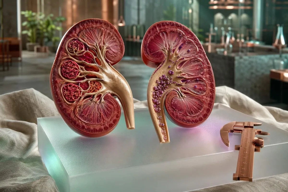
CKD staging is a two-axis risk system: filtration tells one story, urine albumin tells another. I explain how doctors combine both before calling a kidney result low-risk or high-risk.


- CKD stage is not based on eGFR alone; KDIGO uses cause, eGFR category, and urine ACR category together.
- eGFR G3a means 45-59 ml/min/1.73 m², while G3b means 30-44 ml/min/1.73 m².
- Urine ACR A1 is below 30 mg/g, A2 is 30-300 mg/g, and A3 is above 300 mg/g.
- Albuminuria can move a patient with eGFR 65 from low concern to high cardiovascular and kidney risk.
- CKD diagnosis usually requires abnormal eGFR or kidney damage markers for at least 3 months.
- Kidney disease symptoms often appear late, commonly when eGFR falls below 30 ml/min/1.73 m².
- Creatinine-based eGFR can be misleading in very muscular, frail, pregnant, amputee, or recently exercising patients.
- CKD stage 3 is not one risk group; CKD stage 3a A1 is very different from stage 3b A3.
- Urgent review is needed for rapidly falling eGFR, potassium above 6.0 mmol/L, severe swelling, confusion, or breathlessness.
Why CKD staging needs eGFR plus urine ACR
Chronic kidney disease stages are not based on eGFR alone because filtration and kidney leakage measure different types of risk. A person with eGFR 70 and urine ACR 450 mg/g may be at higher long-term kidney and heart risk than someone with eGFR 52 and ACR 5 mg/g.
As of July 12, 2026, the standard clinical language for CKD is CGA staging: cause, GFR category, and albuminuria category. If you only look at eGFR, you miss the patients whose kidneys still filter reasonably well but leak albumin, which is often an earlier vascular injury signal.
Kantesti is an AI blood test interpretation platform that reads kidney results in context, pairing creatinine-derived eGFR with urine ACR, electrolytes, diabetes markers, medication history, and previous results. If you need the plain-English version of filtration first, our guide to what eGFR means is a useful companion.
I’m Thomas Klein, MD, and in clinical review the most common patient misunderstanding I see is this: a normal-looking eGFR is taken as proof that the kidneys are fine. In diabetes, hypertension, autoimmune disease, and some inherited kidney disorders, the urine may start showing albumin years before eGFR drops below 60 ml/min/1.73 m².
A urine albumin-creatinine ratio, or ACR, is usually reported in mg/g in the United States and mg/mmol in the UK and many other countries. For a deeper patient guide on this exact test, see our article on urine ACR testing.
eGFR stages: what G1 to G5 really mean
eGFR stages classify kidney filtration from G1 to G5, with G3 starting below 60 ml/min/1.73 m². The number estimates how much plasma your kidneys filter per minute, adjusted to a standard body surface area of 1.73 m².
G1 means eGFR is 90 ml/min/1.73 m² or higher, and G2 means 60-89 ml/min/1.73 m². These categories are not CKD by themselves unless there is another marker of kidney damage, such as ACR above 30 mg/g for more than 3 months.
G3 is split because outcomes differ: G3a is 45-59 ml/min/1.73 m² and G3b is 30-44 ml/min/1.73 m². I often see patients told they have CKD stage 3 without being told which half; that missing letter changes follow-up, prescribing caution, and sometimes referral.
The CKD-EPI creatinine equation was developed to estimate GFR more accurately than older equations across routine adult lab ranges (Levey et al., 2009). Still, equation accuracy is imperfect, which is why measured clearance and cystatin C sometimes matter; our normal GFR guide explains those differences.
A single eGFR of 58 after dehydration, high-intensity exercise, or a large meat meal is not the same as persistent eGFR 58 across 3 months. Most nephrologists want persistence, trajectory, and urine findings before assigning a stable CKD category.
Urine ACR categories: the risk signal eGFR can miss
Urine ACR measures albumin leakage from the kidney filtering units, and it changes CKD risk even when eGFR looks acceptable. KDIGO 2024 classifies ACR as A1 below 30 mg/g, A2 from 30-300 mg/g, and A3 above 300 mg/g.
Albumin is a blood protein that should mostly stay in the circulation, not pass into urine. When ACR rises above 30 mg/g, the kidney filter is letting through more albumin than expected, and that finding predicts kidney progression and cardiovascular events.
The KDIGO 2024 CKD guideline recommends using both GFR and albuminuria categories for prognosis, not eGFR alone (KDIGO, 2024). This is why a patient with ACR 600 mg/g needs a different conversation than a patient with ACR 8 mg/g, even if both have eGFR 62.
A dipstick that reads trace or 1+ protein is not the same as a properly quantified ACR. Our patient guide on protein in urine explains why concentrated urine, exercise, fever, and menstruation can distort simpler urine screens.
Kantesti AI interprets ACR by checking units, timing, diabetes markers, blood pressure-related medicines, and whether the result was repeated. ACR variability is real: in practice, I treat a first A2 result as a prompt to repeat and investigate, not as a reason to panic.
How eGFR and ACR combine into CKD risk categories
CKD risk category comes from the intersection of eGFR stage and ACR stage. A low eGFR with A1 albuminuria may carry moderate risk, while a preserved eGFR with A3 albuminuria may carry high or very high risk.
Think of eGFR as filtration capacity and ACR as filter integrity. One measures how much work the kidney is doing; the other measures whether the filter barrier is leaking protein.
The CKD Prognosis Consortium meta-analysis in The Lancet found that lower eGFR and higher albuminuria independently predicted mortality and kidney outcomes (Matsushita et al., 2010). That independent effect is the reason modern staging does not let eGFR do all the talking.
A practical example: eGFR 68 with ACR 420 mg/g is not stage G2 low-risk CKD; it is G2-A3, a pattern that should trigger cause-finding and aggressive risk reduction. If the urine also contains red cells, our guide to blood in urine explains why glomerular causes rise on the list.
In our review workflow, I give more attention to a rising ACR slope than to a one-point eGFR wobble. ACR increasing from 45 to 180 mg/g over 12 months tells me the kidney environment has changed, even if eGFR only moved from 76 to 72.
Why CKD stage 3 is not one diagnosis
CKD stage 3 covers eGFR 30-59 ml/min/1.73 m², but stage 3a and 3b behave differently. The ACR category then splits risk again, so CKD stage 3a A1 is not clinically equivalent to CKD stage 3b A3.
Stage 3a means eGFR 45-59, and many older adults in this range remain stable for years, especially with ACR below 30 mg/g. Stage 3b means eGFR 30-44, where drug dosing, anemia screening, acidosis, phosphate, and potassium deserve closer attention.
I once reviewed labs for a 71-year-old cyclist with eGFR 54 and ACR 4 mg/g, unchanged for 5 years. That is a different clinical story from a 48-year-old with eGFR 54, ACR 950 mg/g, low albumin, and ankle swelling.
Kantesti is an AI biomarker interpretation platform that treats CKD stage 3 as a pattern, not a label. If creatinine-based eGFR conflicts with body habitus or clinical context, our guide to cystatin C recheck explains when a second filtration marker can reduce misclassification.
The threshold of 60 ml/min/1.73 m² is useful because complication risk rises below it, but it is not a cliff edge. A stable eGFR of 59 with ACR 3 is usually less concerning than a falling eGFR of 74 to 61 over 6 months with ACR rising to 220.
Why kidney disease symptoms often appear late
Kidney disease symptoms often appear late because the kidneys have large functional reserve. Many patients feel well until eGFR falls below 30 ml/min/1.73 m², and some do not feel clearly unwell until kidney failure range below 15.
The kidney has millions of filtering units, and remaining nephrons can increase their workload when others are injured. That compensation is useful biologically, but it hides early CKD from the patient’s day-to-day body signals.
Symptoms such as fatigue, itching, nausea, restless legs, poor appetite, swelling, and breathlessness are non-specific. I have seen eGFR 28 discovered during a routine test because the patient’s only complaint was needing an afternoon nap, while another patient with eGFR 42 had dramatic swelling because albumin loss was heavy.
Swelling is especially tricky because it can come from kidney disease, heart disease, liver disease, low albumin, venous disease, or medication effects. Our article on swelling lab clues walks through the albumin, urine, BNP, and kidney patterns doctors often compare.
Late symptoms are one reason I prefer trend-based interpretation over symptom-based reassurance. If eGFR falls from 82 to 61 over 18 months or ACR rises from 12 to 95 mg/g, waiting for symptoms is simply too slow.
When creatinine makes eGFR misleading
Creatinine-based eGFR can be misleading when creatinine production is unusual. Very muscular people, frail older adults, amputees, pregnant patients, high-meat meals, creatine supplements, and hard exercise can all distort the estimate.
Creatinine is made from muscle metabolism, so the same creatinine value can mean different things in different bodies. A creatinine of 1.2 mg/dL may be ordinary for a muscular 34-year-old man and concerning for a 78-year-old woman with low muscle mass.
Exercise can cause a temporary creatinine rise, especially after endurance events, strength training, or dehydration. If your eGFR dipped after a race or intense gym session, our guide to creatinine after exercise explains why a 48-72 hour recheck can change the picture.
Diet matters more than people think. A large cooked-meat meal can transiently raise serum creatinine, and creatine supplementation can increase creatinine generation without necessarily damaging the kidney.
When the story and the eGFR do not match, cystatin C, repeat testing, urine ACR, urinalysis, and sometimes measured clearance help. I rarely label a low-risk athletic patient with new CKD from one isolated creatinine-based eGFR result.
What to repeat before accepting a new CKD stage
CKD generally requires abnormal kidney structure or function for at least 3 months. Before accepting a new chronic stage, repeat eGFR, urine ACR, urinalysis, blood pressure, medication review, and recent illness context should be checked.
One abnormal kidney panel can reflect dehydration, infection, NSAID use, contrast exposure, urinary obstruction, or acute kidney injury. The word chronic should not be used casually after one draw unless prior records already show persistence.
A repeat test is often reasonable within 1-2 weeks if the change is large or unexpected, and again at 3 months if the patient is stable. Our guide on repeating abnormal labs gives a practical framework for deciding what needs fast retesting versus routine follow-up.
For urine ACR, I prefer an early-morning sample when possible because posture, exercise, fever, and acute illness can raise albumin transiently. Two abnormal ACR results out of 3 specimens over 3-6 months are more persuasive than one borderline value.
Do not ignore the boring details: fasting status, hydration, lab unit changes, and previous creatinine method can all alter interpretation. If your renal panel was drawn after eating, heavy exercise, or poor fluid intake, that context belongs next to the number.
Diabetes and blood pressure are the common accelerators
Diabetes and high blood pressure are the two most common drivers of CKD progression in many adult populations. ACR often rises before eGFR falls in diabetic kidney disease, which is why urine testing matters even when creatinine looks fine.
In diabetes, small-vessel injury can make the kidney filter leak albumin long before filtration drops below 60 ml/min/1.73 m². ACR above 30 mg/g in a patient with diabetes should prompt repeat testing and risk-factor tightening, not a wait-and-see attitude.
Blood pressure injures kidneys through pressure transmission into delicate filtering structures. In clinical practice, home blood pressure averages above 130/80 mmHg often matter more than a single calm clinic reading of 124/76.
Dietary sodium, sleep apnea, weight gain, alcohol intake, and missed medicines can all raise kidney risk through blood pressure. Our DASH diet guide explains which lab markers patients often recheck after 8-12 weeks of blood-pressure-focused diet changes.
The nuance is that treatment can make eGFR dip at first. ACE inhibitors, ARBs, and SGLT2 inhibitors may cause an early eGFR fall of around 10-30%, yet still protect kidneys long term when monitored correctly.
Medicines and supplements that shift kidney numbers
Kidney lab results can shift after common medicines, including ACE inhibitors, ARBs, diuretics, NSAIDs, SGLT2 inhibitors, lithium, trimethoprim, and some supplements. The direction, timing, and potassium level decide whether the change is expected or unsafe.
An ACE inhibitor or ARB can raise creatinine modestly after starting because pressure inside the kidney filter falls. A creatinine rise up to about 30% may be acceptable in many situations, but potassium above 5.5 mmol/L or a larger rise needs prompt review.
NSAIDs are the medicines patients underestimate most. Ibuprofen, naproxen, and similar drugs can reduce kidney blood flow, especially when combined with dehydration, diuretics, ACE inhibitors, or ARBs.
Kantesti's neural network flags medication-timing patterns when a potassium or creatinine shift appears after a blood pressure medicine change. Our guide to potassium after BP medicines explains why rechecking potassium in 1-2 weeks is common after dose changes.
Supplements are not automatically safe because they are natural. High-dose vitamin C can raise oxalate risk in susceptible patients, creatine can complicate creatinine interpretation, and potassium-containing salt substitutes can be risky when eGFR is below 45.
Diet advice depends on ACR, potassium and phosphate
Diet for CKD should be individualized by eGFR, ACR, potassium, bicarbonate, phosphate, diabetes status, and body weight. A blanket low-protein or low-potassium diet can be unnecessary, and sometimes harmful, in early low-risk CKD.
Protein advice depends on stage and nutrition status. Many stable non-dialysis CKD patients are advised around 0.8 g/kg/day of protein, but frail older adults, athletes, and people losing weight need more careful individualization.
Potassium restriction is not automatic. A patient with eGFR 58 and potassium 4.4 mmol/L may not need to avoid beans, lentils, or fruit, while someone with eGFR 28 and potassium 5.8 mmol/L needs a different plan.
Our guide to kidney disease diet focuses on lab-guided food choices rather than fear lists. Phosphate deserves similar nuance; our article on high phosphate causes explains why kidney function, parathyroid hormone, supplements, and processed foods all matter.
ACR also changes dietary urgency because albuminuria signals vascular stress. In a patient with ACR 600 mg/g, sodium reduction and blood-pressure control may do more for kidney outcomes than obsessing over a single banana.
How Kantesti reads kidney panels without overcalling disease
Kantesti AI interprets kidney panels by comparing eGFR, creatinine, urea or BUN, electrolytes, urine ACR, prior trends, age, sex, medicines, and unit conventions. The goal is pattern recognition with clinical caution, not replacing a nephrologist.
Kantesti is an AI-powered blood test analysis tool used by more than 2M people across 127 countries, so unit conversion is not a small detail for us. BUN in mg/dL, urea in mmol/L, creatinine in µmol/L, and ACR in mg/mmol can all refer to the same biology while looking unfamiliar to patients.
Our clinical checks are designed to reduce false alarm language when a result is borderline, transient, or internally inconsistent. The methods behind this workflow are described in our medical validation material.
Thomas Klein, MD reviews kidney content with the same caution I use in clinic: a single eGFR value should trigger questions before labels. Our AI technology guide explains how structured extraction and trend comparison work across uploaded lab reports.
Kantesti AI also maps kidney markers against broader panels, including hemoglobin, bicarbonate, calcium, phosphate, albumin, HbA1c, lipids, and inflammatory clues. For readers who want a wider marker list, our biomarkers guide covers 15,000+ lab markers.
When to seek urgent care or nephrology review
Urgent kidney review is needed when eGFR drops quickly, potassium is dangerously high, urine output falls sharply, severe swelling appears, or symptoms such as confusion, chest pain, breathlessness, or persistent vomiting occur. Chronic staging should never delay acute assessment.
A potassium level above 6.0 mmol/L is often treated as urgent, especially with weakness, palpitations, ECG changes, or kidney impairment. Mild potassium elevations around 5.2-5.5 mmol/L still deserve medication and diet review, but they are not the same emergency.
A fast creatinine rise can indicate acute kidney injury rather than stable CKD. If creatinine doubles over days to weeks, or eGFR falls by more than 25% after a new medicine, clinicians usually reassess volume status, obstruction, urine findings, and drug exposure.
High urea or BUN can reflect kidney impairment, dehydration, gastrointestinal protein load, steroids, or bleeding into the gut. Our article on high BUN risk explains why the BUN-to-creatinine pattern changes the differential diagnosis.
Nephrology referral thresholds vary by country and health system, but eGFR below 30, ACR above 300 mg/g, suspected glomerulonephritis, persistent hematuria with proteinuria, and rapid progression usually justify specialist input. I would rather refer one month early than explain one year later why we missed a treatable kidney process.
Research notes and records that help future kidney care
Kidney records are most useful when they preserve dates, units, lab methods, medicines, illness context, blood pressure, and urine findings. A graph of eGFR and ACR over 2-5 years is often more clinically useful than a folder of isolated abnormal flags.
Keep the original PDF when possible because unit conversions and reference ranges can be lost in screenshots. Our health history tracker gives a practical list of what to store after each draw, including medication changes and home blood pressure averages.
For patients comparing BUN, urea, creatinine, and hydration markers, our research-style guide to the BUN/creatinine ratio is a useful technical companion. The ratio is not a CKD stage, but it can point toward dehydration, protein load, catabolic stress, or reduced kidney clearance.
Kantesti research publications include methodology-adjacent work, though they are not CKD staging guidelines. Kantesti LTD. (2026). Nipah Virus Blood Test: Early Detection & Diagnosis Guide 2026. Zenodo. https://doi.org/10.5281/zenodo.18487418. ResearchGate: https://www.researchgate.net/. Academia.edu: https://www.academia.edu/.
Kantesti LTD. (2026). B Negative Blood Type, LDH Blood Test & Reticulocyte Count Guide. Figshare. https://doi.org/10.6084/m9.figshare.31333819. ResearchGate: https://www.researchgate.net/. Academia.edu: https://www.academia.edu/. Our physician oversight is described through the Medical Advisory Board, because kidney interpretation should be cautious, documented, and reviewable.
Frequently Asked Questions
What are the chronic kidney disease stages?
Chronic kidney disease stages use eGFR categories G1 to G5 plus urine albumin categories A1 to A3. G1 is eGFR 90 ml/min/1.73 m² or higher, G2 is 60-89, G3a is 45-59, G3b is 30-44, G4 is 15-29, and G5 is below 15. ACR then modifies risk: A1 is below 30 mg/g, A2 is 30-300 mg/g, and A3 is above 300 mg/g. A proper CKD label usually requires the abnormality to persist for at least 3 months.
Can you have CKD with a normal eGFR?
Yes, CKD can be present with normal or near-normal eGFR if there are persistent markers of kidney damage. A urine ACR of 30 mg/g or higher for at least 3 months can indicate kidney damage even when eGFR is above 60 ml/min/1.73 m². This pattern is common in early diabetic kidney disease, hypertension-related kidney injury, and some glomerular conditions. That is why eGFR alone is not enough for CKD staging.
What does CKD stage 3 mean?
CKD stage 3 means eGFR is persistently between 30 and 59 ml/min/1.73 m². Stage 3a is 45-59, while stage 3b is 30-44, and the distinction matters because complication risk rises as eGFR approaches 30. Urine ACR further changes risk: stage 3a A1 is usually much lower risk than stage 3b A3. Most patients with CKD stage 3 need blood pressure review, medication safety checks, urine ACR monitoring, and periodic screening for anemia and mineral changes.
At what eGFR do kidney disease symptoms start?
Kidney disease symptoms often do not appear until eGFR falls below about 30 ml/min/1.73 m², though this varies widely. Fatigue, itching, nausea, swelling, poor appetite, restless legs, and breathlessness are more common in advanced CKD, especially near eGFR below 15. Some patients with heavy albuminuria develop swelling earlier despite a higher eGFR. Waiting for symptoms is unsafe because early CKD can be completely silent.
How often should eGFR and urine ACR be checked?
Many adults with diabetes, hypertension, cardiovascular disease, or known CKD should have eGFR and urine ACR checked at least once per year. Higher-risk patients, such as those with eGFR below 45 or ACR above 300 mg/g, often need monitoring every 3-6 months depending on treatment and progression. After starting or increasing ACE inhibitors, ARBs, diuretics, or SGLT2 inhibitors, creatinine and potassium are commonly rechecked within 1-2 weeks. Your clinician may adjust timing based on age, medicines, and prior trend speed.
Can dehydration lower eGFR temporarily?
Yes, dehydration can temporarily lower eGFR by raising creatinine and reducing kidney blood flow. A borderline eGFR such as 58 ml/min/1.73 m² may improve after hydration and recovery from illness, especially if the previous baseline was above 70. Recent exercise, fever, vomiting, diuretics, NSAIDs, and contrast exposure can also affect a single result. Persistent abnormal results over at least 3 months are more meaningful for CKD staging than one isolated low eGFR.
Can I give a urine ACR sample while I am menstruating?
Menstrual blood can contaminate the sample and may make urine protein or albumin results harder to interpret. If testing is not urgent, collect the sample after bleeding has stopped; otherwise, tell the clinician or laboratory so an unexpected result can be repeated.
Get AI-Powered Blood Test Analysis Today
Join over 2 million users worldwide who trust Kantesti for instant, accurate lab test analysis. Upload your blood test results and receive comprehensive interpretation of 15,000+ biomarkers in seconds.
📚 Referenced Research Publications
Klein, T., Mitchell, S., & Weber, H. (2026). Nipah Virus Blood Test: Early Detection & Diagnosis Guide 2026. Kantesti AI Medical Research.
Klein, T., Mitchell, S., & Weber, H. (2026). B Negative Blood Type, LDH Blood Test & Reticulocyte Count Guide. Kantesti AI Medical Research.
📖 External Medical References
KDIGO CKD Work Group (2024). KDIGO 2024 Clinical Practice Guideline for the Evaluation and Management of Chronic Kidney Disease. Kidney International.
⚕️ Medical Disclaimer
This article is for educational purposes only and does not constitute medical advice. Always consult a qualified healthcare provider for diagnosis and treatment decisions.
E-E-A-T Trust Signals
Experience
Physician-led clinical review of lab interpretation workflows.
Expertise
Laboratory medicine focus on how biomarkers behave in clinical context.
Authoritativeness
Written by Dr. Thomas Klein with review by Dr. Sarah Mitchell and Prof. Dr. Hans Weber.
Trustworthiness
Evidence-based interpretation with clear follow-up pathways to reduce alarm.