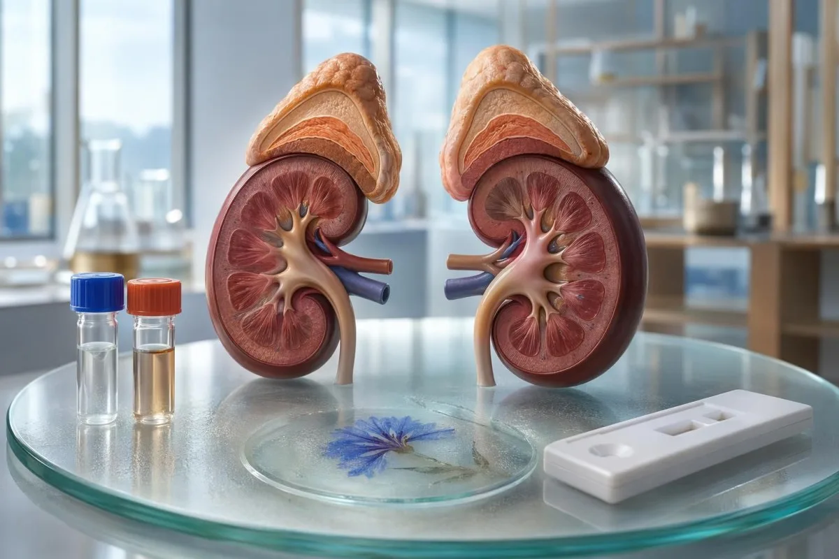
Fatigue, salt craving, low blood pressure and darker skin make more sense when you connect them to cortisol, aldosterone, sodium and ACTH. The pattern matters more than any single symptom.


- Addison disease is primary adrenal insufficiency, where the adrenal cortex cannot make enough cortisol and often aldosterone.
- Morning cortisol below about 3 µg/dL, or 83 nmol/L, strongly raises concern for adrenal insufficiency when symptoms fit.
- ACTH is usually high in primary adrenal insufficiency, often more than 2 times the upper limit of the lab range.
- Low sodium Addison disease usually reflects aldosterone loss, not simply drinking too much water or sweating heavily.
- Salt craving plus standing dizziness is more suspicious when sodium is below 135 mmol/L or blood pressure drops on standing.
- Potassium may rise above 5.0 mmol/L in primary adrenal insufficiency, but a hemolyzed sample can falsely elevate it.
- Darkened skin occurs because high ACTH comes from the same precursor pathway as melanocyte-stimulating hormone.
- Cosyntropin testing checks whether cortisol rises adequately after synthetic ACTH, usually at baseline, 30 minutes and 60 minutes.
- Adrenal crisis can cause vomiting, confusion, severe weakness, low blood pressure, low sodium and low glucose, and needs emergency care.
- Everyday stress does not usually produce the Addison pattern of low cortisol, high ACTH, low sodium, high renin and low aldosterone.
Why fatigue, salt craving and darker skin cluster together
Addison disease symptoms cluster because the adrenal cortex cannot make enough cortisol and often aldosterone: fatigue and weight loss reflect cortisol deficiency, salt craving and low blood pressure reflect sodium wasting, and darkened skin reflects very high ACTH. Doctors suspect primary adrenal insufficiency when morning cortisol is low, ACTH is high, sodium is low, potassium may be high, and renin rises with low aldosterone. Ordinary stress does not usually cause this pattern.
In clinic, the clue is rarely one symptom. A tired person who craves salty food, feels faint when standing, has sodium of 130 mmol/L and has darker gum lines is a very different case from someone exhausted after a bad work month. Low cortisol patterns are most useful when read beside electrolytes and blood pressure.
Kantesti is an AI blood test analyzer that can read cortisol, sodium, potassium, glucose and kidney markers together, rather than treating each flagged result as a separate little drama. As Thomas Klein, MD, I have seen patients dismissed as anxious for 6 to 18 months before someone finally checked an 8 am cortisol and ACTH.
As of July 12, 2026, the safest patient-level rule is simple: do not self-diagnose Addison disease from fatigue alone. If symptoms come with repeated sodium below 135 mmol/L, unexplained weight loss, standing systolic blood pressure drop of 20 mmHg or more, or skin darkening in scars and mouth creases, ask for medical review promptly and read more about our clinical team on About Us.
The cortisol clue: fatigue that does not behave like burnout
Low cortisol in Addison disease causes a heavy, physical fatigue that often worsens with illness, fasting or standing. A random afternoon cortisol can mislead, but an 8 am cortisol below about 3 µg/dL, or 83 nmol/L, is strongly suspicious when symptoms match.
Cortisol normally peaks in the early morning, often between 6 am and 9 am, then falls through the day. The 2016 Endocrine Society guideline recommends morning cortisol and ACTH as first-line tests when primary adrenal insufficiency is suspected, followed by dynamic testing when needed (Bornstein et al., 2016). Our separate cortisol range guide explains why the clock time on the report matters.
What makes Addison fatigue different is the loss of stress reserve. Patients may say they were coping until a stomach bug, long flight or dental infection tipped them into profound weakness; that history tells me more than a single tiredness score ever could. In ordinary burnout, sodium and potassium are usually normal, and ACTH is not persistently high.
A morning cortisol above 15 to 18 µg/dL, roughly 414 to 497 nmol/L depending on assay, usually makes adrenal insufficiency less likely. The grey zone, often 3 to 15 µg/dL, is where clinicians disagree and where stimulation testing is often more honest than guessing. If the story sounds like exhaustion from sleep debt, see our burnout lab mimics guide before chasing rare diagnoses.
Low sodium Addison disease: salt craving and blood pressure
Low sodium Addison disease happens because aldosterone deficiency makes the kidneys waste sodium and water. Serum sodium below 135 mmol/L is hyponatremia, and values below 130 mmol/L with dizziness, vomiting or confusion deserve urgent clinical attention.
Aldosterone tells the distal nephron to keep sodium and excrete potassium. When aldosterone falls, patients can lose salt despite drinking normally; the result is lower blood volume, low blood pressure and a craving for salty foods that feels oddly specific. Our low blood pressure labs article covers other causes, including anemia and medication effects.
Simple dehydration often pushes sodium upward or leaves it normal, especially after sweating without enough water. Addison disease more often produces a low or low-normal sodium because the hormonal signal for salt retention is missing. In my experience, patients notice they need salty soup, pickles or electrolyte drinks long before the sodium finally drops below the lab range.
Orthostatic hypotension is a practical bedside clue: a fall in systolic blood pressure of at least 20 mmHg or diastolic pressure of at least 10 mmHg within 3 minutes of standing is abnormal. If that finding occurs with sodium of 128 mmol/L, potassium of 5.4 mmol/L and morning nausea, I would not call it just poor hydration.
ACTH and darkened skin: why pigmentation can be a lab clue
Darkened skin in Addison disease comes from persistently high ACTH, not from sun exposure alone. ACTH is produced from POMC, a precursor that also generates melanocyte-stimulating signals, so pigmentation often appears in scars, skin creases, gums and pressure points.
ACTH is the pituitary hormone that shouts at the adrenal gland to make cortisol. In primary adrenal insufficiency, the adrenal gland cannot respond, so ACTH keeps rising; values are often more than 2 times the upper limit of normal, and sometimes far higher. Kantesti's neural network treats high ACTH plus low cortisol as a pattern, not as two unrelated flags.
The pigmentation clue is easy to miss in darker skin tones, where patients may notice new darkening of palmar creases, old scars, elbows, nipples, gums or the inside of the cheeks rather than a general tan. I ask about photographs from 6 to 12 months earlier, because slow change is hard to see day by day. Our skin symptom labs guide discusses when skin findings deserve lab context.
Secondary adrenal insufficiency usually has low or inappropriately normal ACTH, so it does not typically cause the classic hyperpigmentation pattern. That distinction is clinically useful: darkened mucosa with low cortisol points more toward primary adrenal failure than pituitary suppression. Still, pigmentation has many causes, including iron overload, pregnancy, medicines and normal familial variation.
Potassium, bicarbonate and urea: the quiet lab cluster
Primary adrenal insufficiency often raises potassium and lowers bicarbonate because aldosterone loss reduces potassium and acid excretion. A potassium level above 5.0 mmol/L can support the Addison pattern, but a hemolyzed tube can falsely raise potassium and must be checked.
The classic electrolyte pattern is low sodium, high potassium and mild metabolic acidosis, often with bicarbonate or CO2 below 22 mmol/L. Not every patient has the full triad, especially early in autoimmune adrenalitis, but seeing sodium 131 mmol/L, potassium 5.6 mmol/L and CO2 19 mmol/L gets my attention quickly.
Kidney markers add another layer. Urea or BUN can rise when low aldosterone causes volume depletion, while creatinine may stay only mildly changed; the ratio can look prerenal rather than intrinsic kidney disease. For a deeper kidney-fluid pattern, our BUN creatinine guide is a useful companion.
Do not overcall Addison disease from one high potassium result. Hemolysis during collection can push potassium up by 0.5 to 2.0 mmol/L, and severe thrombocytosis can also distort serum potassium. If the report mentions hemolysis, compare it with our potassium draw errors checklist before assuming endocrine disease.
Morning cortisol and ACTH: first adrenal insufficiency labs
The first adrenal insufficiency labs are usually 8 am cortisol, plasma ACTH, sodium, potassium, glucose and kidney function. A low morning cortisol with high ACTH is the biochemical doorway into suspected primary adrenal insufficiency.
Timing is not a detail here; it is the test. Cortisol drawn at 4 pm can be low in a healthy person, while cortisol drawn around 8 am should be near its daily peak. The 2016 Endocrine Society guideline advises confirmatory corticotropin testing when the diagnosis is uncertain, but also allows immediate treatment in severe illness after drawing baseline samples if possible (Bornstein et al., 2016).
ACTH handling is fussy. Many labs want plasma ACTH collected in a chilled EDTA tube, transported cold and processed quickly because the peptide can degrade. A falsely low ACTH can blur the primary-versus-secondary distinction, which is why collection conditions matter almost as much as the number.
Kantesti AI reads these markers in context with reference units, time of draw and coexisting electrolytes across 15,000+ biomarkers in our biomarker reference guide. A cortisol of 4.2 µg/dL at 8 am means something different from 4.2 µg/dL at midnight, and a good interpretation should say so plainly.
Cosyntropin testing: why timing changes the answer
Cosyntropin testing checks whether the adrenal cortex can raise cortisol after synthetic ACTH. The standard test often uses 250 µg cosyntropin with cortisol measured at baseline, 30 minutes and sometimes 60 minutes.
Older textbooks used a stimulated cortisol cutoff around 18 µg/dL, or 500 nmol/L. Many modern assays read lower, so some centres use cutoffs closer to 14 to 15 µg/dL, depending on the platform; this is one of those areas where the lab method really matters. Bancos et al. described this assay-related problem in modern adrenal insufficiency diagnosis (Bancos et al., 2015).
Primary adrenal insufficiency usually fails the test because the adrenal cortex is damaged. Early secondary adrenal insufficiency can occasionally pass because the glands have not yet atrophied, which is why doctors interpret the result with ACTH, history and steroid exposure. A neat number without the story can be falsely reassuring.
Kantesti is an AI blood test interpretation platform that uses unit harmonisation and assay-aware context, explained in our AI technology guide. In practical terms, our platform will not treat nmol/L and µg/dL as interchangeable scribbles; conversion errors in cortisol can change the clinical interpretation.
Renin and aldosterone: the salt-wasting signature
High renin with low or inappropriately normal aldosterone is a strong salt-wasting clue in primary adrenal insufficiency. Renin rises because the kidneys sense low effective circulating volume and try to stimulate aldosterone production.
In untreated Addison disease, plasma renin activity may be clearly elevated while aldosterone is low, especially if sodium is low and potassium is high. This is more specific than salt craving alone, because renin is the body's own volume alarm. For background on sampling positions and medication interference, see our renin testing guide.
Aldosterone interpretation depends on posture, sodium intake, time of day and medicines such as ACE inhibitors, ARBs, diuretics and mineralocorticoid blockers. A patient who stopped salt for 2 weeks before testing can push renin up for reasons unrelated to Addison disease. Our aldosterone pattern guide focuses on the opposite problem, high aldosterone, but the pre-test variables are still useful.
Kantesti is an AI biomarker interpretation platform that can place renin and aldosterone beside sodium, potassium, creatinine and blood pressure notes. I still want a clinician to confirm the diagnosis, but pattern recognition can prevent a lonely abnormal sodium from being ignored.
How doctors separate Addison disease from dehydration
Doctors separate Addison disease from dehydration by checking whether the salt-water problem corrects with fluids and whether hormonal markers fit. Dehydration usually has a clear fluid-loss story, while Addison disease often shows low sodium, high renin and persistent orthostatic symptoms.
A single low sodium after vomiting can be simple fluid disturbance. Repeated sodium of 129 to 133 mmol/L with normal kidney function, unexplained nausea and a long history of salt craving is harder to dismiss. Urine testing may help; our urine osmolality clues guide explains why concentrated urine during hyponatremia can point away from plain water loss.
The bedside detail I like is recovery speed. A dehydrated runner often improves within hours after fluids and food, and the next basic metabolic panel may normalise. A patient with Addison disease may feel temporarily better after salty fluids, then slide back because the missing aldosterone signal has not been replaced.
Dizziness has a long differential: anemia, arrhythmia, vestibular disease, hypoglycemia, medications and pregnancy all show up in real life. If the dizziness is worst on standing and paired with sodium below 135 mmol/L, glucose below 70 mg/dL, or weight loss of 5 kg over a few months, use our dizziness lab guide to organise the discussion.
When Addison disease becomes an emergency
Addison disease becomes an emergency when cortisol deficiency causes shock, severe vomiting, confusion, dangerous hyponatremia or hypoglycemia. Suspected adrenal crisis is treated urgently, often with 100 mg intravenous or intramuscular hydrocortisone plus fluids.
Adrenal crisis can look like sepsis, food poisoning, influenza or a fainting episode. The difference is that the body cannot mount a cortisol response to stress, so blood pressure and glucose can fall despite fluid intake. Charmandari, Nicolaides and Chrousos described adrenal insufficiency as a condition where acute illness can rapidly unmask limited adrenal reserve (Charmandari et al., 2014).
Red flags include severe weakness, repeated vomiting, abdominal pain, confusion, fainting, systolic blood pressure below 90 mmHg, sodium often below 130 mmol/L and glucose below 70 mg/dL. Not every crisis has all of these, which is why clinicians treat first when the risk is high. Our hypoglycemia warning signs article explains why low glucose can be especially dangerous in children and lean adults.
If someone already has diagnosed adrenal insufficiency and cannot keep tablets down, they usually need their emergency injection plan and urgent care. I tell patients not to negotiate with persistent vomiting; 6 hours of missed steroid during gastroenteritis can be enough to become unsafe. This is one of the few endocrine situations where over-waiting is the bigger mistake.
Autoimmune partners doctors often screen for next
Autoimmune adrenalitis is the most common cause of Addison disease in many high-income countries, and doctors often screen for thyroid disease, type 1 diabetes, celiac disease and B12-related autoimmunity. A positive 21-hydroxylase antibody supports autoimmune adrenal destruction.
In adults, autoimmune adrenalitis can smoulder for years before cortisol production finally fails. 21-hydroxylase antibodies may appear before full adrenal failure, although the exact progression rate varies by age, genetics and other autoimmune disease. Kantesti trend review can be useful when sodium drifts from 140 to 134 mmol/L over several reports instead of dropping dramatically overnight.
The thyroid deserves special care. Starting levothyroxine in unrecognised adrenal insufficiency can increase cortisol clearance and occasionally worsen symptoms, so clinicians often check adrenal status first when the history fits. For related patterns, see our thyroid disease testing and TPO antibody patterns guides.
B12 deficiency can mimic adrenal fatigue with weakness, dizziness and brain fog, but the lab pattern is different: macrocytosis, high methylmalonic acid or low active B12 may appear instead of high ACTH. Type 1 diabetes can also complicate the picture because recurrent unexplained hypoglycemia may be the first sign of cortisol deficiency in a person using insulin.
Medicines and lab traps that mimic or hide the pattern
Medicines can either mimic adrenal insufficiency or hide it by changing cortisol production, binding proteins or ACTH signalling. Steroid tablets, injections, creams, inhalers and joint injections can suppress ACTH and create secondary adrenal insufficiency.
A patient may say they are not on steroids, then remember a shoulder injection 5 weeks ago, a high-dose inhaler, or a potent skin cream used daily. These exposures can lower ACTH, which is not the Addison pattern, but the symptoms can overlap. Low eosinophils can also reflect steroid or cortisol effects; our cortisol and steroid effects guide explains that CBC clue.
Some medicines reduce cortisol synthesis or increase cortisol breakdown. Ketoconazole, etomidate, mitotane and some anti-seizure or tuberculosis medicines can matter, and opioids can suppress the hypothalamic-pituitary-adrenal axis in susceptible patients. Bancos et al. emphasised medication history as a central part of adrenal insufficiency diagnosis because labs alone cannot reconstruct exposure (Bancos et al., 2015).
Supplements marketed for adrenal fatigue are a separate trap. Some contain hidden steroids or licorice-like compounds that alter blood pressure and potassium, and ashwagandha can affect thyroid tests in some patients. Before mixing products with unexplained low cortisol symptoms, read our adrenal supplement safety guide and bring the bottles to your clinician.
What to ask your clinician after a suspicious panel
After a suspicious panel, ask whether the pattern fits primary adrenal insufficiency and whether an 8 am cortisol, ACTH, sodium, potassium, glucose, renin and aldosterone should be checked. Bring symptom timing, blood pressure readings and medication history.
The most useful question is not, do I have Addison disease? It is, does this exact cluster need adrenal testing? I would write down weight change over 3 to 6 months, salt craving, morning nausea, fainting episodes, infections, steroid exposure and any sodium values below 135 mmol/L.
If you are stable, clinicians often prefer drawing cortisol and ACTH before starting steroids, because treatment can blur the diagnosis. If you are very unwell, safety wins and treatment should not wait for perfect paperwork. Use a concise doctor visit checklist so the appointment does not get swallowed by vague fatigue.
A home blood pressure log can be surprisingly persuasive. Record lying and standing readings after 1 and 3 minutes, plus pulse, for 3 mornings; a consistent 20 mmHg systolic drop is more informative than saying you feel woozy. Bring the actual lab reports, not screenshots cropped around the red flags.
What AI can flag and what still needs a doctor
AI can flag an Addison disease-like pattern, but only a clinician can diagnose adrenal insufficiency, order dynamic testing and decide on steroid treatment. Pattern recognition is useful because low cortisol, low sodium and high ACTH are easy to miss when reports arrive separately.
Kantesti can highlight combinations such as sodium 130 mmol/L, potassium 5.5 mmol/L, glucose 62 mg/dL, morning cortisol 2.4 µg/dL and ACTH above range as a possible urgent endocrine pattern. Our role is to make the pattern visible and explain what to discuss next, not to prescribe hydrocortisone or decide whether you need hospital care.
Kantesti is an AI-powered blood test analysis tool used by 2M+ people across 127+ countries, and our clinical standards are described in the clinical validation process. We also design for multilingual interpretation because adrenal insufficiency warnings should not depend on a patient reading English medical jargon perfectly.
As Thomas Klein, MD, I see the best results when AI pattern checks and physician judgement work together. A dangerous symptom always beats a reassuring app screen: fainting, confusion, severe vomiting, chest pain or very low blood pressure needs urgent care. Our Medical Advisory Board reviews medical content so patient-facing explanations stay cautious where the evidence is not tidy.
Frequently Asked Questions
What are the early symptoms of Addison disease?
Early Addison disease symptoms often include persistent fatigue, weight loss, nausea, salt craving, muscle aches and dizziness on standing. The pattern becomes more suspicious when sodium is below 135 mmol/L, morning cortisol is low and ACTH is high. Darkening of scars, gums or skin creases can appear before a crisis, but it is not present in every patient. Fatigue alone is common and does not diagnose Addison disease.
What labs suggest Addison disease?
Labs that suggest Addison disease include an 8 am cortisol below about 3 µg/dL or 83 nmol/L, ACTH above the reference range, sodium below 135 mmol/L, potassium above 5.0 mmol/L, high renin and low or inappropriately normal aldosterone. A cosyntropin stimulation test is often used to confirm whether cortisol rises adequately. Doctors interpret these values with symptoms, medication history and sample timing because afternoon cortisol and mishandled ACTH samples can mislead.
Is low sodium from Addison disease different from dehydration?
Low sodium from Addison disease is usually driven by aldosterone deficiency, which makes the kidneys waste sodium and water. Simple dehydration often has a clear fluid-loss trigger and may show normal or high sodium, while Addison disease may show sodium below 135 mmol/L with high renin, low blood pressure and salt craving. Repeated sodium values around 128-133 mmol/L with standing dizziness should not be dismissed as ordinary dehydration without a medical review.
Can stress cause the same cortisol labs as Addison disease?
Everyday stress does not usually cause the Addison disease pattern of low morning cortisol with high ACTH and low sodium. Acute illness and major stress usually raise cortisol, sometimes above 18 µg/dL, because the body is trying to maintain blood pressure and glucose. Burnout can cause profound fatigue, but it does not typically produce high renin, low aldosterone, potassium above 5.0 mmol/L or ACTH more than 2 times the upper limit.
Why does Addison disease cause darkened skin?
Addison disease can cause darkened skin because primary adrenal failure makes the pituitary release more ACTH. ACTH comes from the POMC precursor pathway, which is linked to melanocyte-stimulating signals that increase pigment production. The darkening often appears in scars, gum lines, palmar creases, elbows and pressure points rather than as an even suntan. Secondary adrenal insufficiency usually has low or normal ACTH, so this pigmentation pattern is less typical.
When should Addison disease symptoms be treated as an emergency?
Addison disease symptoms are an emergency when severe weakness, repeated vomiting, confusion, fainting, systolic blood pressure below 90 mmHg, sodium below 130 mmol/L or glucose below 70 mg/dL occurs. Suspected adrenal crisis is usually treated urgently with hydrocortisone, often 100 mg by intravenous or intramuscular route, plus fluids. A person with known adrenal insufficiency who cannot keep steroid tablets down should follow their emergency plan and seek urgent care.
Can Addison disease affect periods, sex drive, or pregnancy?
Untreated adrenal insufficiency can disrupt menstrual cycles, lower libido, and make pregnancy riskier because the body cannot respond normally to illness or physical stress. With replacement therapy and pre-pregnancy planning with endocrinology and obstetric teams, many people can have a healthy pregnancy.
Get AI-Powered Blood Test Analysis Today
Join over 2 million users worldwide who trust Kantesti for instant, accurate lab test analysis. Upload your blood test results and receive comprehensive interpretation of 15,000+ biomarkers in seconds.
📚 Referenced Research Publications
Klein, T., Mitchell, S., & Weber, H. (2026). Diarrhea After Fasting, Black Specks in Stool & GI Guide 2026. Kantesti AI Medical Research.
Klein, T., Mitchell, S., & Weber, H. (2026). Women's Health Guide: Ovulation, Menopause & Hormonal Symptoms. Kantesti AI Medical Research.
📖 External Medical References
⚕️ Medical Disclaimer
This article is for educational purposes only and does not constitute medical advice. Always consult a qualified healthcare provider for diagnosis and treatment decisions.
E-E-A-T Trust Signals
Experience
Physician-led clinical review of lab interpretation workflows.
Expertise
Laboratory medicine focus on how biomarkers behave in clinical context.
Authoritativeness
Written by Dr. Thomas Klein with review by Dr. Sarah Mitchell and Prof. Dr. Hans Weber.
Trustworthiness
Evidence-based interpretation with clear follow-up pathways to reduce alarm.