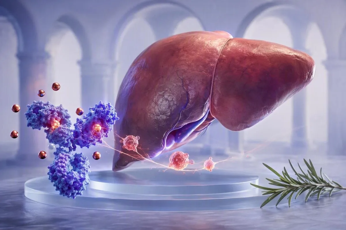
Madal transferriini tulemus võib peegeldada maksa reaktsiooni põletikule, mitte rauavarude vähenemist. Seerumi raua, ferriini, CRP ja transferriini koos lugemine hoiab ära levinud vead.


- Negatiivne akuutfaasi valk: Transferriini tase langeb tavaliselt infektsiooni, autoimmuunhaiguste aktiivsuse, operatsiooni ja muude põletikuliste seisundite korral, isegi kui organismi rauavarud on piisavad.
- Tüüpiline täiskasvanu vahemik: Enamik laboreid kasutab transferriini võrdlusvahemikku umbes 200-360 mg/dl (2,0-3,6 g/l), kuid kohalikud analüüsi vahemikud on prioriteetsed.
- TSAT arvutus: Transferriini küllastus võrdub seerumi rauasisalduse jagamisel TIBC-ga ja korrutamisel 100-ga; madal TIBC võib muuta küllastuse vähem ebanormaalseks kui oodati.
- Ferritiini hoiatus: Ferriini tase tõuseb põletiku korral, seega ferriini tase alla 30 ng/ml tugevalt toetab rauavaegust, samas kui väärtus 100 ng/ml ei välista seda usaldusväärselt, kui CRP on kõrgenenud.
- Kasulik kaasnev test: Lahustuv transferriini retseptor on tavaliselt põletiku mõjust vähem mõjutatud kui ferriin või transferriin ja võib aidata segatüüpi aneemiat selgitada.
- Uusanalüüsi ajastus: Mitte-kriitiliste kõrvalekallete korral annab raua uuringute kordamine umbes 2–4 nädalat pärast lühiajalise haiguse möödumist sageli esinduslikuma lähtetaseme.
- Ärge ravige ennast pimedalt: Raualisaid võivad olla kasulikud tõestatud puuduse korral, kuid võivad olla sobimatud rauakülluse, aktiivse maksahaiguse või peamiselt põletikust põhjustatud aneemia korral.
Miks transferriini test langeb põletiku ajal?
Põletik alandab transferriini, kuna transferriin on negatiivne akuutfaasi valk. Tsütokiinid, eriti interleukiin-6, annavad maksale signaali vähendada transferriini tootmist, samal ajal kui hepitsidiin piirab raua vabanemist plasmasse; see võib põhjustada madalat transferriini ja madalat seerumi rauda, ilma et see tõestaks rauavarude ammendumist.
Lühiversioon on see, et keha muudab immuunaktiveerimise ajal ajutiselt raua vähem kättesaadavaks. Hepcidin vähendab ferriportiin-vahendatud raua väljavoolu enterotsüütidest ja makrofaagidest, nii et seerumi raud langeb sageli 24–48 tunni jooksul; maks võib samaaegselt vähendada transferriini sünteesi. See muster on peremehe kaitsereaktsioon, mitte otsene mõõtmine toidulisandi raua tarbimisest.
Oma kliinilises praktikas võib bakteriaalse kopsupõletiku järgselt inimesel, kelle CRP on 68 mg/L, olla seerumi raud 22 µg/dL ja transferriin 165 mg/dL, kuid ferriini 240 ng/mL. Selle üksiku mustri nimetamine “rauakülluseks” või rauavaeguse kindlaks väljajätmiseks oleksid mõlemad vead. Rauauuringud juhendavad selgitab alusmõisteid üksikasjalikumalt.
Kantesti on AI vereanalüüsi analüsaator mis loeb transferriini koos CRP, ferriini, täisvereloendi indeksite, maksa markerite ja varasemate tulemustega, selle asemel et pidada ühte madalat väärtust diagnoosiks. Dr. Thomas Kleini praktiline reegel on lihtne: palaviku, ägeda faasi või taastumise ajal saadud transferriini tulemus tuleks enne raviotsuse tegemist märgistada kui kontekstipõhine.
Akuutfaasi suund on oluline
CRP, ferriin, fibrinogeen ja haptoglobiin tõusevad üldiselt positiivsete akuutfaasi valkudena, samas kui transferriin ja albumiin võivad langeda. CRP üle 10 mg/L muudab üksiku ferriini väärtuse tõlgendamise oluliselt raskemaks, kuigi ükski universaalne CRP künnis ei saa iga patsiendi tulemust parandada.
Millised on transferriini ja TIBC normaalsed vahemikud?
Täiskasvanute transferriini tavaline vahemik on umbes 200–360 mg/dL ja TIBC vahemik on umbes 250–450 µg/dL. Need vahemikud erinevad laborimeetodi, soo, raseduse staatuse ja kohalike aruandlusühikute järgi, seega tuleks kasutada teie enda tulemuse kõrval olevat vahemikku.
Transferriini mõõdetakse tavaliselt otseselt mg/dL või g/L, samas kui kogu raua seonduv võime ehk TIBC, hindab, kui palju rauda saadaolev transferriin suudab siduda. Paljud laborid tuletavad TIBC transferriinist, selle asemel et seda eraldi mõõta; ligikaudne teisendus on TIBC µg/dL võrdub transferriiniga mg/dL korrutatud 1,25-ga. See seos on kasulik orienteerumiseks, mitte labori enda arvutuse ületamiseks.
Transferriini küllastus, lühendatult TSAT, arvutatakse seerumi raua jagamisel TIBC-ga korrutatud 100-ga. TSAT lähedal 20-45% on paljudes täiskasvanute laborites tüüpiline; väärtused alla 20% viitavad ringleva raua piirangule, kuid need ei erista iseenesest absoluutset rauavaegust põletikust tingitud raua sekvestreerimisest. Madal transferriini küllastus käsitleb seda eristust.
Mõned Euroopa laborid annavad transferriini kohta aru g/L, kus 2,0–3,6 g/L vastab laias laastus 200–360 mg/dL. Tulemus võib pärast viirusnakkust jääda veidi alla normvahemiku ja korduvatel uuringutel normaliseeruda, samas kui püsivad väärtused alla 150 mg/dL väärivad maksa sünteesiprobleemide, valgukao, olulise põletiku või alatoitumise sihipärast otsimist.
Kuidas raua uuringuid tulemuste kogumina tõlgendada?
Rauauuringud on kõige usaldusväärsemad, kui seerumi raud, transferriin või TIBC, TSAT, ferritiin ja põletikumarkerit tõlgendatakse koos. Seerumi raud muutub päeva jooksul märkimisväärselt ja võib samal inimesel proovide vahel langeda üle 30%.
Klassikaline komplikatsioonideta rauavaegus põhjustab tavaliselt madalat seerumi rauda, kõrget transferriini või TIBC, TSAT alla 16% ja ferritiini alla 30 ng/mL. Kõrge TIBC tekib seetõttu, et maks suurendab transferriini tootmist, kui raua kättesaadavus on madal. Punaste vereliblede jaotuslaiuse kasv võib ilmneda enne hemoglobiini langust; vaata meie juhendit RDW muutused pärast rauateraapiat ajakulu kohta.
Põletikuanemia põhjustab sagedamini madalat seerumi rauda, madalat või normaalset transferriini, madalat TSAT ja normaalse või kõrge ferritiini. Weiss ja Goodnough kirjeldasid seda kui rauapiiranguga erütropoeesi, mis on põhjustatud rauakättesaadavuse häiretest, mitte tingimata salvestatud raua puudumisest (Weiss & Goodnough, 2005). Need kaks seisundit võivad sageli esineda koos, eriti põletikulise soolehaiguse, reumatoidartriidi, kroonilise neeruhaiguse ja vähktõve korral.
Vähem ilmne lõks on aritmeetika: kui seerumi raud on 30 µg/dL ja TIBC on langenud 180 µg/dL-ni, on TSAT 17%. Kui TIBC oleks 360 µg/dL, annaks sama seerumi raud 8%. Alumine nimetaja võib varjata, kui vähe rauda ringleb, mistõttu madal seerumi raud vajab konteksti, mitte refleksretsepti.
Praktiline segamusteri vihje
Ferritiin vahemikus 30–100 ng/mL, TSAT alla 16% ja CRP üle 10 mg/L on sageli hall ala, mitte rahustav. Selles olukorras võivad arstid kasutada lahustuvat transferriini retseptorit, retikulotsüütide hemoglobiini sisaldust, trendandmeid või raviprotokolli, mis on kohandatud alusprobleemile.
Miks ferriini tase võib olla normaalne, kui raud on madal
Ferritiin on nii rauahoidla valk kui ka positiivne ägeda faasi reaktant, seega võib põletik seda tõsta sõltumata salvestatud rauast. Ferritiin alla 30 ng/mL toetab enamikul täiskasvanutel tugevalt rauavaegust, kuid kõrgem väärtus ei saa usaldusväärselt välistada puudust, kui CRP või ESR on tõusnud.
Maailma Terviseorganisatsiooni (WHO) 2020. aasta ferritiini juhendis soovitatakse mõõta põletikumarkereid koos ferritiiniga, kui infektsioon või põletik on levinud. Põletikuga täiskasvanutel soovitatakse WHO poolt ferritiini alla 70 µg/L kui võimalikku rauavaegust; see on populatsioonipõhine künnis, mitte individuaalse kliinilise hindamise asendus (WHO, 2020).
Näen sageli patsiente, kes on mures ferritiini 180 ng/mL pärast pärast põletikulist ägedust, arvates, et nende rauavarud peavad olema külluslikud. Mõnikord ongi. Siiski võib ferritiin tõusta pärast pingutavat treeningut, maksa rakkude kahjustust, ainevahetushaigust, alkoholitarvitamist ja immuunaktiivsust, seega number ei ole iseseisev inventuurikontroll. Ferritiin ja CRP on kasulik kaasnev arutelu.
Kantesti on AI vereanalüüsi tulemuste tõlgendamise platvorm mis märgib madala transferriini, madala TSAT ja kõrgenenud CRP vastuolulist kombinatsiooni kui võimalikku põletikuga seotud rauapiirangut. Meie andmestik ei asenda diagnostilist uurimistööd, kuid see kordab pidevalt arsti vana õppetundi: ferritiin käitub erinevalt, kui patsient on aktiivselt haige.
Ferritiini väärtused, mis vajavad teistsugust vestlust
Ferritiin üle 1000 ng/mL nõuab õigeaegset meditsiinilist läbivaatust, kuna raske põletik, märkimisväärne maksakahjustus, rauaküllastussündroomid ja mitmed muud seisundid võivad seda taset põhjustada. Kiireloomulisus sõltub sümptomitest, maksaensüümidest, transferriini küllastumisest ja muutuste kiirusest, mitte ainult ferritiini numbrist.
Millised testid selgitavad rauavaegust põletiku ajal?
Lahustuv transferriini retseptor ja retikulotsüütide hemoglobiini sisaldus võivad aidata tuvastada rauapiirangulist erütrotsüütide tootmist, kui ferritiin ja transferriin ei ühti. Lahustuv transferriini retseptor üldiselt tõuseb koos rakkude rauavajadusega ja on ägeda põletiku poolt vähem moonutatud kui ferritiin.
Lahustuv transferriini retseptor, või sTfR, peegeldab transferriini retseptori ekspressiooni erütroidsetest eelkäijatest. See tõuseb tavaliselt absoluutse rauavaeguse korral ja on sageli normaalne puhta põletikuanemia korral; siiski ei ole analüüsi võrdlusvahemikud standardiseeritud ning hemolüüs või kõrge erütropoeesi aktiivsus võib seda tõsta. See on üks neist valdkondadest, kus täpne laborimeetod on olulisem kui veebipõhised piirväärtused.
Retikulotsüütide hemoglobiini sisaldus, mida sõltuvalt analüsaatorist nimetatakse CHr või Ret-He, peegeldab hiljuti toodetud punalibledes umbes eelnenud 2-4 päeva jooksul saadaval olnud rauda. Väärtused alla umbes 28-30 pg võivad toetada rauapiirangulist erütropoeesi, kuigi kohalikud piirväärtused ja neeruhaiguse protokollid erinevad. Lahustuva transferriini retseptori testimine selgitab, kuhu see sobib.
Luuüdi rauavärvimine jääb ajalooliseks referentsmeetodiks, kuid seda on harva vaja üksnes rutiinse ambulatoorse rauapaneeli lahendamiseks. Dr Thomas Klein eelistab üldiselt proovide kordamist pärast taastumist, verejooksu ja dieedi ajaloo ülevaatamist ning sTfR või retikulotsüütide mõõtmist, kui vastus muudab tõeliselt ravi; lisatestid ilma otsuseta kipuvad looma müra.
sTfR-ferritiini indeks
Mõned spetsialistid arvutavad sTfR/log ferritiini indeksi, mis võib parandada eristamist põletikulistes tingimustes. Piirväärtused erinevad analüüsi järgi oluliselt, sageli ligikaudu 1,0–3,2, seega tuleks tulemust tõlgendada labori valideeritud meetodi järgi, mitte laenatud interneti piirväärtuse järgi.
Millal tuleks transferriini testi korrata?
Transferriini testi on kõige parem korrata pärast lühiajalise nakkuse, palaviku või suure põletikulise sündmuse möödumist, tavaliselt 2-4 nädala pärast, kui olukord ei ole kiireloomuline. Hommikune kogumine ja ühtlased eeltesti tingimused vähendavad seerumi raua ja TSATi vältimatut varieeruvust.
Seerumi raual on päevane varieeruvus ja see võib olla päeva lõpus madalam, samas kui hiljutine suukaudne raud võib seda ajutiselt tõsta. Paljud arstid tellivad hommikuse proovi pärast umbes 8-12 tundi kestnud öist paastu, kui nad vajavad selget võrdlust, kuigi paast ei ole kohustuslik iga rauauuringu puhul. Järgige telliva arsti juhiseid, mitte ärge lõpetage ettenähtud ravimite võtmist omal käel.
48 tundi pärast maratoni, hambaraviprotseduuri, vaktsineerimist või ägedat hingamisteede haigust võetud transferriini tulemus võib peegeldada ägeda faasi reaktsiooni, mitte püsivat rauaprofiili. Kehaline koormus võib samuti muuta CK-d, plasma mahtu ja ferritiini; kestvussportlased võivad leida meie jooksja rauauuringute juhendi kasulikuks.
Kantesti trendianalüüs võrdleb kuupäevi, mitte ainult võrdluslippe, ja võib esile tõsta langust 310 mg/dl-lt 180 mg/dl-ni, mis langeb kokku CRP tõusuga 1 mg/l-lt 52 mg/l-ni. See on kliiniliselt informatiivsem kui kumbki eraldiseisev tulemus. Vaadake üle TIBC testi ettevalmistus enne planeeritud kordustesti korraldamist.
Millal mitte oodata kordustesti
Ärge lükake meditsiinilist läbivaatust kordustesti jaoks edasi, kui esineb tugev õhupuudus, valu rinnus, minestamine, must väljaheide, tugev pidev verejooks, kollatõbi või kiiresti süvenev nõrkus. Hemoglobiin alla 80 g/l (8 g/dl) on sageli kliiniliselt oluline, kuid kiireloomulisus sõltub sümptomitest, languse kiirusest, raseduse staatusest ja südame-veresoonkonna haigustest.
Kuidas CRP, ESR ja hepdiin selgitavad madalat transferriini taset?
CRP ja ESR ei mõõda rauavarusid, kuid need näitavad, kas põletik võib transferriini testi moonutada. CRP tõuseb ja langeb tundide kuni päevade jooksul, samas kui ESR võib jääda kõrgenenuks nädalaid, kuna seda mõjutavad fibrinogeen, aneemia, vanus ja immunoglobuliinid.
Interleukiin-6 stimuleerib maksa hepcidini tootmist ja hepcidiin seondub ferroportiiniga, põhjustades raua ekspordi vähenemist makrofaagidest ja soolerakkudest. Ganz ja Nemethi ülevaade kirjeldab seda rada kui keskset põletikuanemiale ja rauapuudusest tingitud erütropoeesile (Ganz & Nemeth, 2012). Tulemuseks võib olla madal seerumi raua sisaldus päeva jooksul, samal ajal kui transferriini tase langeb sama süsteemse vastusena.
CRP alla 5 mg/L loetakse sageli normi piiridesse kuuluvaks, kuigi laborid erinevad. CRP 40 mg/L ei määra põletiku põhjust, kuid see peaks tegema arsti ettevaatlikumaks ainult ferriini või transferriini alusel rauavaeguse diagnoosimisel. Kõrge ESR-i põhjused selgitab, miks ESR on aeglasem ja vähem spetsiifiline.
Kantesti AI tõlgendab transferriini tulemusi, võrreldes CRP, ferriini, albumiini, valgete vereliblede arvu ja hiljutiste trendide suunda. See ei ole nakkuse või autoimmuunhaiguse diagnoosimise mootor; see on struktureeritud viip, mis küsib, kas rauapaneel võeti bioloogiliselt ebastabiilsel hetkel.
Miks hepcidini rutiinselt ei mõõdeta
Hepcidini analüüsid on tavapärases praktikas endiselt piiratud kättesaadavuse, standardimise ja analüüsi aja tõttu. Hepcidini väärtus võib olla informatiivne spetsialistide uuringutes või harvadel aneemia juhtudel, kuid seerumi ferriin, TSAT, CRP, neerufunktsioon ja verepildi näitajad jäävad praktilisteks esmaseks tööriistadeks.
Kuidas põletikust tingitud aneemia erineb rauavaegusest?
Absoluutne rauavaegus tähendab, et kogu keha raua hulk on ebapiisav, samas kui põletikuanemia tähendab, et raud on olemas, kuid punaliblede tootmiseks halvasti kättesaadav. Mõlemad võivad põhjustada väsimust, madalat TSAT-i ja langevat hemoglobiini ning sageli esinevad koos.
Komplitseerimata rauavaeguse korral on ferriin tavaliselt madal ja transferriin tõuseb sageli üle 360 mg/dL, kuna sidumismahutavus suureneb. Põletikuanemia korral langeb transferriin tavaliselt alla 200 mg/dL, ferriin on sageli üle 100 ng/mL ja CRP võib olla kõrgenenud. Kumbki muster ei ole absoluutne; krooniline neeruhaigus ja maksahaigus on eriti altid kattuma.
Hemoglobiin ja MCV võivad rauapiirangust maha jääda. Isikul võib olla ferriini 18 ng/mL, TSAT 14% ja normaalne hemoglobiin 132 g/L, eriti vaeguse alguses; vastupidi, põletik võib põhjustada aneemiat normaalse MCV-ga 82-100 fL. Mida hemoglobiin tähendab aitab asetada CBC rauauuringute kõrvale.
Põhjus, miks me muretseme madala transferriini ja madala albumiini koosmõju pärast, on see, et koos viitavad need kas tugevamale põletikus koormusele, kahjustunud maksa sünteesile või valgukaotusele, samas kui madal transferriin üksi pärast külmetushaigust on sageli mööduv. Arst peaks üle vaatama neerufunktsiooni, uriinivalku, maksaanalüüse, toitumise, ravimid, verejooksu ajaloo ja trajektoori vähemalt kahe proovi võtmise korral.
Ravi ei ole vahetatav
Suukaudne raud parandab sageli absoluutset puudust, kuid sellel võib olla piiratud mõju, kui põletik hoiab hepcidini kõrgel. Valitud seisundites, nagu krooniline neeruhaigus või aktiivne põletikuline soolehaigus, võivad arstid kasutada intravenoosset rauda või ravida esmalt põletikulist tegurit; lähenemisviis sõltub diagnoosist, sümptomitest, hemoglobiinist ja ohutuse kaalutlustest.
Miks madal transferriini tase võib moonutada transferriini küllastust?
Madal transferriin võib muuta transferriini küllastatuse vähem madalaks, kuna TSAT kasutab selle nimetajana TIBC-d. Tavaline või veidi madal TSAT ei tähenda alati, et raua kohaletoimetamine on piisav, kui TIBC on põletiku või maksa düsfunktsiooni tõttu pärsitud.
Võtke arvesse seerumi raua 36 µg/dL. Kui TIBC on 360 µg/dL, on TSAT 10%; kui TIBC on 180 µg/dL, on TSAT 20%. Teine tulemus tundub matemaatiliselt vähem murettekitav, kuid mõlemad proovid sisaldavad sama madalat ringlevat rauda. Sellepärast peaksid arstid kontrollima toorväärtusi, mitte ainult protsenti.
Vastupidine probleem ilmneb tähelepanuväärse maksakahjustuse või ägeda hepatotsellulaarse kahjustuse korral: transferriini tootmine võib langeda ja seerumi raud võib muutunud ainevahetuse tõttu tõusta, põhjustades tõusnud TSAT-i. Püsivalt üle 45% tõusev TSAT väärib rauakuhjumise hindamist õiges kontekstis, kuid seda ei tohiks kasutada päriliku hemokromatoosi diagnoosimiseks ägeda maksahaiguse ajal. Lugege meie tsirroosi vereanalüüsi vihjeid laiema maksa konteksti saamiseks.
Kantesti on AI-põhine vereanalüüsi analüüsitööriist kasutatakse 127+ riigis, seega normaliseerib see ühikud enne küllastatuse arvutamist ja märgib tulemused, mis võivad olla matemaatiliselt ebastabiilsed, kuna TIBC on ebatavaliselt madal. Väljund peaks toetama, mitte asendama arsti, kes teab, kas patsiendil on palavik, hepatiit, nefrootiline sündroom või hiljutine rauravi.
Ärge kasutage TSAT-i vedelikubilansi markerina
Dehüdratsioon võib kontsentreerida mitmeid seerumimõõtmisi, kuid ei loo sõltuvat rauasisalduse mustrit. Kui albumiin, hematokrit, uurea ja naatrium viitavad vähenenud plasma mahule, võib paneeli kordamine pärast normaalse niisutustase saavutamist olla mõistlik enne piiripealsele TSAT-ile tähenduse omistamist.
Mis veel põhjustab madalat transferriini taset peale põletiku?
Madal tserotransfereiin esineb ka vähenenud maksa sünteesi, neerude või soolestiku kaudu valgu kadu, ebapiisava valgu-energia tarbimise ja harva kaasasündinud häirete korral. Põletik on tavaline, kuid see ei tohiks kunagi olla üldine seletus ilma ülejäänud paneeli kontrollimata.
Maks sünteesib tserotransfereiin, nii et madal tserotransfereiin koos suurenenud bilirubiini, INR, AST, ALT või madala albumiiniga võib viidata maksa haigusele, mitte rauastatus. Raske maksapuudulikkus võib vähendada tserotransfereiin alla 150 mg/dL. Maksa paneeli tulemused aitavad kindlaks teha, kas see selgitus on usutav.
Nefrootilise tasemega uriini valgu kadu võib koos albumiini ja teiste valkudega eemaldada tserotransfereiin. Uriini albumiin-kreatiniini suhe üle 300 mg/g ehk 30 mg/mmol on raskelt suurenenud albuminuuria ja nõuab neerule keskendunud hindamist; uriiniriba valgusu enkel ei ole täieliku vastuse jaoks piisav. Vaadake meie juhendit uriinis oleva valgu kohta.
Halb toitumine, malabsorptsioon või raske kataboolne haigus võib vähendada tserotransfereiin, kuigi tserotransfereiin on liiga põletikutundlik, et seda üksi toitumise markerina kasutada. Kaasasündinud atransferrinemia on erandlikult haruldane ja ilmneb tavaliselt palju varem elu jooksul raske aneemia ja paradoksaalse süsteemse rauakoormusega. See ebatavaline kombinatsioon vajab spetsialisti hematoloogi hinnangut.
Ravimite ja hormoonide mõju
Östrogeeni ekspositsioon ja rasedus võivad suurendada tserotransfereiin, samas kui androgeenid võivad seda veidi vähendada. Need nihked on tavaliselt väiksemad kui olulise põletiku või maksahaiguse mõjud, kuid need võivad selgitada piiripealset tulemust, kui ülejäänud rauapaneel on stabiilne.
Kuidas rasedus ja menstruaalverekaotus muudavad transferriini taset?
Rasedus suurendab sageli tserotransfereiin ja TIBC, samas kui rauavajadus suureneb kõige järsemalt teisel ja kolmandal trimestril. Seetõttu väärib madal tserotransfereiin raseduse ajal erilist tähelepanu põletikule, maksafunktsioonile, valgu kadu ja laboratoorsele kontekstile.
Plasma maht raseduse ajal laieneb ja östrogeen suurendab tserotransfereiin sünteesi, seega TIBC tõuseb sageli mitte-raseda naise normist kõrgemale. Ferriin langeb samuti tavaliselt raseduse edenedes; ferriini alla 30 µg/L kasutatakse tavaliselt rauavarude ammendumise tuvastamiseks raseduse ajal, kuigi kohalikud emadusjuhised võivad erineda. Ferriin trimestri järgi annab praktilised vahemikud.
Tugev menstruaalverejooks on absoluutse rauavaeguse tavaline põhjus, eriti kui ferriin on alla 30 ng/mL ja tserotransfereiin on tõusnud, mitte langenud. Kuid autoimmuunhaiguse ja tugevate perioodidega inimene võib kogeda nii verekaotuse puudust kui ka põletikku; “normaalne” ferriin 75 ng/mL ei lahenda küsimust, kui CRP on 24 mg/L.
Uus madal tserotransfereiin koos kõrge vererõhu, turse, proteinuuria, peavalu, parema ülakõhuvalu või ebanormaalsete maksatestide korral raseduse ajal vajab kiiret sünnitusabi hindamist. See ei ole preeklampsia diagnoosimise viis, kuid laiem valgu- ja maksa muster võib olla palju olulisem kui ainult rauamõõtmine.
Kuidas tõlgendatakse transferriini neerukao ja kroonilise haiguse korral?
Krooniline neeruhaigus põhjustab sageli funktsionaalset rauavaegust, kuna põletik ja vähenenud erütropoetiin piiravad punaliblede tootmiseks kasutatavat rauda. Selles olukorras toetavad TSAT alla 20% ja ferriin alla 100 ng/mL sageli rauavaegust enne dialüüsi, kuigi ravi lävendid erinevad juhendi ja kliinilise olukorra järgi.
KDIGO juhend on ajalooliselt kasutanud rauaproovi lähenemisviisi paljudel täiskasvanud CKD patsientidel, kui TSAT on 30% või madalam ja ferriin 500 ng/mL või madalam, tingimusel et kliiniline eesmärk on hemoglobiini tõstmine või erütropoeesi stimuleeriva ravi vähendamine. Need on ravi kaalutlused, mitte rauavarude universaalsed definitsioonid, ja 500 ng/mL lagi on sageli valesti mõistetud.
Patsiendil eGFR 28 mL/min/1.73 m², hemoglobiin 96 g/L, TSAT 16%, ferriin 220 ng/mL ja CRP 12 mg/L võib olla funktsionaalne puudus mitte-madala ferriini taseme korral. Suukaudne raua imendumine võib olla vähenenud ja arstid peavad hindama ka B12, foolhapet, varjatud kadu, erütropoetiini kasutamist ja neerude seisundit. Kroonilise neeruhaiguse staadiumi määramine pakub kasulikku taustteavet.
Kantesti tehisintellekt saab paigutada transferriini neerutervise pikaajalisse konteksti, kuid ei saa kindlaks teha, kas sobiv on intravenoosne rauaravi, erütropoeesi stimuleeriv ravi või eriarsti poole pöördumine. See otsus eeldab sümptomite, vererõhu, infektsioonistaatuse, ravimite ülevaate ja täielike neeruandmete arvestamist.
Millised anamneesi detailid muudavad transferriini tulemuse kasulikumaks?
Kõige kasulikum transferriini tõlgendus hõlmab sümptomeid, hiljutist haigestumist, menstruatsiooni- või seedetrakti verekaotust, toitumist, alkoholi tarvitamist, ravimeid, kehalist koormust ja varasemaid tulemusi. Üksik proov on hetkepilt; kaks võrreldavat proovi näitavad sageli, kas muutus on füsioloogiline, põletikuline või püsiv.
Küsige, kas verevõtule eelnenud 14 päeva jooksul esines palavikku, vaktsineerimist, vigastust, haiglaravi, hambaravi, intensiivset treeningut või teadaolevat ägenemist. Samuti pange kirja rauatabletid, multivitamiinid, prootonpumba inhibiitorid, mittesteroidsed põletikuvastased ravimid, antikoagulandid ja hiljutised muutused toitumises. Need üksikasjad võivad mõjutada raua imendumist või varjatud verekaotuse tõenäosust.
Transferriini muutus väärtuselt 290 väärtusele 205 mg/dL on olulisem, kui CRP tõusis väärtuselt 2 väärtusele 35 mg/L ja albumiin langes väärtuselt 44 väärtusele 37 g/L. Seevastu 10 mg/dL suurune muutus stabiilse CRP ja samasuguse proovivõtu ajastuse korral võib jääda bioloogilise ja analüütilise varieeruvuse piiresse. Vereanalüüside võrdlemine selgitab, miks väikesed muutused ei ole alati tegelikud muutused.
Kantesti mitmekeelsed haigusloo tööriistad võimaldavad inimestel salvestada tulemuse kõrvale konteksti, sealhulgas haigestumise kuupäevi ja muutusi toidulisandite tarvitamises, 75+ keeles. See ajalugu muudab arsti hinnangu ohutumaks; meie pikaajaline vereanalüüsi juhis loetleb praktilised üksikasjad, mida tasub säilitada.
Sümptomid ei ole spetsiifilised
Väsimus, vähenenud koormustaluvus, juuste väljalangemine, rahutud jalad ja keskendumisraskused võivad esineda rauapuuduse, põletiku, kilpnäärmehaiguse, unehäirete, depressiooni ja paljude muude seisundite korral. Sümptomid peaksid ajendama asjakohast hindamist, kuid nende põhjal ei saa kindlaks teha, kas madal transferriin viitab rauavarude ammendumisele.
Millal vajab madal transferriini tulemus kiiret läbivaatamist?
Madal transferriinitase vajab kiiret arstlikku hindamist, kui sellega kaasnevad oluline aneemia, ikterus, turse, rohke verejooks, must väljaheide, seletamatu kaalulangus või viited neeru- või maksafunktsiooni häirele. Tulemus ise on harva erakorraline, kuid selle taga olev seisund võib seda mõnikord olla.
Samal päeval on mõistlik pöörduda hindamisele rindkerevalu, minestamise, tugeva õhupuuduse, segasusseisundi, musta tõrvataolise väljaheite, verise oksendamise või kiiresti süveneva turse korral. Hemoglobiinisisaldus alla 70 g/L (7 g/dL), bilirubiinisisaldus üle 50 µmol/L koos ikterusega või ootamatult kõrge INR vajab individuaalset kliinilist otsust ja võib nõuda kiiret hindamist. Ärge püüdke neid seisundeid korrigeerida üksnes käsimüügist saadava rauaga.
Stabiilse, kergelt langenud transferriinitaseme (185 mg/dL) korral, kui hemoglobiinisisaldus ja maksaanalüüsid on normis ning CRP on dokumenteeritud hingamisteede infektsiooni ajal 18 mg/L, on kordusanalüüside tegemine pärast paranemist sageli mõistlik. Kui muutus püsib üle 6–8 nädala, laiendavad arstid tavaliselt hindamist maksaproovide, uriinivalgu, toitumisanamneesi, põletikulise haiguse aktiivsuse ja verejooksuallikate suhtes.
Meie kliinilist sisu vaadatakse läbi koostöös Meditsiininõukogu, -ga ning Kantesti tõlgendusloogika on dokumenteeritud meie meditsiinilise valideerimise raamistik. Seisuga 5. september 2026 on kõige ohutum sõnum endiselt muutumatu: madal transferriinitulemus on vihje raua transpordi ja süsteemse füsioloogia kohta, mitte iseseisev diagnoos.
Korduma kippuvad küsimused
Kas põletik võib põhjustada madalat transferriini ilma rauapuuduseta?
Jah. Põletik võib vähendada transferriini isegi siis, kui rauavarud on piisavad, kuna transferriin on negatiivne akuutfaasi valk, mida toodab maks. CRP üle 10 mg/L-ga inimestel võib põletikust tingitud rauasemosteerimise tõttu olla madal transferriin, madal seerumi raua sisaldus ja ferritiin üle 100 ng/mL, mitte puhas rauavaegus. Rauavaegus võib siiski esineda, eriti kui TSAT on alla 20% või ferritiin on alla 30 ng/mL. Arst peaks tõlgendama kogu pilti ja põletiku põhjust.
Kas madal transferriin tähendab madalat rauasisaldust?
Madal transferriini tase ei tähenda automaatselt keha madalat rauasisaldust. Madal transferriini tase esineb sageli põletiku, maksahaiguse, neeruvalgukao ja ebapiisava valgudieedi korral, samas kui klassikaline rauavaegus tõstab transferriini tavaliselt üle umbes 360 mg/dL. Alla 50 µg/dL seerumi raud võib esineda nii rauavaeguse kui ka põletiku korral, seetõttu on vaja ferritiini, CRP, TIBC, TSAT ja vereanalüüsi. Transferriini tulemus alla 200 mg/dL tuleks tõlgendada koos labori referentsväärtuste ja kliinilise anamneesiga.
Kas madal transferriin võib muuta transferriini küllastatuse normaalseks?
Jah. Transferriini küllastus on seerumi raud jagatud TIBC-ga korrutatud 100-ga, nii et madal transferriinist põhjustatud madal TIBC tekitab väiksema nimetaja. Näiteks seerumi raud 36 µg/dL annab TSAT-i 10%, kui TIBC on 360 µg/dL, kuid 20%, kui TIBC on 180 µg/dL. See tähendab, et piiripealne TSAT võib alahinnata rauapiirangut, kui transferriin on allasurutud. Arstid peaksid kontrollima nii protsenti kui ka tooreid seerumi raua ja TIBC väärtusi.
Milline ferriinitase kinnitab rauavaegust põletiku ajal?
Ferriit tase alla 30 ng/mL või µg/L viitab enamikul täiskasvanutest tugevalt rauapuudusele, sealhulgas paljudel inimestel kerge põletikuga. WHO 2020. aasta suunis soovitab, et ferriit alla 70 µg/L võib viidata rauapuudusele täiskasvanutel koos põletiku tunnustega, kuid see piirväärtus ei ole absoluutne igale indiviidile. Ferriit võib tõusta koos CRP taseme tõusu, maksa kahjustuse ja ainevahetushaigustega, seetõttu nõuavad 30–100 ng/mL vahelised väärtused sageli TSAT, CRP ja mõnikord lahustuva transferriini retseptori testimist. Ferriit üle 100 ng/mL ei välista alati rauapuudust, kui esineb põletikuline aktiivsus.
Kui kaua pärast haigust peaksin kordama transferriini testi?
Vähemtähtsate mööduvate haiguste korral on sageli otstarbekas korrata transferriini testi umbes 2–4 nädalat pärast sümptomite ja palaviku taandumist, kui puuduvad kiireloomulised tunnused. CRP võib normaliseeruda mõne päevaga, kuid ESR, ferritiini, albumiini ja transferriini taseme normaliseerumine võib võtta kauem aega. Korduvaks analüüsiks kasutage sarnaseid tingimusi, ideaalis hommikust proovi ja võimalusel sama laborit. Ärge lükake edasi läbivaatust, kui hemoglobiinitase langeb, verejooks jätkub või maksa- ja neerude näitajad on ebanormaalsed.
Kas ma peaksin võtma rauapreparaate madala transferriini korral?
Madal transferriinitase iseenesest ei ole põhjus rauapreparaatide alustamiseks. Suukaudset rauda kaalutakse tavaliselt absoluutse rauavaeguse korral, nagu ferritiin alla 30 ng/mL, madal TSAT, sobivad sümptomid või selge kaotuse allikas; annus ja manustamissagedus tuleb individuaalselt määrata. Põletikuanemiat korral võib raud halvasti imenduda või olla kättesaamatu, kuna heptsidiini tase on kõrge, ja alusprobleemi ravimine võib olla olulisem. Raud võib olla kahjulik või eksitav rauakuhjumise häirete ja mõnede maksahaiguste korral, seega kinnitage probleemiga arsti poole.
Hangi AI-toega vereanalüüsi analüüs juba täna
Liitu enam kui 2 miljoni kasutajaga üle maailma, kes usaldavad Kantesti-d kohese ja täpse laborianalüüsi jaoks. Laadi üles oma vereanalüüsi tulemused ja saad põhjaliku tõlgenduse 15,000+ biomarkerite kohta sekunditega.
📚 Viidatud teaduspublikatsioonid
Klein, T., Mitchell, S., & Weber, H. (2026). Kantesti vereanalüüsi tõlgendamise mootori automatiseeritud tehniline võrdlusuuring, mis põhineb eelregistreeritud rubriigil, 100 000 sünteetilise testjuhtumi peal. Kantesti AI Medical Research.
Klein, T., Mitchell, S., & Weber, H. (2026). Kliinilise valideerimise raamistik v2.0 (meditsiinilise valideerimise leht). Kantesti AI Medical Research.
📖 Välised meditsiinilised viited
Maailma Terviseorganisatsioon (2020). WHO juhend ferritiini kontsentratsioonide kasutamiseks rauaseisundi hindamisel üksikisikutel ja populatsioonides. Maailma Terviseorganisatsioon.
⚕️ Meditsiiniline lahtiütlus
Käesolev artikkel on mõeldud üksnes hariduslikel eesmärkidel ega kujuta endast meditsiinilist nõuannet. Diagnoosi ja ravivalikute otsuste tegemiseks konsulteeri alati kvalifitseeritud tervishoiutöötajaga.
E-E-A-T usaldussignaalid
Kogemus
Arsti juhitud kliiniline ülevaade labori tõlgendamise töövoogudest.
Ekspertiis
Laborimeditsiin keskendub sellele, kuidas biomarkerid käituvad kliinilises kontekstis.
Autoriteetsus
Kirjutanud dr Thomas Klein, ülevaade: dr Sarah Mitchell ja prof dr Hans Weber.
Usaldusväärsus
Tõenduspõhine tõlgendus selgete edasiste sammudega, et vähendada ärevust.