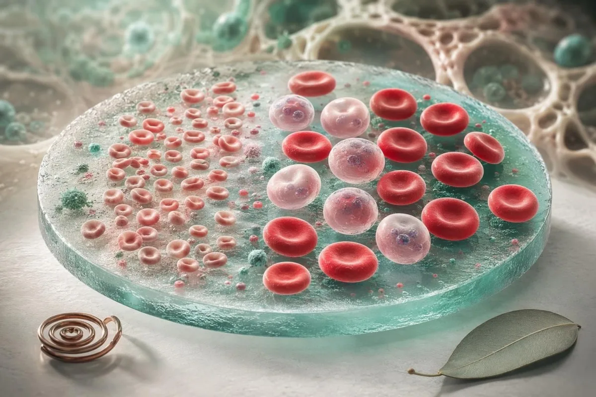
Varajane RDW-i tõus pärast rauavaeguse ravi peegeldab sageli taastumist, mitte ebaõnnestumist. Oluline küsimus on, kas kõrval selle liiguvad õiges suunas hemoglobiin, retikulotsüüdid, MCV ja rauavarud.


- Kõrge RDW tõuseb sageli esimese 1–3 nädala jooksul pärast tõhusat rauaravi, sest vanad mikrotsüüdid esinevad koosäsäsäsäsäsäsäsäsäsäsäsäsäsäsäsäsäsäsäsäsäsäsäsäsäsäsäsäsäsäsäsäsäsäsäsäsäsäsäsäsäsäsäsäsäsäsäsäsäsäsäsäsäsäsäsäsäsäsäsäsäsäsäsäsäsäsäsäsäsäsäsäsäsäsäsäsäsäsäsäsäsäsäsäsäsäsäsäsäsäsäsäsäsäsäsäsäsäsäsäsäsäsäsäsäsäsäsäsäsäsäsäsäsäsäsäsäsäsäsäsäsäsäsäsäsäsäsäsäsäsäsäsäsäsäsäsäsäsäsäsäsäsäsäsäsäsäsäsäsäsäsäsäsäsäsäsäsäsäsäsäsäsäsäsäsäsäsäsäsäsäsäsäsäsäsäsäsäsäsäsäsäsäsäsäsäsäsäsäsäsäsäsäsäsäsäsäsäsäsäsäsäsäsäsäsäsäsäsäsäsäsäsäsäsäsäsäsäsäsäsäsäsäsäsäsäsäsäsäsäsäsäsäsäsäsäsäsäsäsäsäsäsäsäsäsäsäsäsäsäsäsäsäsäsäsäsäsäsäsäsäsäsäsäsäsäsäsäsäsäsäsäsäsäsäsäsäsäsäsäsäsäsäsäsäsäsäsäsäsäsäsäsäsäsäsäsäsäsäsäsäsäsäsäsäsäsäsäsäsäsäsäsäsäsäsäsäsäsäsäsäsäsäsäsäsäsäsäsäsäsäsäsäsäsäsäsäsäsäsäsäsäsäsäsäsäsäsäsäsäsäsäsäsäsäsäsäsäsäsäsäsäsäsäsäsäsäsäsäsäsäsäsäsäsäsäsäsäsäsäsäsäsäsäsäsäsäsäsäsäsäsäsäsäsäsäsäsäsäsäsäsäsäsäsäsäsäsäsäsäsäsäsäsäsäsäsäsäsäsäsäsäsäsäsäsäsäsäsäsäsäsäsäsäsäsäsäsäsäsäsäsäsäsäsäsäsäsäsäsäsäsäsäsäsäsäsäsäsäsäsäsäsäsäsäsäsäsäsäsäsäsäsäsäsäsäsäsäsäsäsäsäsäsäsäsäsäsäsäsäsäsäsäsäsäsäsäsäsäsäsäsäsäsäsäsäsäsäsäsäsäsäsäsäsäsäsäsäsäsäsäsäsäsäsäsäsäsäsäsäsäsäsäsäsäsäsäsäsäsäsäsäsäsäsäsäsäsäsäsäsäsäsäsäsäsäsäsäsäsäsäsäsäsäsäsäsäsäsäsäsäsäsäsäsäsäsäsäsäsäsäsäsäsäsäsäsäsäsäsäsäsäsäsäsäsäsäsäsäsäsäsäsäsäsäsäsäsäsäsäsäsäsäsäsäsäsäsäsäsäsäsäsäsäsäsäsäsäsäsäsäsäsäsäsäsäsäsäsäsäsäsäsäsäsäsäsäsäsäsäsäsäsäsäsäsäsäsäsäsäsäsäsäsäsäsäsäsäsäsäsäsäsäsäsäsäsäsäsäsäsäsäsäsäsäsäsäsäsäsäsäsäsäsäsäsäsäsäsäsäsäsäsäsäsäsäsäsäsäsäsäsäsäsäsäsäsäsäsäsäsäsäsäsäsäsäsäsäsäsäsäsäsäsäsäsäsäsäsäsäsäsäsäsäsäsäsäsäsäsäsäsäsäsäsäsäsäsäsäsäsäsäsäsäsäsäsäsäsäsäsäsäsäsäsäsäsäsäsäsäsäsäsäsäsäsäsäsäsäsäsäsäsäsäsäsäsäsäsäsäsäsäsäsäsäsäsäsäsäsäsäsäsäsäsäsäsäsäsäsäsäsäsäsäsäsäsäsäsäsäsäsäsäsäsäsäsäsäsäsäsäsäsäsäsäsäsäsäsäsäsäsäsäsäsäsäsäsäsäsäsäsäsäsäsäsäsäsäsäsäsäsäsäsäsäsäsäsäsäsäsäsäsäsäsäsäsäsäsäsäsäsäsäsäsäsäsäsäsäsäsäsäsäsäsäsäsäsäsäsäsäsäsäsäsäsäsäsäsäsäsäsäsäsäsäsäsäsäsäsäsäsäsäsäsäsäsäsäsäsäsäsäsäsäsäsäsäsäsäsäsäsäsäsäsäsäsäsäsäsäsäsäsäsäsäsäsäsäsäsäsäsäsäsäsäsäsäsäsäsäsäsäsäsäsäsäsäsäsäsäsäsäsäsäsäsäsäsäsäsäsäsäsäsäsäsäsäsäsäsäsäsäsäsäsäsäsäsäsäsäsäsäsäsäsäsäsäsäsäsäsäsäsäsäsäsäsäsäsäsäsäsäsäsäsäsäsäsäsäsäsäsäsäsäsäsäsäsäsäsäsäsäsäsäsäsäsäsäsäsäsäsäsäsäsäsäsäsäsäsäsäsäsäsäsäsäsäsäsäsäsäsäsäsäsäsäsäsäsäsäsäsäsäsäsäsäsäsäsäsäsäsäsäsäsäsäsäsäsäsäsäsäsäsäsäsäsäsäsäsäsäsäsäsäsäsäsäsäsäsäsäsäsäsäsäsäsäsäsäsäsäsäsäsäsäsäsäsäsäsäsäsäsäsäsäsäsäsäsäsäsäsäsäsäsäsäsäsäsäsäsäsäsäsäsäsäsäsäsäsäsäsäsäsäsäsäsäsäsäsäsäsäsäsäsäsäsäsäsäsäsäsäsäsäsäsäsäsäsäsäsäsäsäsäsäsäsäsäsäsäsäsäsäsäsäsäsäsäsäsäsäsäsäsäsäsäsäsäsäsäsäsäsäsäsäsäsäsäsäsäsäsäsäsäsäsäsäsäsäsäsäsäsäsäsäsäsäsäsäsäsäsäsäsäsäsäsäsäsäsäsäsäsäsäsäsäsäsäsäsäsäsäsäsäsäsäsäsäsäsäsäsäsäsäsäsäsäsäsäsäsäsäsäsäsäsäsäsäsäsäsäsäsäsäsäsäsäsäsäsäsäsäsäsäsäsäsäsäsäsäsäsäsäsäsäsäsäsäsäsäsäsäsäsäsäsäsäsäsäsäsäsäsäsäsäsäsäsäsäsäsäsäsäsäsäsäsäsäsäsäsäsäsäsäsäsäsäsäsäsäsäsäsäsäsäsäsäsäsäsäsäsäsäsäsäsäsäsäsäsäsäsäsäsäsäsäsäsäsäsäsäsäsäsäsäsäsäsäsäsäsäsäsäsäsäsäsäsäsäsäsäsäsäsäsäsäsäsäsäsäsäsäsäsäsäsäsäsäsäsäsäsäsäsäsäsäsäsäsäsäsäsäsäsäsäsäsäsäsäsäsäsäsäsäsäsäsäsäsäsäsäsäsäsäsäsäsäsäsäsäsäsäsäsäsäsäsäsäsäsäsäsäsäsäsäsäsäsäsäsäsäsäsäsäsäsäsäsäsäsäsäsäsäsäsäsäsäsäsäsäsäsäsäsäsäsäsäsäsäsäsäsäsäsäsäsäsäsäsäsäsäsäsäsäsäsäsäsäsäsäsäsäsäsäsäsäsäsäsäsäsäsäsäsäsäsäsäsäsäsäsäsäsäsäsäsäsäsäsäsäsäsäsäsäsäsäsäsäsäsäsäsäsäsäsäsäsäsäsäsäsäsäsäsäsäsäsäsäsäsäsäsäsäsäsäsäsäsäsäsäsäsäsäsäsäsäsäsäsäsäsäsäsäsäsäsäsäsäsäsäsäsäsäsäsäsäsäsäsäsäsäsäsäsäsäsäsäsäsäsäsäsäsäsäsäsäsäsäsäsäsäsäsäsäsäsäsäsäsäsäsäsäsäsäsäsäsäsäsäsäsäsäsäsäsäsäsäsäsäsäsäsäsäsäsäsäsäsäsäsäsäsäsäsäsäsäsäsäsäsäsäsäsäsäsäsäsäsäsäsäsäsäsäsäsäsäsäsäsäsäsäsäsäsäsäsäsäsäsäsäsäsäsäsäsäsäsäsäsäsäsäsäsäsäsäsäsäsäsäsäsäsäsäsäsäsäsäsäsäsäsäsäsäsäsäsäsäsäsäsäsäsäsäsäsäsäsäsäsäsäsäsäsäsäsäsäsäsäsäsäsäsäsäsäsäsäsäsäsäsäsäsäsäsäsäsäsäsäsäsäsäsäsäsäsäsäsäsäsäsäsäsäsäsäsäsäsäsäsäsäsäsäsäsäsäsäsäsäsäsäsäsäsäsäsäsäsäsäsäsäsäsäsäsäsäsäsäsäsäsäsäsäsäsäsäsäsäsäsäsäsäsäsäsäsäsäsäsäsäsäsäsäsäsäsäsäsäsäsäsäsäsäsäsäsäsäsäsäsäsäsäsäsäsäsäsäsäsäsäsäsäsäsäsäsäsäsäsäsäsäsäsäsäsäsäsäsäsäsäsäsäsäsäsäsäsäsäsäsäsäsäsäsäsäsäsäsäsäsäsäsäsäsäsäsäsäsäsäsäsäsäsäsäsäsäsäsäsäsäsäsäsäsäsäsäsäsäsäsäsäsäsäsäsäsäsäsäsäsäsäsäsäsäsäsäsäsäsäsäsäsäsäsäsäsäsäsäsäsäsäsäsäsäsäsäsäsäsäsäsäsäsäsäsäsäsäsäsäsäsäsäsäsäsäsäsäsäsäsäsäsäsäsäsäsäsäsäsäsäsäsäsäsäsäsäsäsäsäsäsäsäsäsäsäsäsäsäsäsäsäsäsäsäsäsäsäsäsäsäsäsäsäsäsäsäsäsäsäsäsäsäsäsäsäsäsäsäsäsäsäsäsäsäsäsäsäsäsäsäsäsäsäsäsäsäsäsäsäsäsäsäsäsäsäsäsäsäsäsäsäsäsäsäsäsäsäsäsäsäsäsäsäsäsäsäsäsäsäsäsäsäsäsäsäsäsäsäsäsäsäsäsäsäsäsäsäsäsäsäsäsäsäsäsäsäsäsäsäsäsäsäsäsäsäsäsäsäsäsäsäsäsäsäsäsäsäsäsäsäsäsäsäsäsäsäsäsäsäsäsäsäsäsäsäsäsäsäsäsäsäsäsäsäsäsäsäsäsäsäsäsäsäsäsäsäsäsäsäsäsäsäsäsäsäsäsäsäsäsäsäsäsäsäsäsäsäsäsäsäsäsäsäsäsäsäsäsäsäsäsäsäsäsäsäsäsäsäsäsäsäsäsäsäsäsäsäsäsäsäsäsäsäsäsäsäsäsäsäsäsäsäsäsäsäsäsäsäsäsäsäsäsäsäsäsäsäsäsäsäsäsäsäsäsäsäsäsäsäsäsäsäsäsäsäsäsäsäsäsäsäsäsäsäsäsäsäsäsäsäsäsäsäsäsäsäsäsäsäsäsäsäsäsäsäsäsäsäsäsäsäsäsäsäsäsäsäsäsäsäsäsäsäsäsäsäsäsäsäsäsäsäsäsäsäsäsäsäsäsäsäsäsäsäsäsäsäsäsäsäsäsäsäsäsäsäsäsäsäsäsäsäsäsäsäsäsäsäsäsäsäsäsäsäsäsäsäsäsäsäsäsäsäsäsäsäsäsäsäsäsäsäsäsäsäsäsäsäsäsäsäsäsäsäsäsäsäsäsäsäsäsäsäsäsäsäsäsäsäsäsäsäsäsäsäsäsäsäsäsäsäsäsäsäsäsäsäsäsäsäsäsäsäsäsäsäsäsäsäsäsäsäsäsäsäsäsäsäsäsäsäsäsäsäsäsäsäsäsäsäsäsäsäsäsäsäsäsäsäsäsäsäsäsäsäsäsäsäsäsäsäsäsäsäsäsäsäsäsäsäsäsäsäsäsäsäsäsäsäsäsäsäsäsäsäsäsäsäsäsäsäsäsäsäsäsäsäsäsäsäsäsäsäsäsäsäsäsäsäsäsäsäsäsäsäsäsäsäsäsäsäsäsäsäsäsäsäsäsäsäsäsäsäsäsäsäsäsäsäsäsäsäsäsäsäsäsäsäsäsäsäsäsäsäsäsäsäsäsäsäsäsäsäsäsäsäsäsäsäsäsäsäsäsäsäsäsäsäsäsäsäsäsäsäsäsäsäsäsäsäsäsäsäsäsäsäsäsäsäsäsäsäsäsäsäsäsäsäsäsäsäsäsäsäsäsäsäsäsäsäsäsäsäsäsäsäsäsäsäsäsäsäsäsäsäsäsäsäsäsäsäsäsäsäsäsäsäsäsäsäsäsäsäsäsäsäsäsäsäsäsäsäsäsäsäsäsäsäsäsäsäsäsäsäsäsäsäsäsäsäsäsäsäsäsäsäsäsäsäsäsäsäsäsäsäsäsäsäsäsäsäsäsäsäsäsäsäsäsäsäsäsäsäsäsäsäsäsäsäsäsäsäsäsäsäsäsäsäsäsäsäsäsäsäsäsäsäsäsäsäsäsäsäsäsäsäsäsäsäsäsäsäsäsäsäsäsäsäsäsäsäsäsäsäsäsäsäsäsäsäsäsäsäsäsäsäsäsäsäsäsäsäsäsäsäsäsäsäsäsäsäsäsäsäsäsäsäsäsäsäsäsäsäsäsäsäsäsäsäsäsäsäsäsäsäsäsäsäsäsäsäsäsäsäsäsäsäsäsäsäsäsäsäsäsäsäsäsäsäsäsäsäsäsäsäsäsäsäsäsäsäsäsäsäsäsäsäsäsäsäsäsäsäsäsäsäsäsäsäsäsäsäsäsäsäsäsäsäsäsäsäsäsäsäsäsäsäsäsäsäsäsäsäsäsäsäsäsäsäsäsäsäsäsäsäsäsäsäsäsäsäsäsäsäsäsäsäsäsäsäsäsäsäsäsäsäsäsäsäsäsäsäsäsäsäsäsäsäsäsäsäsäsäsäsäsäsäsäsäsäsäsäsäsäsäsäsäsäsäsäsäsäsäsäsäsäsäsäsäsäsäsäsäsäsäsäsäsäsäsäsäsäsäsäsäsäsäsäsäsäsäsäsäsäsäsäsäsäsäsäsäsäsäsäsäsäsäsäsäsäsäsäsäsäsäsäsäsäsäsäsäsäsäsäsäsäsäsäsäsäsäsäsäsäsäsäsäsäsäsäsäsäsäsäsäsäsäsäsäsäsäsäsäsäsäsäsäsäsäsäsäsäsäsäsäsäsäsäsäsäsäsäsäsäsäsäsäsäsäsäsäsäsäsäsäsäsäsäsäsäsäsäsäsäsäsäsäsäsäsäsäsäsäsäsäsäsäsäsäsäsäsäsäsäsäsäsäsäsäsäsäsäsäsäsäsäsäsäsäsäsäsäsäsäsäsäsäsäsäsäsäsäsäsäsäsäsäsäsäsäsäsäsäsäsäsäsäsäsäsäsäsäsäsäsäsäsäsäsäsäsäsäsäsäsäsäsäsäsäsäsäsäsäsäsäsäsäsäsäsäsäsäsäsäsäsäsäsäsäsäsäsäsäsäsäsäsäsäsäsäsäsäsäsäsäsäsäsäsäsäsäsäsäsäsäsäsäsäsäsäsäsäsäsäsäsäsäsäsäsäsäsäsäsäsäsäsäsäsäsäsäsäsäsäsäsäsäsäsäsäsäsäsäsäsäsäsäsäsäsäsäsäsäsäsäsäsäsäsäsäsäsäsäsäsäsäsäsäsäsäsäsäsäsäsäsäsäsäsäsäsäsäsäsäsäsäsäsäsäsäsäsäsäsäsäsäsäsäsäsäsäsäsäsäsäsäsäsäsäsäsäsäsäsäsäsäsäsäsäsäsäsäsäsäsäsäsäsäsäsäsäsäsäsäsäsäsäsäsäsäsäsäsäsäsäsäsäsäsäsäsäsäsäsäsäsäsäsäsäsäsäsäsäsäsäsäsäsäsäsäsäsäsäsäsäsäsäsäsäsäsäsäsäsäsäsäsäsäsäsäsäsäsäsäsäsäsäsäsäsäsäsäsäsäsäsäsäsäsäsäsäsäsäsäsäsäsäsäsäsäsäsäsäsäsäsäsäsäsäsäsäsäsäsäsäsäsäsäsäsäsäsäsäsäsäsäsäsäsäsäsäsäsäsäsäsäsäsäsäsäsäsäsäsäsäsäsäsäsäsäsäsäsäsäsäsäsäsäsäsäsäsäsäsäsäsäsäsäsäsäsäsäsäsäsäsäsäsäsäsäsäsäsäsäsäsäsäsäsäsäsäsäsäsäsäsäsäsäsäsäsäsäsäsäsäsäsäsäsäsäsäsäsäsäsäsäsäsäsäsäsäsäsäsäsäsäsäsäsäsäsäsäsäsäsäsäsäsäsäsäsäsäsäsäsäsäsäsäsäsäsäsäsäsäsäsäsäsäsäsäsäsäsäsäsäsäsäsäsäsäsäsäsäsäsäsäsäsäsäsäsäsäsäsäsäsäsäsäsäsäsäsäsäsäsäsäsäsäsäsäsäsäsäsäsäsäsäsäsäsäsäsäsäsäsäsäsäsäsäsäsäsäsäsäsäsäsäsäsäsäsäsäsäsäsäsäsäsäsäsäsäsäsäsäsäsäsäsäsäsäsäsäsäsäsäsäsäsäsäsäsäsäsäsäsäsäsäsäsäsäsäsäsäsäsäsäsäsäsäsäsäsäsäsäsäsäsäsäsäsäsäsäsäsäsäsäsäsäsäsäsäsäsäsäsäsäsäsäsäsäsäsäsäsäsäsäsäsäsäsäsäsäsäsäsäsäsäsäsäsäsäsäsäsäsäsäsäsäsäsäsäsäsäsäsäsäsäsäsäsäsäsäsäsäsäsäsäsäsäsäsäsäsäsäsäsäsäsäsäsäsäsäsäsäsäsäsäsäsäsäsäsäsäsäsäsäsäsäsäsäsäsäsäsäsäsäsäsäsäsäsäsäsäsäsäsäsäsäsäsäsäsäsäsäsäsäsäsäsäsäsäsäsäsäsäsäsäsäsäsäsäsäsäsäsäsäsäsäsäsäsäsäsäsäsäsäsäsäsäsäsäsäsäsäsäsäsäsäsäsäsäsäsäsäsäsäsäsäsäsäsäsäsäsäsäsäsäsäsäsäsäsäsäsäsäsäsäsäsäsäsäsäsäsäsäsäsäsäsäsäsäsäsäsäsäsäsäsäsäsäsäsäsäsäsäsäsäsäsäsäsäsäsäsäsäsäsäsäsäsäsäsäsäsäsäsäsäsäsäsäsäsäsäsäsäsäsäsäsäsäsäsäsäsäsäsäsäsäsäsäsäsäsäsäsäsäsäsäsäsäsäsäsäsäsäsäsäsäsäsäsäsäsäsäsäsäsäsäsäsäsäsäsäsäsäsäsäsäsäsäsäsäsäsäsäsäsäsäsäsäsäsäsäsäsäsäsäsäsäsäsäsäsäsäsäsäsäsäsäsäsäsäsäsäsäsäsäsäsäsäsäsäsäsäsäsäsäsäsäsäsäsäsäsäsäsäsäsäsäsäsäsäsäsäsäsäsäsäsäsäsäsäsäsäsäsäsäsäsäsäsäsäsäsäsäsäsäsäsäsäsäsäsäsäsäsäsäsäsäsäsäsäsäsäsäsäsäsäsäsäsäsäsäsäsäsäsäsäsäsäsäsäsäsäsäsäsäsäsäsäsäsäsäsäsäsäsäsäsäsäsäsäsäsäsäsäsäsäsäsäsäsäsäsäsäsäsäsäsäsäsäsäsäsäsäsäsäsäsäsäsäsäsäsäsäsäsäsäsäsäsäsäsäsäsäsäsäsäsäsäsäsäsäsäsäsäsäsäsäsäsäsäsäsäsäsäsäsäsäsäsäsäsäsäsäsäsäsäsäsäsäsäsäsäsäsäsäsäsäsäsäsäsäsäsäsäsäsäsäsäsäsäsäsäsäsäsäsäsäsäsäsäsäsäsäsäsäsäsäsäsäsäsäsäsäsäsäsäsäsäsäsäsäsäsäsäsäsäsäsäsäsäsäsäsäsäsäsäsäsäsäsäsäsäsäsäsäsäsäsäsäsäsäsäsäsäsäsäsäsäsäsäsäsäsäsäsäsäsäsäsäsäsäsäsäsäsäsäsäsäsäsäsäsäsäsäsäsäsäsäsäsäsäsäsäsäsäsäsäsäsäsäsäsäsäsäsäsäsäsäsäsäsäsäsäsäsäsäsäsäsäsäsäsäsäsäsäsäsäsäsäsäsäsäsäsäsäsäsäsäsäsäsäsäsäsäsäsäsäsäsäsäsäsäsäsäsäsäsäsäsäsäsäsäsäsäsäsäsäsäsäsäsäsäsäsäsäsäsäsäsäsäsäsäsäsäsäsäsäsäsäsäsäsäsäsäsäsäsäsäsäsäsäsäsäsäsäsäsäsäsäsäsäsäsäsäsäsäsäsäsäsäsäsäsäsäsäsäsäsäsäsäsäsäsäsäsäsäsäsäsäsäsäsäsäsäsäsäsäsäsäsäsäsäsäsäsäsäsäsäsäsäsäsäsäsäsäsäsäsäsäsäsäsäsäsäsäsäsäsäsäsäsäsäsäsäsäsäsäsäsäsäsäsäsäsäsäsäsäsäsäsäsäsäsäsäsäsäsäsäsäsäsäsäsäsäsäsäsäsäsäsäsäsäsäsäsäsäsäsäsäsäsäsäsäsäsäsäsäsäsäsäsäsäsäsäsäsäsäsäsäsäsäsäsäsäsäsäsäsäsäsäsäsäsäsäsäsäsäsäsäsäsäsäsäsäsäsäsäsäsäsäsäsäsäsäsäsäsäsäsäsäsäsäsäsäsäsäsäsäsäsäsäsäsäsäsäsäsäsäsäsäsäsäsäsäsäsäsäsäsäsäsäsäsäsäsäsäsäsäsäsäsäsäsäsäsäsäsäsäsäsäsäsäsäsäsäsäsäsäsäsäsäsäsäsäsäsäsäsäsäsäsäsäsäsäsäsäsäsäsäsäsäsäsäsäsäsäsäsäsäsäsäsäsäsäsäsäsäsäsäsäsäsäsäsäsäsäsäsäsäsäsäsäsäsäsäsäsäsäsäsäsäsäsäsäsäsäsäsäsäsäsäsäsäsäsäsäsäsäsäsäsäsäsäsäsäsäsäsäsäsäsäsäsäsäsäsäsäsäsäsäsäsäsäsäsäsäsäsäsäsäsäsäsäsäsäsäsäsäsäsäsäsäsäsäsäsäsäsäsäsäsäsäsäsäsäsäsäsäsäsäsäsäsäsäsäsäsäsäsäsäsäsäsäsäsäsäsäsäsäsäsäsäsäsäsäsäsäsäsäsäsäsäsäsäsäsäsäsäsäsäsäsäsäsäsäsäsäsäsäsäsäsäsäsäsäsäsäsäsäsäsäsäsäsäsäsäsäsäsäsäsäsäsäsäsäsäsäsäsäsäsäsäsäsäsäsäsäsäsäsäsäsäsäsäsäsäsäsäsäsäsäsäsäsäsäsäsäsäsäsäsäsäsäsäsäsäsäsäsäsäsäsäsäsäsäsäsäsäsäsäsäsäsäsäsäsäsäsäsäsäsäsäsäsäsäsäsäsäsäsäsäsäsäsäsäsäsäsäsäsäsäsäsäsäsäsäsäsäsäsäsäsäsäsäsäsäsäsäsäsäsäsäsäsäsäsäsäsäsäsäsäsäsäsäsäsäsäsäsäsäsäsäsäsäsäsäsäsäsäsäsäsäsäsäsäsäsäsäsäsäsäsäsäsäsäsäsäsäsäsäsäsäsäsäsäsäsäsäsäsäsäsäsäsäsäsäsäsäsäsäsäsäsäsäsäsäsäsäsäsäsäsäsäsäsäsäsäsäsäsäsäsäsäsäsäsäsäsäsäsäsäsäsäsäsäsäsäsäsäsäsäsäsäsäsäsäsäsäsäsäsäsäsäsäsäsäsäsäsäsäsäsäsäsäsäsäsäsäsäsäsäsäsäsäsäsäsäsäsäsäsäsäsäsäsäsäsäsäsäsäsäsäsäsäsäsäsäsäsäsäsäsäsäsäsäsäsäsäsäsäsäsäsäsäsäsäsäsäsäsäsäsäsäsäsäsäsäsäsäsäsäsäsäsäsäsäsäsäsäsäsäsäsäsäsäsäsäsäsäsäsäsäsäsäsäsäsäsäsäsäsäsäsäsäsäsäsäsäsäsäsäsäsäsäsäsäsäsäsäsäsäsäsäsäsäsäsäsäsäsäsäsäsäsäsäsäsäsäsäsäsäsäsäsäsäsäsäsäsäsäsäsäsäsäsäsäsäsäsäsäsäsäsäsäsäsäsäsäsäsäsäsäsäsäsäsäsäsäsäsäsäsäsäsäsäsäsäsäsäsäsäsäsäsäsäsäsäsäsäsäsäsäsäsäsäsäsäsäsäsäsäsäsäsäsäsäsäsäsäsäsäsäsäsäsäsäsäsäsäsäsäsäsäsäsäsäsäsäsäsäsäsäsäsäsäsäsäsäsäsäsäsäsäsäsäsäsäsäsäsäsäsäsäsäsäsäsäsäsäsäsäsäsäsäsäsäsäsäsäsäsäsäsäsäsäsäsäsäsäsäsäsäsäsäsäsäsäsäsäsäsäsäsäsäsäsäsäsäsäsäsäsäsäsäsäsäsäsäsäsäsäsäsäsäsäsäsäsäsäsäsäsäsäsäsäsäsäsäsäsäsäsäsäsäsäsäsäsäsäsäsäsäsäsäsäsäsäsäsäsäsäsäsäsäsäsäsäsäsäsäsäsäsäsäsäsäsäsäsäsäsäsäsäsäsäsäsäsäsäsäsäsäsäsäsäsäsäsäsäsäsäsäsäsäsäsäsäsäsäsäsäsäsäsäsäsäsäsäsäsäsäsäsäsäsäsäsäsäsäsäsäsäsäsäsäsäsäsäsäsäsäsäsäsäsäsäsäsäsäsäsäsäsäsäsäsäsäsäsäsäsäsäsäsäsäsäsäsäsäsäsäsäsäsäsäsäsäsäsäsäsäsäsäsäsäsäsäsäsäsäsäsäsäsäsäsäsäsäsäsäsäsäsäsäsäsäsäsäsäsäsäsäsäsäsäsäsäsäsäsäsäsäsäsäsäsäsäsäsäsäsäsäsäsäsäsäsäsäsäsäsäsäsäsäsäsäsäsäsäsäsäsäsäsäsäsäsäsäsäsäsäsäsäsäsäsäsäsäsäsäsäsäsäsäsäsäsäsäsäsäsäsäsäsäsäsäsäsäsäsäsäsäsäsäsäsäsäsäsäsäsäsäsäsäsäsäsäsäsäsäsäsäsäsäsäsäsäsäsäsäsäsäsäsäsäsäsäsäsäsäsäsäsäsäsäsäsäsäsäsäsäsäsäsäsäsäsäsäsäsäsäsäsäsäsäsäsäsäsäsäsäsäsäsäsäsäsäsäsäsäsäsäsäsäsäsäsäsäsäsäsäsäsäsäsäsäsäsäsäsäsäsäsäsäsäsäsäsäsäsäsäsäsäsäsäsäsäsäsäsäsäsäsäsäsäsäsäsäsäsäsäsäsäsäsäsäsäsäsäsäsäsäsäsäsäsäsäsäsäsäsäsäsäsäsäsäsäsäsäsäsäsäsäsäsäsäsäsäsäsäsäsäsäsäsäsäsäsäsäsäsäsäsäsäsäsäsäsäsäsäsäsäsäsäsäsäsäsäsäsäsäsäsäsäsäsäsäsäsäsäsäsäsäsäsäsäsäsäsäsäsäsäsäsäsäsäsäsäsäsäsäsäsäsäsäsäsäsäsäsäsäsäsäsäsäsäsäsäsäsäsäsäsäsäsäsäsäsäsäsäsäsäsäsäsäsäsäsäsäsäsäsäsäsäsäsäsäsäsäsäsäsäsäsäsäsäsäsäsäsäsäsäsäsäsäsäsäsäsäsäsäsäsäsäsäsäsäsäsäsäsäsäsäsäsäsäsäsäsäsäsäsäsäsäsäsäsäsäsäsäsäsäsäsäsäsäsäsäsäsäsäsäsäsäsäsäsäsäsäsäsäsäsäsäsäsäsäsäsäsäsäsäsäsäsäsäsäsäsäsäsäsäsäsäsäsäsäsäsäsäsäsäsäsäsäsäsäsäsäsäsäsäsäsäsäsäsäsäsäsäsäsäsäsäsäsäsäsäsäsäsäsäsäsäsäsäsäsäsäsäsäsäsäsäsäsäsäsäsäsäsäsäsäsäsäsäsäsäsäsäsäsäsäsäsäsäsäsäsäsäsäsäsäsäsäsäsäsäsäsäsäsäsäsäsäsäsäsäsäsäsäsäsäsäsäsäsäsäsäsäsäsäsäsäsäsäsäsäsäsäsäsäsäsäsäsäsäsäsäsäsäsäsäsäsäsäsäsäsäsäsäsäsäsäsäsäsäsäsäsäsäsäsäsäsäsäsäsäsäsäsäsäsäsäsäsäsäsäsäsäsäsäsäsäsäsäsäsäsäsäsäsäsäsäsäsäsäsäsäsäsäsäsäsäsäsäsäsäsäsäsäsäsäsäsäsäsäsäsäsäsäsäsäsäsäsäsäsäsäsäsäsäsäsäsäsäsäsäsäsäsäsäsäsäsäsäsäsäsäsäsäsäsäsäsäsäsäsäsäsäsäsäsäsäsäsäsäsäsäsäsäsäsäsäsäsäsäsäsäsäsäsäsäsäsäsäsäsäsäsäsäsäsäsäsäsäsäsäsäsäsäsäsäsäsäsäsäsäsäsäsäsäsäsäsäsäsäsäsäsäsäsäsäsäsäsäsäsäsäsäsäsäsäsäsäsäsäsäsäsäsäsäsäsäsäsäsäsäsäsäsäsäsäsäsäsäsäsäsäsäsäsäsäsäsäsäsäsäsäsäsäsäsäsäsäsäsäsäsäsäsäsäsäsäsäsäsäsäsäsäsäsäsäsäsäsäsäsäsäsäsäsäsäsäsäsäsäsäsäsäsäsäsäsäsäsäsäsäsäsäsäsäsäsäsäsäsäsäsäsäsäsäsäsäsäsäsäsäsäsäsäsäsäsäsäsäsäsäsäsäsäsäsäsäsäsäsäsäsäsäsäsäsäsäsäsäsäsäsäsäsäsäsäsäsäsäsäsäsäsäsäsäsäsäsäsäsäsäsäsäsäsäsäsäsäsäsäsäsäsäsäsäsäsäsäsäsäsäsäsäsäsäsäsäsäsäsäsäsäsäsäsäsäsäsäsäsäsäsäsäsäsäsäsäsäsäsäsäsäsäsäsäsäsäsäsäsäsäsäsäsäsäsäsäsäsäsäsäsäsäsäsäsäsäsäsäsäsäsäsäsäsäsäsäsäsäsäsäsäsäsäsäsäsäsäsäsäsäsäsäsäsäsäsäsäsäsäsäsäsäsäsäsäsäsäsäsäsäsäsäsäsäsäsäsäsäsäsäsäsäsäsäsäsäsäsäsäsäsäsäsäsäsäsäsäsäsäsäsäsäsäsäsäsäsäsäsäsäsäsäsäsäsäsäsäsäsäsäsäsäsäsäsäsäsäsäsäsäsäsäsäsäsäsäsäsäsäsäsäsäsäsäsäsäsäsäsäsäsäsäsäsäsäsäsäsäsäsäsäsäsäsäsäsäsäsäsäsäsäsäsäsäsäsäsäsäsäsäsäsäsäsäsäsäsäsäsäsäsäsäsäsäsäsäsäsäsäsäsäsäsäsäsäsäsäsäsäsäsäsäsäsäsäsäsäsäsäsäsäsäsäsäsäsäsäsäsäsäsäsäsäsäsäsäsäsäsäsäsäsäsäsäsäsäsäsäsäsäsäsäsäsäsäsäsäsäsäsäsäsäsäsäsäsäsäsäsäsäsäsäsäsäsäsäsäsäsäsäsäsäsäsäsäsäsäsäsäsäsäsäsäsäsäsäsäsäsäsäsäsäsäsäsäsäsäsäsäsäsäsäsäsäsäsäsäsäsäsäsäsäsäsäsäsäsäsäsäsäsäsäsäsäsäsäsäsäsäsäsäsäsäsäsäsäsäsäsäsäsäsäsäsäsäsäsäsäsäsäsäsäsäsäsäsäsäsäsäsäsäsäsäsäsäsäsäsäsäsäsäsäsäsäsäsäsäsäsäsäsäsäsäsäsäsäsäsäsäsäsäsäsäsäsäsäsäsäsäsäsäsäsäsäsäsäsäsäsäsäsäsäsäsäsäsäsäsäsäsäsäsäsäsäsäsäsäsäsäsäsäsäsäsäsäsäsäsäsäsäsäsäsäsäsäsäsäsäsäsäsäsäsäsäsäsäsäsäsäsäsäsäsäsäsäsäsäsäsäsäsäsäsäsäsäsäsäsäsäsäsäsäsäsäsäsäsäsäsäsäsäsäsäsäsäsäsäsäsäsäsäsäsäsäsäsäsäsäsäsäsäsäsäsäsäsäsäsäsäsäsäsäsäsäsäsäsäsäsäsäsäsäsäsäsäsäsäsäsäsäsäsäsäsäsäsäsäsäsäsäsäsäsäsäsäsäsäsäsäsäsäsäsäsäsäsäsäsäsäsäsäsäsäsäsäsäsäsäsäsäsäsäsäsäsäsäsäsäsäsäsäsäsäsäsäsäsäsäsäsäsäsäsäsäsäsäsäsäsäsäsäsäsäsäsäsäsäsäsäsäsäsäsäsäsäsäsäsäsäsäsäsäsäsäsäsäsäsäsäsäsäsäsäsäsäsäsäsäsäsäsäsäsäsäsäsäsäsäsäsäsäsäsäsäsäsäsäsäsäsäsäsäsäsäsäsäsäsäsäsäsäsäsäsäsäsäsäsäsäsäsäsäsäsäsäsäsäsäsäsäsäsäsäsäsäsäsäsäsäsäsäsäsäsäsäsäsäsäsäsäsäsäsäsäsäsäsäsäsäsäsäsäsäsäsäsäsäsäsäsäsäsäsäsäsäsäsäsäsäsäsäsäsäsäsäsäsäsäsäsäsäsäsäsäsäsäsäsäsäsäsäsäsäsäsäsäsäsäsäsäsäsäsäsäsäsäsäsäsäsäsäsäsäsäsäsäsäsäsäsäsäsäsäsäsäsäsäsäsäsäsäsäsäsäsäsäsäsäsäsäsäsäsäsäsäsäsäsäsäsäsäsäsäsäsäsäsäsäsäsäsäsäsäsäsäsäsäsäsäsäsäsäsäsäsäsäsäsäsäsäsäsäsäsäsäsäsäsäsäsäsäsäsäsäsäsäsäsäsäsäsäsäsäsäsäsäsäsäsäsäsäsäsäsäsäsäsäsäsäsäsäsäsäsäsäsäsäsäsäsäsäsäsäsäsäsäsäsäsäsäsäsäsäsäsäsäsäsäsäsäsäsäsäsäsäsäsäsäsäsäsäsäsäsäsäsäsäsäsäsäsäsäsäsäsäsäsäsäsäsäsäsäsäsäsäsäsäsäsäsäsäsäsäsäsäsäsäsäsäsäsäsäsäsäsäsäsäsäsäsäsäsäsäsäsäsäsäsäsäsäsäsäsäsäsäsäsäsäsäsäsäsäsäsäsäsäsäsäsäsäsäsäsäsäsäsäsäsäsäsäsäsäsäsäsäsäsäsäsäsäsäsäsäsäsäsäsäsäsäsäsäsäsäsäsäsäsäsäsäsäsäsäsäsäsäsäsäsäsäsäsäsäsäsäsäsäsäsäsäsäsäsäsäsäsäsäsäsäsäsäsäsäsäsäsäsäsäsäsäsäsäsäsäsäsäsäsäsäsäsäsäsäsäsäsäsäsäsäsäsäsäsäsäsäsäsäsäsäsäsäsäsäsäsäsäsäsäsäsäsäsäsäsäsäsäsäsäsäsäsäsäsäsäsäsäsäsäsäsäsäsäsäsäsäsäsäsäsäsäsäsäsäsäsäsäsäsäsäsäsäsäsäsäsäsäsäsäsäsäsäsäsäsäsäsäsäsäsäsäsäsäsäsäsäsäsäsäsäsäsäsäsäsäsäsäsäsäsäsäsäsäsäsäsäsäsäsäsäsäsäsäsäsäsäsäsäsäsäsäsäsäsäsäsäsäsäsäsäsäsäsäsäsäsäsäsäsäsäsäsäsäsäsäsäsäsäsäsäsäsäsäsäsäsäsäsäsäsäsäsäsäsäsäsäsäsäsäsäsäsäsäsäsäsäsäsäsäsäsäsäsäsäsäsäsäsäsäsäsäsäsäsäsäsäsäsäsäsäsäsäsäsäsäsäsäsäsäsäsäsäsäsäsäsäsäsäsäsäsäsäsäsäsäsäsäsäsäsäsäsäsäsäsäsäsäsäsäsäsäsäsäsäsäsäsäsäsäsäsäsäsäsäsäsäsäsäsäsäsäsäsäsäsäsäsäsäsäsäsäsäsäsäsäsäsäsäsäsäsäsäsäsäsäsäsäsäsäsäsäsäsäsäsäsäsäsäsäsäsäsäsäsäsäsäsäsäsäsäsäsäsäsäsäsäsäsäsäsäsäsäsäsäsäsäsäsäsäsäsäsäsäsäsäsäsäsäsäsäsäsäsäsäsäsäsäsäsäsäsäsäsäsäsäsäsäsäsäsäsäsäsäsäsäsäsäsäsäsäsäsäsäsäsäsäsäsäsäsäsäsäsäsäsäsäsäsäsäsäsäsäsäsäsäsäsäsäsäsäsäsäsäsäsäsäsäsäsäsäsäsäsäsäsäsäsäsäsäsäsäsäsäsäsäsäsäsäsäsäsäsäsäsäsäsäsäsäsäsäsäsäsäsäsäsäsäsäsäsäsäsäsäsäsäsäsäsäsäsäsäsäsäsäsäsäsäsäsäsäsäsäsäsäsäsäsäsäsäsäsäsäsäsäsäsäsäsäsäsäsäsäsäsäsäsäsäsäsäsäsäsäsäsäsäsäsäsäsäsäsäsäsäsäsäsäsäsäsäsäsäsäsäsäsäsäsäsäsäsäsäsäsäsäsäsäsäsäsäsäsäsäsäsäsäsäsäsäsäsäsäsäsäsäsäsäsäsäsäsäsäsäsäsäsäsäsäsäsäsäsäsäsäsäsäsäsäsäsäsäsäsäsäsäsäsäsäsäsäsäsäsäsäsäsäsäsäsäsäsäsäsäsäsäsäsäsäsäsäsäsäsäsäsäsäsäsäsäsäsäsäsäsäsäsäsäsäsäsäsäsäsäsäsäsäsäsäsäsäsäsäsäsäsäsäsäsäsäsäsäsäsäsäsäsäsäsäsäsäsäsäsäsäsäsäsäsäsäsäsäsäsäsäsäsäsäsäsäsäsäsäsäsäsäsäsäsäsäsäsäsäsäsäsäsäsäsäsäsäsäsäsäsäsäsäsäsäsäsäsäsäsäsäsäsäsäsäsäsäsäsäsäsäsäsäsäsäsäsäsäsäsäsäsäsäsäsäsäsäsäsäsäsäsäsäsäsäsäsäsäsäsäsäsäsäsäsäsäsäsäsäsäsäsäsäsäsäsäsäsäsäsäsäsäsäsäsäsäsäsäsäsäsäsäsäsäsäsäsäsäsäsäsäsäsäsäsäsäsäsäsäsäsäsäsäsäsäsäsäsäsäsäsäsäsäsäsäsäsäsäsäsäsäsäsäsäsäsäsäsäsäsäsäsäsäsäsäsäsäsäsäsäsäsäsäsäsäsäsäsäsäsäsäsäsäsäsäsäsäsäsäsäsäsäsäsäsäsäsäsäsäsäsäsäsäsäsäsäsäsäsäsäsäsäsäsäsäsäsäsäsäsäsäsäsäsäsäsäsäsäsäsäsäsäsäsäsäsäsäsäsäsäsäsäsäsäsäsäsäsäsäsäsäsäsäsäsäsäsäsäsäsäsäsäsäsäsäsäsäsäsäsäsäsäsäsäsäsäsäsäsäsäsäsäsäsäsäsäsäsäsäsäsäsäsäsäsäsäsäsäsäsäsäsäsäsäsäsäsäsäsäsäsäsäsäsäsäsäsäsäsäsäsäsäsäsäsäsäsäsäsäsäsäsäsäsäsäsäsäsäsäsäsäsäsäsäsäsäsäsäsäsäsäsäsäsäsäsäsäsäsäsäsäsäsäsäsäsäsäsäsäsäsäsäsäsäsäsäsäsäsäsäsäsäsäsäsäsäsäsäsäsäsäsäsäsäsäsäsäsäsäsäsäsäsäsäsäsäsäsäsäsäsäsäsäsäsäsäsäsäsäsäsäsäsäsäsäsäsäsäsäsäsäsäsäsäsäsäsäsäsäsäsäsäsäsäsäsäsäsäsäsäsäsäsäsäsäsäsäsäsäsäsäsäsäsäsäsäsäsäsäsäsäsäsäsäsäsäsäsäsäsäsäsäsäsäsäsäsäsäsäsäsäsäsäsäsäsäsäsäsäsäsäsäsäsäsäsäsäsäsäsäsäsäsäsäsäsäsäsäsäsäsäsäsäsäsäsäsäsäsäsäsäsäsäsäsäsäsäsäsäsäsäsäsäsäsäsäsäsäsäsäsäsäsäsäsäsäsäsäsäsäsäsäsäsäsäsäsäsäsäsäsäsäsäsäsäsäsäsäsäsäsäsäsäsäsäsäsäsäsäsäsäsäsäsäsäsäsäsäsäsäsäsäsäsäsäsäsäsäsäsäsäsäsäsäsäsäsäsäsäsäsäsäsäsäsäsäsäsäsäsäsäsäsäsäsäsäsäsäsäsäsäsäsäsäsäsäsäsäsäsäsäsäsäsäsäsäsäsäsäsäsäsäsäsäsäsäsäsäsäsäsäsäsäsäsäsäsäsäsäsäsäsäsäsäsäsäsäsäsäsäsäsäsäsäsäsäsäsäsäsäsäsäsäsäsäsäsäsäsäsäsäsäsäsäsäsäsäsäsäsäsäsäsäsäsäsäsäsäsäsäsäsäsäsäsäsäsäsäsäsäsäsäsäsäsäsäsäsäsäsäsäsäsäsäsäsäsäsäsäsäsäsäsäsäsäsäsäsäsäsäsäsäsäsäsäsäsäsäsäsäsäsäsäsäsäsäsäsäsäsäsäsäsäsäsäsäsäsäsäsäsäsäsäsäsäsäsäsäsäsäsäsäsäsäsäsäsäsäsäsäsäsäsäsäsäsäsäsäsäsäsäsäsäsäsäsäsäsäsäsäsäsäsäsäsäsäsäsäsäsäsäsäsäsäsäsäsäsäsäsäsäsäsäsäsäsäsäsäsäsäsäsäsäsäsäsäsäsäsäsäsäsäsäsäsäsäsäsäsäsäsäsäsäsäsäsäsäsäsäsäsäsäsäsäsäsäsäsäsäsäsäsäsäsäsäsäsäsäsäsäsäsäsäsäsäsäsäsäsäsäsäsäsäsäsäsäsäsäsäsäsäsäsäsäsäsäsäsäsäsäsäsäsäsäsäsäsäsäsäsäsäsäsäsäsäsäsäsäsäsäsäsäsäsäsäsäsäsäsäsäsäsäsäsäsäsäsäsäsäsäsäsäsäsäsäsäsäsäsäsäsäsäsäsäsäsäsäsäsäsäsäsäsäsäsäsäsäsäsäsäsäsäsäsäsäsäsäsäsäsäsäsäsäsäsäsäsäsäsäsäsäsäsäsäsäsäsäsäsäsäsäsäsäsäsäsäsäsäsäsäsäsäsäsäsäsäsäsäsäsäsäsäsäsäsäsäsäsäsäsäsäsäsäsäsäsäsäsäsäsäsäsäsäsäsäsäsäsäsäsäsäsäsäsäsäsäsäsäsäsäsäsäsäsäsäsäsäsäsäsäsäsäsäsäsäsäsäsäsäsäsäsäsäsäsäsäsäsäsäsäsäsäsäsäsäsäsäsäsäsäsäsäsäsäsäsäsäsäsäsäsäsäsäsäsäsäsäsäsäsäsäsäsäsäsäsäsäsäsäsäsäsäsäsäsäsäsäsäsäsäsäsäsäsäsäsäsäsäsäsäsäsäsäsäsäsäsäsäsäsäsäsäsäsäsäsäsäsäsäsäsäsäsäsäsäsäsäsäsäsäsäsäsäsäsäsäsäsäsäsäsäsäsäsäsäsäsäsäsäsäsäsäsäsäsäsäsäsäsäsäsäsäsäsäsäsäsäsäsäsäsäsäsäsäsäsäsäsäsäsäsäsäsäsäsäsäsäsäsäsäsäsäsäsäsäsäsäsäsäsäsäsäsäsäsäsäsäsäsäsäsäsäsäsäsäsäsäsäsäsäsäsäsäsäsäsäsäsäsäsäsäsäsäsäsäsäsäsäsäsäsäsäsäsäsäsäsäsäsäsäsäsäsäsäsäsäsäsäsäsäsäsäsäsäsäsäsäsäsäsäsäsäsäsäsäsäsäsäsäsäsäsäsäsäsäsäsäsäsäsäsäsäsäsäsäsäsäsäsäsäsäsäsäsäsäsäsäsäsäsäsäsäsäsäsäsäsäsäsäsäsäsäsäsäsäsäsäsäsäsäsäsäsäsäsäsäsäsäsäsäsäsäsäsäsäsäsäsäsäsäsäsäsäsäsäsäsäsäsäsäsäsäsäsäsäsäsäsäsäsäsäsäsäsäsäsäsäsäsäsäsäsäsäsäsäsäsäsäsäsäsäsäsäsäsäsäsäsäsäsäsäsäsäsäsäsäsäsäsäsäsäsäsäsäsäsäsäsäsäsäsäsäsäsäsäsäsäsäsäsäsäsäsäsäsäsäsäsäsäsäsäsäsäsäsäsäsäsäsäsäsäsäsäsäsäsäsäsäsäsäsäsäsäsäsäsäsäsäsäsäsäsäsäsäsäsäsäsäsäsäsäsäsäsäsäsäsäsäsäsäsäsäsäsäsäsäsäsäsäsäsäsäsäsäsäsäsäsäsäsäsäsäsäsäsäsäsäsäsäsäsäsäsäsäsäsäsäsäsäsäsäsäsäsäsäsäsäsäsäsäsäsäsäsäsäsäsäsäsäsäsäsäsäsäsäsäsäsäsäsäsäsäsäsäsäsäsäsäsäsäsäsäsäsäsäsäsäsäsäsäsäsäsäsäsäsäsäsäsäsäsäsäsäsäsäsäsäsäsäsäsäsäsäsäsäsäsäsäsäsäsäsäsäsäsäsäsäsäsäsäsäsäsäsäsäsäsäsäsäsäsäsäsäsäsäsäsäsäsäsäsäsäsäsäsäsäsäsäsäsäsäsäsäsäsäsäsäsäsäsäsäsäsäsäsäsäsäsäsäsäsäsäsäsäsäsäsäsäsäsäsäsäsäsäsäsäsäsäsäsäsäsäsäsäsäsäsäsäsäsäsäsäsäsäsäsäsäsäsäsäsäsäsäsäsäsäsäsäsäsäsäsäsäsäsäsäsäsäsäsäsäsäsäsäsäsäsäsäsäsäsäsäsäsäsäsäsäsäsäsäsäsäsäsäsäsäsäsäsäsäsäsäsäsäsäsäsäsäsäsäsäsäsäsäsäsäsäsäsäsäsäsäsäsäsäsäsäsäsäsäsäsäsäsäsäsäsäsäsäsäsäsäsäsäsäsäsäsäsäsäsäsäsäsäsäsäsäsäsäsäsäsäsäsäsäsäsäsäsäsäsäsäsäsäsäsäsäsäsäsäsäsäsäsäsäsäsäsäsäsäsäsäsäsäsäsäsäsäsäsäsäsäsäsäsäsäsäsäsäsäsäsäsäsäsäsäsäsäsäsäsäsäsäsäsäsäsäsäsäsäsäsäsäsäsäsäsäsäsäsäsäsäsäsäsäsäsäsäsäsäsäsäsäsäsäsäsäsäsäsäsäsäsäsäsäsäsäsäsäsäsäsäsäsäsäsäsäsäsäsäsäsäsäsäsäsäsäsäsäsäsäsäsäsäsäsäsäsäsäsäsäsäsäsäsäsäsäsäsäsäsäsäsäsäsäsäsäsäsäsäsäsäsäsäsäsäsäsäsäsäsäsäsäsäsäsäsäsäsäsäsäsäsäsäsäsäsäsäsäsäsäsäsäsäsäsäsäsäsäsäsäsäsäsäsäsäsäsäsäsäsäsäsäsäsäsäsäsäsäsäsäsäsäsäsäsäsäsäsäsäsäsäsäsäsäsäsäsäsäsäsäsäsäsäsäsäsäsäsäsäsäsäsäsäsäsäsäsäsäsäsäsäsäsäsäsäsäsäsäsäsäsäsäsäsäsäsäsäsäsäsäsäsäsäsäsäsäsäsäsäsäsäsäsäsäsäsäsäsäsäsäsäsäsäsäsäsäsäsäsäsäsäsäsäsäsäsäsäsäsäsäsäsäsäsäsäsäsäsäsäsäsäsäsäsäsäsäsäsäsäsäsäsäsäsäsäsäsäsäsäsäsäsäsäsäsäsäsäsäsäsäsäsäsäsäsäsäsäsäsäsäsäsäsäsäsäsäsäsäsäsäsäsäsäsäsäsäsäsäsäsäsäsäsäsäsäsäsäsäsäsäsäsäsäsäsäsäsäsäsäsäsäsäsäsäsäsäsäsäsäsäsäsäsäsäsäsäsäsäsäsäsäsäsäsäsäsäsäsäsäsäsäsäsäsäsäsäsäsäsäsäsäsäsäsäsäsäsäsäsäsäsäsäsäsäsäsäsäsäsäsäsäsäsäsäsäsäsäsäsäsäsäsäsäsäsäsäsäsäsäsäsäsäsäsäsäsäsäsäsäsäsäsäsäsäsäsäsäsäsäsäsäsäsäsäsäsäsäsäsäsäsäsäsäsäsäsäsäsäsäsäsäsäsäsäsäsäsäsäsäsäsäsäsäsäsäsäsäsäsäsäsäsäsäsäsäsäsäsäsäsäsäsäsäsäsäsäsäsäsäsäsäsäsäsäsäsäsäsäsäsäsäsäsäsäsäsäsäsäsäsäsäsäsäsäsäsäsäsäsäsäsäsäsäsäsäsäsäsäsäsäsäsäsäsäsäsäsäsäsäsäsäsäsäsäsäsäsäsäsäsäsäsäsäsäsäsäsäsäsäsäsäsäsäsäsäsäsäsäsäsäsäsäsäsäsäsäsäsäsäsäsäsäsäsäsäsäsäsäsäsäsäsäsäsäsäsäsäsäsäsäsäsäsäsäsäsäsäsäsäsäsäsäsäsäsäsäsäsäsäsäsäsäsäsäsäsäsäsäsäsäsäsäsäsäsäsäsäsäsäsäsäsäsäsäsäsäsäsäsäsäsäsäsäsäsäsäsäsäsäsäsäsäsäsäsäsäsäsäsäsäsäsäsäsäsäsäsäsäsäsäsäsäsäsäsäsäsäsäsäsäsäsäsäsäsäsäsäsäsäsäsäsäsäsäsäsäsäsäsäsäsäsäsäsäsäsäsäsäsäsäsäsäsäsäsäsäsäsäsäsäsäsäsäsäsäsäsäsäsäsäsäsäsäsäsäsäsäsäsäsäsäsäsäsäsäsäsäsäsäsäsäsäsäsäsäsäsäsäsäsäsäsäsäsäsäsäsäsäsäsäsäsäsäsäsäsäsäsäsäsäsäsäsäsäsäsäsäsäsäsäsäsäsäsäsäsäsäsäsäsäsäsäsäsäsäsäsäsäsäsäsäsäsäsäsäsäsäsäsäsäsäsäsäsäsäsäsäsäsäsäsäsäsäsäsäsäsäsäsäsäsäsäsäsäsäsäsäsäsäsäsäsäsäsäsäsäsäsäsäsäsäsäsäsäsäsäsäsäsäsäsäsäsäsäsäsäsäsäsäsäsäsäsäsäsäsäsäsäsäsäsäsäsäsäsäsäsäsäsäsäsäsäsäsäsäsäsäsäsäsäsäsäsäsäsäsäsäsäsäsäsäsäsäsäsäsäsäsäsäsäsäsäsäsäsäsäsäsäsäsäsäsäsäsäsäsäsäsäsäsäsäsäsäsäsäsäsäsäsäsäsäsäsäsäsäsäsäsäsäsäsäsäsäsäsäsäsäsäsäsäsäsäsäsäsäsäsäsäsäsäsäsäsäsäsäsäsäsäsäsäsäsäsäsäsäsäsäsäsäsäsäsäsäsäsäsäsäsäsäsäsäsäsäsäsäsäsäsäsäsäsäsäsäsäsäsäsäsäsäsäsäsäsäsäsäsäsäsäsäsäsäsäsäsäsäsäsäsäsäsäsäsäsäsäsäsäsäsäsäsäsäsäsäsäsäsäsäsäsäsäsäsäsäsäsäsäsäsäsäsäsäsäsäsäsäsäsäsäsäsäsäsäsäsäsäsäsäsäsäsäsäsäsäsäsäsäsäsäsäsäsäsäsäsäsäsäsäsäsäsäsäsäsäsäsäsäsäsäsäsäsäsäsäsäsäsäsäsäsäsäsäsäsäsäsäsäsäsäsäsäsäsäsäsäsäsäsäsäsäsäsäsäsäsäsäsäsäsäsäsäsäsäsäsäsäsäsäsäsäsäsäsäsäsäsäsäsäsäsäsäsäsäsäsäsäsäsäsäsäsäsäsäsäsäsäsäsäsäsäsäsäsäsäsäsäsäsäsäsäsäsäsäsäsäsäsäsäsäsäsäsäsäsäsäsäsäsäsäsäsäsäsäsäsäsäsäsäsäsäsäsäsäsäsäsäsäsäsäsäsäsäsäsäsäsäsäsäsäsäsäsäsäsäsäsäsäsäsäsäsäsäsäsäsäsäsäsäsäsäsäsäsäsäsäsäsäsäsäsäsäsäsäsäsäsäsäsäsäsäsäsäsäsäsäsäsäsäsäsäsäsäsäsäsäsäsäsäsäsäsäsäsäsäsäsäsäsäsäsäsäsäsäsäsäsäsäsäsäsäsäsäsäsäsäsäsäsäsäsäsäsäsäsäsäsäsäsäsäsäsäsäsäsäsäsäsäsäsäsäsäsäsäsäsäsäsäsäsäsäsäsäsäsäsäsäsäsäsäsäsäsäsäsäsäsäsäsäsäsäsäsäsäsäsäsäsäsäsäsäsäsäsäsäsäsäsäsäsäsäsäsäsäsäsäsäsäsäsäsäsäsäsäsäsäsäsäsäsäsäsäsäsäsäsäsäsäsäsäsäsäsäsäsäsäsäsäsäsäsäsäsäsäsäsäsäsäsäsäsäsäsäsäsäsäsäsäsäsäsäsäsäsäsäsäsäsäsäsäsäsäsäsäsäsäsäsäsäsäsäsäsäsäsäsäsäsäsäsäsäsäsäsäsäsäsäsäsäsäsäsäsäsäsäsäsäsäsäsäsäsäsäsäsäsäsäsäsäsäsäsäsäsäsäsäsäsäsäsäsäsäsäsäsäsäsäsäsäsäsäsäsäsäsäsäsäsäsäsäsäsäsäsäsäsäsäsäsäsäsäsäsäsäsäsäsäsäsäsäsäsäsäsäsäsäsäsäsäsäsäsäsäsäsäsäsäsäsäsäsäsäsäsäsäsäsäsäsäsäsäsäsäsäsäsäsäsäsäsäsäsäsäsäsäsäsäsäsäsäsäsäsäsäsäsäsäsäsäsäsäsäsäsäsäsäsäsäsäsäsäsäsäsäsäsäsäsäsäsäsäsäsäsäsäsäsäsäsäsäsäsäsäsäsäsäsäsäsäsäsäsäsäsäsäsäsäsäsäsäsäsäsäsäsäsäsäsäsäsäsäsäsäsäsäsäsäsäsäsäsäsäsäsäsäsäsäsäsäsäsäsäsäsäsäsäsäsäsäsäsäsäsäsäsäsäsäsäsäsäsäsäsäsäsäsäsäsäsäsäsäsäsäsäsäsäsäsäsäsäsäsäsäsäsäsäsäsäsäsäsäsäsäsäsäsäsäsäsäsäsäsäsäsäsäsäsäsäsäsäsäsäsäsäsäsäsäsäsäsäsäsäsäsäsäsäsäsäsäsäsäsäsäsäsäsäsäsäsäsäsäsäsäsäsäsäsäsäsäsäsäsäsäsäsäsäsäsäsäsäsäsäsäsäsäsäsäsäsäsäsäsäsäsäsäsäsäsäsäsäsäsäsäsäsäsäsäsäsäsäsäsäsäsäsäsäsäsäsäsäsäsäsäsäsäsäsäsäsäsäsäsäsäsäsäsäsäsäsäsäsäsäsäsäsäsäsäsäsäsäsäsäsäsäsäsäsäsäsäsäsäsäsäsäsäsäsäsäsäsäsäsäsäsäsäsäsäsäsäsäsäsäsäsäsäsäsäsäsäsäsäsäsäsäsäsäsäsäsäsäsäsäsäsäsäsäsäsäsäsäsäsäsäsäsäsäsäsäsäsäsäsäsäsäsäsäsäsäsäsäsäsäsäsäsäsäsäsäsäsäsäsäsäsäsäsäsäsäsäsäsäsäsäsäsäsäsäsäsäsäsäsäsäsäsäsäsäsäsäsäsäsäsäsäsäsäsäsäsäsäsäsäsäsäsäsäsäsäsäsäsäsäsäsäsäsäsäsäsäsäsäsäsäsäsäsäsäsäsäsäsäsäsäsäsäsäsäsäsäsäsäsäsäsäsäsäsäsäsäsäsäsäsäsäsäsäsäsäsäsäsäsäsäsäsäsäsäsäsäsäsäsäsäsäsäsäsäsäsäsäsäsäsäsäsäsäsäsäsäsäsäsäsäsäsäsäsäsäsäsäsäsäsäsäsäsäsäsäsäsäsäsäsäsäsäsäsäsäsäsäsäsäsäsäsäsäsäsäsäsäsäsäsäsäsäsäsäsäsäsäsäsäsäsäsäsäsäsäsäsäsäsäsäsäsäsäsäsäsäsäsäsäsäsäsäsäsäsäsäsäsäsäsäsäsäsäsäsäsäsäsäsäsäsäsäsäsäsäsäsäsäsäsäsäsäsäsäsäsäsäsäsäsäsäsäsäsäsäsäsäsäsäsäsäsäsäsäsäsäsäsäsäsäsäsäsäsäsäsäsäsäsäsäsäsäsäsäsäsäsäsäsäsäsäsäsäsäsäsäsäsäsäsäsäsäsäsäsäsäsäsäsäsäsäsäsäsäsäsäsäsäsäsäsäsäsäsäsäsäsäsäsäsäsäsäsäsäsäsäsäsäsäsäsäsäsäsäsäsäsäsäsäsäsäsäsäsäsäsäsäsäsäsäsäsäsäsäsäsäsäsäsäsäsäsäsäsäsäsäsäsäsäsäsäsäsäsäsäsäsäsäsäsäsäsäsäsäsäsäsäsäsäsäsäsäsäsäsäsäsäsäsäsäsäsäsäsäsäsäsäsäsäsäsäsäsäsäsäsäsäsäsäsäsäsäsäsäsäsäsäsäsäsäsäsäsäsäsäsäsäsäsäsäsäsäsäsäsäsäsäsäsäsäsäsäsäsäsäsäsäsäsäsäsäsäsäsäsäsäsäsäsäsäsäsäsäsäsäsäsäsäsäsäsäsäsäsäsäsäsäsäsäsäsäsäsäsäsäsäsäsäsäsäsäsäsäsäsäsäsäsäsäsäsäsäsäsäsäsäsäsäsäsäsäsäsäsäsäsäsäsäsäsäsäsäsäsäsäsäsäsäsäsäsäsäsäsäsäsäsäsäsäsäsäsäsäsäsäsäsäsäsäsäsäsäsäsäsäsäsäsäsäsäsäsäsäsäsäsäsäsäsäsäsäsäsäsäsäsäsäsäsäsäsäsäsäsäsäsäsäsäsäsäsäsäsäsäsäsäsäsäsäsäsäsäsäsäsäsäsäsäsäsäsäsäsäsäsäsäsäsäsäsäsäsäsäsäsäsäsäsäsäsäsäsäsäsäsäsäsäsäsäsäsäsäsäsäsäsäsäsäsäsäsäsäsäsäsäsäsäsäsäsäsäsäsäsäsäsäsäsäsäsäsäsäsäsäsäsäsäsäsäsäsäsäsäsäsäsäsäsäsäsäsäsäsäsäsäsäsäsäsäsäsäsäsäsäsäsäsäsäsäsäsäsäsäsäsäsäsäsäsäsäsäsäsäsäsäsäsäsäsäsäsäsäsäsäsäsäsäsäsäsäsäsäsäsäsäsäsäsäsäsäsäsäsäsäsäsäsäsäsäsäsäsäsäsäsäsäsäsäsäsäsäsäsäsäsäsäsäsäsäsäsäsäsäsäsäsäsäsäsäsäsäsäsäsäsäsäsäsäsäsäsäsäsäsäsäsäsäsäsäsäsäsäsäsäsäsäsäsäsäsäsäsäsäsäsäsäsäsäsäsäsäsäsäsäsäsäsäsäsäsäsäsäsäsäsäsäsäsäsäsäsäsäsäsäsäsäsäsäsäsäsäsäsäsäsäsäsäsäsäsäsäsäsäsäsäsäsäsäsäsäsäsäsäsäsäsäsäsäsäsäsäsäsäsäsäsäsäsäsäsäsäsäsäsäsäsäsäsäsäsäsäsäsäsäsäsäsäsäsäsäsäsäsäsäsäsäsäsäsäsäsäsäsäsäsäsäsäsäsäsäsäsäsäsäsäsäsäsäsäsäsäsäsäsäsäsäsäsäsäsäsäsäsäsäsäsäsäsäsäsäsäsäsäsäsäsäsäsäsäsäsäsäsäsäsäsäsäsäsäsäsäsäsäsäsäsäsäsäsäsäsäsäsäsäsäsäsäsäsäsäsäsäsäsäsäsäsäsäsäsäsäsäsäsäsäsäsäsäsäsäsäsäsäsäsäsäsäsäsäsäsäsäsäsäsäsäsäsäsäsäsäsäsäsäsäsäsäsäsäsäsäsäsäsäsäsäsäsäsäsäsäsäsäsäsäsäsäsäsäsäsäsäsäsäsäsäsäsäsäsäsäsäsäsäsäsäsäsäsäsäsäsäsäsäsäsäsäsäsäsäsäsäsäsäsäsäsäsäsäsäsäsäsäsäsäsäsäsäsäsäsäsäsäsäsäsäsäsäsäsäsäsäsäsäsäsäsäsäsäsäsäsäsäsäsäsäsäsäsäsäsäsäsäsäsäsäsäsäsäsäsäsäsäsäsäsäsäsäsäsäsäsäsäsäsäsäsäsäsäsäsäsäsäsäsäsäsäsäsäsäsäsäsäsäsäsäsäsäsäsäsäsäsäsäsäsäsäsäsäsäsäsäsäsäsäsäsäsäsäsäsäsäsäsäsäsäsäsäsäsäsäsäsäsäsäsäsäsäsäsäsäsäsäsäsäsäsäsäsäsäsäsäsäsäsäsäsäsäsäsäsäsäsäsäsäsäsäsäsäsäsäsäsäsäsäsäsäsäsäsäsäsäsäsäsäsäsäsäsäsäsäsäsäsäsäsäsäsäsäsäsäsäsäsäsäsäsäsäsäsäsäsäsäsäsäsäsäsäsäsäsäsäsäsäsäsäsäsäsäsäsäsäsäsäsäsäsäsäsäsäsäsäsäsäsäsäsäsäsäsäsäsäsäsäsäsäsäsäsäsäsäsäsäsäsäsäsäsäsäsäsäsäsäsäsäsäsäsäsäsäsäsäsäsäsäsäsäsäsäsäsäsäsäsäsäsäsäsäsäsäsäsäsäsäsäsäsäsäsäsäsäsäsäsäsäsäsäsäsäsäsäsäsäsäsäsäsäsäsäsäsäsäsäsäsäsäsäsäsäsäsäsäsäsäsäsäsäsäsäsäsäsäsäsäsäsäsäsäsäsäsäsäsäsäsäsäsäsäsäsäsäsäsäsäsäsäsäsäsäsäsäsäsäsäsäsäsäsäsäsäsäsäsäsäsäsäsäsäsäsäsäsäsäsäsäsäsäsäsäsäsäsäsäsäsäsäsäsäsäsäsäsäsäsäsäsäsäsäsäsäsäsäsäsäsäsäsäsäsäsäsäsäsäsäsäsäsäsäsäsäsäsäsäsäsäsäsäsäsäsäsäsäsäsäsäsäsäsäsäsäsäsäsäsäsäsäsäsäsäsäsäsäsäsäsäsäsäsäsäsäsäsäsäsäsäsäsäsäsäsäsäsäsäsäsäsäsäsäsäsäsäsäsäsäsäsäsäsäsäsäsäsäsäsäsäsäsäsäsäsäsäsäsäsäsäsäsäsäsäsäsäsäsäsäsäsäsäsäsäsäsäsäsäsäsäsäsäsäsäsäsäsäsäsäsäsäsäsäsäsäsäsäsäsäsäsäsäsäsäsäsäsäsäsäsäsäsäsäsäsäsäsäsäsäsäsäsäsäsäsäsäsäsäsäsäsäsäsäsäsäsäsäsäsäsäsäsäsäsäsäsäsäsäsäsäsäsäsäsäsäsäsäsäsäsäsäsäsäsäsäsäsäsäsäsäsäsäsäsäsäsäsäsäsäsäsäsäsäsäsäsäsäsäsäsäsäsäsäsäsäsäsäsäsäsäsäsäsäsäsäsäsäsäsäsäsäsäsäsäsäsäsäsäsäsäsäsäsäsäsäsäsäsäsäsäsäsäsäsäsäsäsäsäsäsäsäsäsäsäsäsäsäsäsäsäsäsäsäsäsäsäsäsäsäsäsäsäsäsäsäsäsäsäsäsäsäsäsäsäsäsäsäsäsäsäsäsäsäsäsäsäsäsäsäsäsäsäsäsäsäsäsäsäsäsäsäsäsäsäsäsäsäsäsäsäsäsäsäsäsäsäsäsäsäsäsäsäsäsäsäsäsäsäsäsäsäsäsäsäsäsäsäsäsäsäsäsäsäsäsäsäsäsäsäsäsäsäsäsäsäsäsäsäsäsäsäsäsäsäsäsäsäsäsäsäsäsäsäsäsäsäsäsäsäsäsäsäsäsäsäsäsäsäsäsäsäsäsäsäsäsäsäsäsäsäsäsäsäsäsäsäsäsäsäsäsäsäsäsäsäsäsäsäsäsäsäsäsäsäsäsäsäsäsäsäsäsäsäsäsäsäsäsäsäsäsäsäsäsäsäsäsäsäsäsäsäsäsäsäsäsäsäsäsäsäsäsäsäsäsäsäsäsäsäsäsäsäsäsäsäsäsäsäsäsäsäsäsäsäsäsäsäsäsäsäsäsäsäsäsäsäsäsäsäsäsäsäsäsäsäsäsäsäsäsäsäsäsäsäsäsäsäsäsäsäsäsäsäsäsäsäsäsäsäsäsäsäsäsäsäsäsäsäsäsäsäsäsäsäsäsäsäsäsäsäsäsäsäsäsäsäsäsäsäsäsäsäsäsäsäsäsäsäsäsäsäsäsäsäsäsäsäsäsäsäsäsäsäsäsäsäsäsäsäsäsäsäsäsäsäsäsäsäsäsäsäsäsäsäsäsäsäsäsäsäsäsäsäsäsäsäsäsäsäsäsäsäsäsäsäsäsäsäsäsäsäsäsäsäsäsäsäsäsäsäsäsäsäsäsäsäsäsäsäsäsäsäsäsäsäsäsäsäsäsäsäsäsäsäsäsäsäsäsäsäsäsäsäsäsäsäsäsäsäsäsäsäsäsäsäsäsäsäsäsäsäsäsäsäsäsäsäsäsäsäsäsäsäsäsäsäsäsäsäsäsäsäsäsäsäsäsäsäsäsäsäsäsäsäsäsäsäsäsäsäsäsäsäsäsäsäsäsäsäsäsäsäsäsäsäsäsäsäsäsäsäsäsäsäsäsäsäsäsäsäsäsäsäsäsäsäsäsäsäsäsäsäsäsäsäsäsäsäsäsäsäsäsäsäsäsäsäsäsäsäsäsäsäsäsäsäsäsäsäsäsäsäsäsäsäsäsäsäsäsäsäsäsäsäsäsäsäsäsäsäsäsäsäsäsäsäsäsäsäsäsäsäsäsäsäsäsäsäsäsäsäsäsäsäsäsäsäsäsäsäsäsäsäsäsäsäsäsäsäsäsäsäsäsäsäsäsäsäsäsäsäsäsäsäsäsäsäsäsäsäsäsäsäsäsäsäsäsäsäsäsäsäsäsäsäsäsäsäsäsäsäsäsäsäsäsäsäsäsäsäsäsäsäsäsäsäsäsäsäsäsäsäsäsäsäsäsäsäsäsäsäsäsäsäsäsäsäsäsäsäsäsäsäsäsäsäsäsäsäsäsäsäsäsäsäsäsäsäsäsäsäsäsäsäsäsäsäsäsäsäsäsäsäsäsäsäsäsäsäsäsäsäsäsäsäsäsäsäsäsäsäsäsäsäsäsäsäsäsäsäsäsäsäsäsäsäsäsäsäsäsäsäsäsäsäsäsäsäsäsäsäsäsäsäsäsäsäsäsäsäsäsäsäsäsäsäsäsäsäsäsäsäsäsäsäsäsäsäsäsäsäsäsäsäsäsäsäsäsäsäsäsäsäsäsäsäsäsäsäsäsäsäsäsäsäsäsäsäsäsäsäsäsäsäsäsäsäsäsäsäsäsäsäsäsäsäsäsäsäsäsäsäsäsäsäsäsäsäsäsäsäsäsäsäsäsäsäsäsäsäsäsäsäsäsäsäsäsäsäsäsäsäsäsäsäsäsäsäsäsäsäsäsäsäsäsäsäsäsäsäsäsäsäsäsäsäsäsäsäsäsäsäsäsäsäsäsäsäsäsäsäsäsäsäsäsäsäsäsäsäsäsäsäsäsäsäsäsäsäsäsäsäsäsäsäsäsäsäsäsäsäsäsäsäsäsäsäsäsäsäsäsäsäsäsäsäsäsäsäsäsäsäsäsäsäsäsäsäsäsäsäsäsäsäsäsäsäsäsäsäsäsäsäsäsäsäsäsäsäsäsäsäsäsäsäsäsäsäsäsäsäsäsäsäsäsäsäsäsäsäsäsäsäsäsäsäsäsäsäsäsäsäsäsäsäsäsäsäsäsäsäsäsäsäsäsäsäsäsäsäsäsäsäsäsäsäsäsäsäsäsäsäsäsäsäsäsäsäsäsäsäsäsäsäsäsäsäsäsäsäsäsäsäsäsäsäsäsäsäsäsäsäsäsäsäsäsäsäsäsäsäsäsäsäsäsäsäsäsäsäsäsäsäsäsäsäsäsäsäsäsäsäsäsäsäsäsäsäsäsäsäsäsäsäsäsäsäsäsäsäsäsäsäsäsäsäsäsäsäsäsäsäsäsäsäsäsäsäsäsäsäsäsäsäsäsäsäsäsäsäsäsäsäsäsäsäsäsäsäsäsäsäsäsäsäsäsäsäsäsäsäsäsäsäsäsäsäsäsäsäsäsäsäsäsäsäsäsäsäsäsäsäsäsäsäsäsäsäsäsäsäsäsäsäsäsäsäsäsäsäsäsäsäsäsäsäsäsäsäsäsäsäsäsäsäsäsäsäsäsäsäsäsäsäsäsäsäsäsäsäsäsäsäsäsäsäsäsäsäsäsäsäsäsäsäsäsäsäsäsäsäsäsäsäsäsäsäsäsäsäsäsäsäsäsäsäsäsäsäsäsäsäsäsäsäsäsäsäsäsäsäsäsäsäsäsäsäsäsäsäsäsäsäsäsäsäsäsäsäsäsäsäsäsäsäsäsäsäsäsäsäsäsäsäsäsäsäsäsäsäsäsäsäsäsäsäsäsäsäsäsäsäsäsäsäsäsäsäsäsäsäsäsäsäsäsäsäsäsäsäsäsäsäsäsäsäsäsäsäsäsäsäsäsäsäsäsäsäsäsäsäsäsäsäsäsäsäsäsäsäsäsäsäsäsäsäsäsäsäsäsäsäsäsäsäsäsäsäsäsäsäsäsäsäsäsäsäsäs.
- RDW-CV on tavaliselt märgitud 11,5–14,5%, kuid iga labor seab oma intervalli ja üksik tulemus ei ole hädaolukord.
- Retikulotsüüdid tavaliselt suurenevad 3–7 päeva jooksul, kui luuüdis on rauda saadaval; need on loomulikult suuremad kui küpsed punalibled.
- Haemoglobin response vähemalt 10 g/L umbes 2 nädala jooksul tugevalt toetab reaktsiooni suukaudsele rauale rauavaegusaneemia korral.
- Ferritiin võib olla eksitav nakkuse või põletikulise haiguse korral, kuna see tõuseb akuutfaasi proteiinina; CRP ja transferriini küllastus lisavad konteksti.
- Reassessment on vajalik, kui hemoglobiinitase langeb, sümptomid süvenevad, retikulotsüüdid jäävad madalaks või RDW püsib kõrgena pärast seda, kui CBC on muidu normaliseerunud.
- Suukaudne rauapreparaatide manustamise aeg mõjutab: kaltsium, antatsiidid, tee ja kohv võivad vähendada imendumist, samas kui ülepäevane annustamine aitab mõnel inimesel ravi paremini taluda.
- kiirabi sobib rindkerevalu, minestuse, raske hingelduse korral puhkeolekus, musta tõrvalaadse väljaheite või kiire, jätkuva verekaotuse korral — mitte ainult kõrge RDW korral.
Miks kõrge RDW võib ilmneda enne, kui rauaravi näib olevat edukas
kõrge RDW võib pärast rauaravi esimesena tõusta, sest vereringes on ajutiselt kaks erineva suurusega erütrotsüütide populatsiooni: vanemad rauavaegusmikrotsüüdid ja äsja toodetud rakud, mille maht on normaalsem. seda mustrit täheldatakse kõige sagedamini ligikaudu 7. päevast kuni 3. nädalani, isegi kui hemoglobiin hakkab taastuma. 23. juulil 2026 ei nimetaks ma seda ravi ebaõnnestumiseks ainult RDW põhjal. Kantesti AI on an AI vereanalüüsi analüsaator mis loeb RDW koos hemoglobiiniga, MCV-ga, retikulotsüütidega, ferritiiniga ja kuupäevaga, millal ravi alustati.
RDW mõõdab erütrotsüütide suuruse varieeruvust, mitte seda, kui palju rauda sinu kehas on. RDW-CV arvutatakse rakkude mahtude jaotuse põhjal suhtena MCV-ga, seega võib see suureneda, kui kaks populatsiooni kattuvad — isegi kui kumbki populatsioon iseenesest ei ole murettekitav. Meie täielik RDW juhend selgitab, miks labori märget tuleb lugeda koos kogu CBC-ga.
esimene rahustav märk on tavaliselt retikulotsüütide vastus, mitte madalam RDW. retikulotsüüdid hakkavad ilmuma suuremates kogustes umbes 3–7 päeva pärast tõhusat ravi ning need on ligikaudu 10-15% suuremad kui küpsed erütrotsüüdid. See ajutine suuruse erinevus võib laiendada RDW jaotust enne, kui piisavalt vanu mikrotsüüte on eemaldatud.
oma 15-aastase kliinilise praktika jooksul on patsient, kelle hemoglobiin tõuseb 92-lt 104 g/L-ni ja RDW liigub 16.8%-lt 19.1%-ni 2 nädala jooksul, sageli taastumas täpselt nii, nagu loodeti. Dr Thomas Kleini praktiline reegel on lihtne: RDW suurenemine koos paraneva hemoglobiiniga, tõusvate retikulotsüütidega ja stabiilsete sümptomitega on tavaliselt ülemineku signaal, mitte tagasilöök.
Mida RDW-CV ja RDW-SD tegelikult mõõdavad
RDW-CV-l on täiskasvanu referentsvahemik sageli ligikaudu 11.5-14.5%, samas kui RDW-SD on sageli umbes 39–46 fL, kuid vahemikud erinevad analüsaatori ja labori lõikes. vahemikust kõrgem tulemus tähendab, et erütrotsüütide mahud on oodatust varieeruvamad; see ei määra iseenesest põhjust.
RDW-CV võib MCV madaluse korral tunduda märksa dramaatilisem, sest MCV ilmub selle arvutusse. rauavaeguse korral, kui MCV on 72 fL, võib rakkude mahtude tagasihoidlik absoluutne hajuvus anda kõrgema RDW-CV kui sama hajuvus MCV korral 90 fL. See matemaatiline eripära selgitab osa ärevusest, mis kaasneb rauavaeguse taastumise vereanalüüsiga.
RDW-SD on absoluutne laius, mõõdetuna femtoliitrites, ning sõltub vähem MCV-st. mitte kõik laborid ei raporteeri seda ning seeriate võrdlus on kõige kasulikum, kui iga kord kasutatakse sama meetodit. Kaasnevate näitajate konteksti kohta vt mida CBC sisaldab.
kõrge RDW 18% koos hemoglobiiniga 120 g/L võib olla vähem murettekitav kui RDW 14% koos hemoglobiiniga 78 g/L ja aktiivsete sümptomitega. Aneemia raskusaste, selle kulgemise suund ja rauakaotuse põhjus kaaluvad kliiniliselt rohkem kui RDW märge.
Erütrotsüütide ajatelg pärast rauaravi alustamist
Tõhus suukaudne rauaravi põhjustab sageli retikulotsüütide tõusu 3–7 päeva jooksul, mõõdetava hemoglobiini tõusu 2–4 nädala jooksul ning MCV aeglasema taastumise mitme nädala jooksul. RDW võib selle järjestuse keskel saavutada tipu, sest kõige vanemad mikrotsüüdid püsivad vereringes kuid.
Küpsed erütrotsüüdid ringlevad umbes 120 päeva, seega ei saa CBC üleöö ühtlustuda. Rauaravi muudab täna toodetavaid rakke; see ei muuda 6 nädalat varem valmistatud mikrotsüüte. Segapopulatsioon on eriti hästi nähtav pärast rasket vaegust, kui algne MCV on alla 75 fL.
Vähemalt 10 g/L hemoglobiini tõus 2 nädala järel on kasulik varajane ravivastuse võrdlusalus täiskasvanutel, kes võtavad suukaudset rauda. Briti Gastroenteroloogia Seltsi juhis kasutab seda tõusu tugeva tõendina rauavaegusaneemia ravivastuse kohta (Snook et al., 2021). Kantesti AI võrdleb ajastatud paneele, mitte ei käsitle ühe postravi CBC-d kui otsust.
Kiirus on erinev pärast intravenoosset rauda, raseduse, neeruhaiguse või jätkuva menstruatsioonist tingitud kaotuse korral. EPO-ravi (erütropoeesi stimuleeriv ravi) saaval inimesel võivad retikulotsüütide muutused ilmneda kiiresti, samas kui ravimata tsöliaakia või jätkuv verejooks võib tõusu summutada. The raua uuringute võrdlusjuhend aitab eristada varustuse, transpordi ja säilitamise markereid.
Kuidas vanad mikrotsüüdid ja uued rakud tekitavad RDW suurenemise
RDW tõus pärast rauapreparaate on tavaliselt kahe populatsiooni efekt: väikesed enne ravi olnud mikrotsüüdid püsivad, samal ajal kui uuemad retikulotsüüdid ja normotsüüdid sisenevad vereringesse. Verepreparaadi ülevaade, kui seda tehakse, võib kirjeldada seda kui anisotsütoosi või dimorfset erütrotsüütide populatsiooni.
Retikulotsüüdid on suuremad, sest need on äsja luuüdist vabanenud rakud, mis sisaldavad veel jääk-RNA-d. Nende normaalne küpsemine 1–2 päeva jooksul veres vähendab nende suurust, kuid esialgne sissevool on piisav, et muuta jaotust automaatses analüsaatoris. Seetõttu lisab retikulotsüütide arv tähendust, mida RDW üksi ei suuda anda.
MCV võib jääda madalaks isegi siis, kui rauaravi töötab. MCV 76 fL 2. nädalal võib esineda koos terve retikulotsüütide vastusega ja 12 g/L hemoglobiini tõusuga, sest MCV on kõigi vereringes olevate rakkude keskmine. Lisateavet nende omavahel seotud näitajate kohta leiate meie retikulotsüütide ja hematoloogia markerite juhendist.
Kasulik nüanss: mõned inimesed alustavad ravi normaalse MCV-ga, kuid kõrge RDW-ga, sest defitsiit on kujunenud järk-järgult. Sellises olukorras ei pruugi uued rakud MCV-d peaaegu üldse muuta, samas kui RDW peegeldab endiselt ebaühtlast vanuse ja suuruse segu.
Millised CBC muutused on pärast ravi olulisemad kui RDW
Hemoglobiini suund, retikulotsüütide produktsioon, MCV trend ja sümptomid on pärast rauaravi olulisemad kui RDW. RDW tõus, samal ajal kui hemoglobiin suureneb 10–20 g/L jooksul 2–4 nädala jooksul, on üldiselt rahustavam kui normaalne RDW koos langeva hemoglobiiniga.
Hemoglobiin ütleb tavaliselt, kas hapnikku kandev võime taastub. Täiskasvanute referentsvahemikud varieeruvad, kuid paljud laborid kasutavad ligikaudu 120–160 g/L mitte-rasedatel naistel ja 130–170 g/L meestel. Väärtus võib paraneda märkimisväärselt enne vahemikku jõudmist, seega väärivad tähelepanu protsentuaalne muutus ja sümptomid.
Retikulotsüütide hemoglobiini sisaldus, mida mõned analüsaatorid raporteerivad kui RET-He või CHr, võib reageerida varem kui ferritiin. Madal väärtus viitab sageli rauapiiratud erütropoeesile; tõus 1–2 nädala jooksul viitab sellele, et raud jõuab luuüdisse. Äkilised, ebareaalsed muutused peaksid ajendama labori delta-check’i ülevaadet, eriti kui proovid pärinesid eri laboritest.
Trombotsüüdid võivad samuti rauapuuduse paranedes langeda tagasi algtasemele. Kerge reaktiivne trombotsütoos võib mõnikord kaasneda rauapuudusega, kuigi püsivalt kõrge trombotsüütide arv üle 450 × 10⁹/L vajab omaette hindamist. RDW ei selgita seda leidu ära.
Milline on oodatav RDW muster esimese 12 nädala jooksul
Oodatav taastumismuster on retikulotsüütide tõus 1. nädalal, hemoglobiini paranemine 2–4. nädalaks, ajutiselt kõrge RDW 1–3. nädalal ning RDW järkjärguline kitsenemine järgmise 1–3 kuu jooksul. Individuaalne ajastus sõltub algsest hemoglobiinist, annusest, imendumisest ja jätkuvatest kaotustest.
Algseisus näitab rauapuudus sageli madalat ferritiini, madalat transferriini küllastatust, madalat või langevat MCV-d ning kõrget RDW-d. Seerumi raud üksi võib ajas oluliselt kõikuda sõltuvalt kellaajast ja hiljutistest toidulisanditest, mistõttu on see halb üksikmõõdik progressi hindamiseks. Ferritiin alla 15 µg/L on väga spetsiifiline ammendunud varude suhtes muidu hästi täiskasvanul.
2. nädalaks võib paranev CBC siiski visuaalselt segane välja näha. Olen näinud patsiente, kes lõpetasid 65 mg elementaarse raua igapäevaselt, sest nende RDW tõusis 15.2%-lt 17.6%-ni, kuigi hemoglobiin tõusis 98-lt 110 g/L-ni. Nende mure oli mõistetav, kuid sellel hetkel lõpetamine võib pikendada defitsiiti; meie rauapreparaadi võtmise ajastuse juhend hõlmab ohutumaid kordustestimise intervalle.
8–12 nädala jooksul väärib püsiv kõrge RDW lähemat uurimist, kui hemoglobiin ja MCV on normaliseerunud. Pidev alkoholi tarvitamine, B12- või folaadipuudus, maksahaigus, hemolüüs ja segane aneemia võivad kõik säilitada suuruse varieeruvust. See on ümberhindluse küsimus, mitte automaatne tõestus, et tegu pole rauapuudusega.
Annus, raviskeem ja imendumine võivad taastumismustrit muuta
Enamik täiskasvanuid alustatakse ühe igapäevase suukaudse rauapreparaadiga, mis annab sageli umbes 40–100 mg elementaarset rauda, kuid talutavus ja imendumine määravad, kas see retsept toimib. Rohkemate tablettide võtmine ei pruugi alati tekitada kiiremat hemoglobiini tõusu, sest heptsidiin vähendab pärast annust ajutiselt imendumist.
Raud(II)sulfaat 200 mg sisaldab umbes 65 mg elementaarset rauda, samas kui toote tugevused varieeruvad suuresti sõltuvalt soolast ja riigist. Suurbritannia Gastroenteroloogia Selts soovitab alustada ühe tabletiga päevas raud(II)sulfaadi, fumaraadi või glükonaadi kujul; kui seda ei taluta, on mõistlik alternatiiv kasutada ülepäeviti (Snook et al., 2021).
Tee, kohv, kaltsiumilisandid ja antatsiidid võivad vähendada mitte-heemse raua imendumist, kui neid võetakse annusele liiga lähedal. 2-tunnine vahe on praktiline lähtepunkt, kuigi alati on esikohal arsti poolt määratud ravimid ning mõned inimesed vajavad apteekri nõu. Meie võrdlus raua bisglütsinaadi ja sulfaadiga selgitab, miks talutavus erineb.
Kõhukinnisus, iiveldus ja tume väljaheide on sagedased kõrvaltoimed; must tõrvataoline väljaheide koos nõrkuse või pearinglusega on teistsugune ja vajab kiiret meditsiinilist hindamist. Dr Thomas Klein soovitab patsientidel mitte kasutada RDW-d põhjusena oma annust kahekordistada. Parem on järgida talutavat ajakava kui agressiivset raviskeemi, mis jäetakse 5 päeva järel.
Miks ferritiin ei pruugi kinnitada taastumist, kui RDW on kõrge
Ferritiin võib tõusta infektsiooni, rasvumise, maksahaiguse või põletikuliste seisundite ajal isegi siis, kui luuüdisse kättesaadav raud püsib madal. Kui RDW on pärast ravi kõrge, tuleb ferritiini tõlgendada koos CRP-ga, transferriini küllastatusega, CBC suunistega ja kliinilise kontekstiga.
Ferritiin alla 15 µg/L toetab tugevalt rauavarude puudumist, samas kui väärtused alla 30 µg/L toetavad rutiinses kliinilises praktikas sageli rauapuudust. Aktiivse põletiku korral võib ferritiin alla 100 µg/L ja transferriini küllastatus alla 20% siiski olla kooskõlas rauapiiratud erütropoeesiga, sõltuvalt seisundist.
Transferriini küllastatus alla 20% näitab, et suhteliselt vähe ringlevat rauda on saadaval punaliblede tootmiseks. Kantesti on AI vereanalüüsi tulemuste tõlgendamise platvorm mis hindab ferritiini ja transferriini küllastatust paarina, seejärel kontrollib, kas CRP või maksensüümid võivad talletussignaali moonutada. Vaata meie juhendit ferritiin ja CRP levinud põletikulise mustri kohta.
Intravenoosne raud võib tõsta ferritiini kiiresti pärast infusiooni, seega võib liiga varane varude kontroll olla eksitav. Paljud kliinikud ootavad enne ferritiini kohta püsiva hinnangu andmist vähemalt 4–8 nädalat pärast intravenoosset asendusravi, välja arvatud juhul, kui spetsialist on määranud teistsuguse jälgimisplaani.
Millal püsiv kõrge RDW vajab ümberhindamist, mitte kannatlikkust
Kõrge RDW vajab uuesti hindamist, kui hemoglobiin langeb või ei muutu pärast 2–4 nädalat kestnud juhistele vastavat ravi, retikulotsüüdid on madalad, MCV tõuseb ootamatult või sümptomid progresseeruvad. RDW ise ei ole ohtlik, kuid see võib paljastada aneemia mittetäieliku või vale selgituse.
Hemoglobiini tõus umbes 10 g/L võrra 2. nädalaks peaks käivitama arutelu ravijärgimuse, imendumise, diagnoosi ja jätkuva kaotuse üle. See piir on sõeluuringu signaal, mitte range reegel; raske põletik, krooniline neeruhaigus ja madal algne retikulotsüütide arv võivad muuta tempot. AGA juhis rõhutab rauapuuduse kinnitamist ja tõenäolise allika uurimist, mitte numbreid lõputult ravimist (Ko et al., 2020).
Kõrge MCV üle 100 fL pärast rauaravi alustamist ei ole seletatav ainult raua taastumisega. B12-vitamiini puudus, folaadipuudus, alkohol, hüpotüreoos, ravimite toimed, retikulotsütoos ja luuüdi häired kuuluvad diferentsiaaldiagnoosi. A lahustuva transferriini retseptori analüüsis võib olla kasulik, kui ferritiini on raske tõlgendada.
Hemolüüs võib samuti laiendada RDW-d, sest luuüdi vabastab rohkem retikulotsüüte, samal ajal kui erütrotsüüte eemaldatakse varakult. Kõrge LDH, konjugeerimata bilirubiin ja madal haptoglobiin on vihjed, kuid igal neist võib olla ka teisi põhjuseid. See on üks neist valdkondadest, kus kontekst on olulisem kui number.
Testimisvead, mis võivad muuta RDW pärast rauaravi segaseks
Erinevate laborite CBC-de võrdlemine, vere võtmine ebajärjekindlatel intervallidel ja seerumi raua käsitlemine stabiilse markerina võib panna eeldatava taastumise paistma ebanormaalsena. RDW on analüütiliselt üsna stabiilne, kuid bioloogiline taastumine ja analüüsimeetodid siiski loevad.
Seerumi raud võib muutuda pärast hiljutist tabletti ja varieeruda päeva jooksul, seega kõrge seerumi raua tase ei tõesta, et varud on piisavad. Rauapaneeli puhul paluvad kliinikud patsientidel sageli järgida labori juhiseid paastumise ja toidulisandite osas. Meie TIBC ettevalmistusjuhend selgitab, miks need detailid tõlgendust muudavad.
Hüdreeritus võib muuta hemoglobiini ja hematokriti mõõdukalt plasma mahu muutuste kaudu, ilma et see muudaks erütrotsüütide tootmist. Dehüdreeritud proov võib paista kontsentreeritum; intravenoosse vedeliku manustamise korral võib see paista lahjendatum. RDW-d mõjutab hüdreeritus üldiselt vähem kui hemoglobiini, kuid seda ei tohiks siiski eraldada kliinilistest asjaoludest.
Vere määrdiku (blood film) tegemine ei ole rutiinselt vajalik iga kõrge RDW tulemuse korral. See muutub eriti kasulikuks, kui on viiteid ebanormaalsele raku kujule, seletamatutele tsütopeeniatele, väga kõrgele RDW-le üle 20% või lahknevusele automaatsete näitajate ja kliinilise pildi vahel.
Miks põhjuse ravimine on oluline, kui RDW püsib kõrge
Rauaasendus võib korrigeerida CBC-d ajutiselt, kuid RDW võib jääda kõrgeks või uuesti tõusta, kui menstruatsioonikaotus, seedetrakti kaotus, madal tarbimine, malabsorptsioon või suurenenud vajadus jätkub. Täiskasvanud ei tohiks eeldada, et ainult toitumine selgitab kinnitatud rauavaegusaneemiat, arvestamata kogu anamneesi.
Rohke menstruatsiooniverejooks on sagedane põhjus premenopausaalsetel inimestel, kuid see ei välista tsöliaakiat ega seedetrakti kaotust. Ferritiin alla 30 µg/L väsimuse ja korduva vaeguse kontekstis nõuab põhjustele keskenduvat anamneesi, sh ravimite kasutamine, soole sümptomid, vereloovutus ja toitumispiirangud. Vaata meie ülevaadet madalast ferritiinist ilma tugeva menstruatsioonita.
Rauavaegusaneemiaga meestel ja postmenopausaalsetel naistel soovitatakse seedetrakti hindamist sageli, välja arvatud juhul, kui on olemas ilmne alternatiivne selgitus. AGA juhis toetab paljudel sellistel juhtudel kahesuunalist endoskoopiat, sest varjatud seedetrakti põhjused võivad olla kliiniliselt vaiksed (Ko et al., 2020). Otsus on individuaalne ja peaks arvestama vanust, riski ja sümptomeid.
Rasedus, noorukieas, vastupidavussport, bariatriline kirurgia ja sagedane vereloovutus muudavad erinevate põhjuste tõenäosust. Ferritiini tulemust ei tohi kunagi kasutada uue kõhuvalu, kehakaalu languse, muutunud sooleharjumuse või nähtava seedetrakti verejooksu kõrvalejätmiseks.
Millal korrata rauavaeguse taastumise vereanalüüsi
Lihtsa suukaudse rauaravi korral näitab CBC kordamine umbes 2–4 nädala pärast tavaliselt, kas hemoglobiin reageerib, samal ajal kui ferritiin on sageli kasulikum pärast hemoglobiini korrigeerimist või umbes 8–12 nädala pärast. Testimine iga paari päeva tagant püüab peamiselt müra ja varajase RDW ülemineku.
2-nädalane CBC on eriti kasulik, kui algne hemoglobiin on alla 100 g/L, sümptomid on olulised, tegemist on rasedusega või imendumine on ebakindel. 4-nädalane kontroll on paljudele stabiilsetele täiskasvanutele, kellel on kerge aneemia ja hea taluvus. The aneemia mustri juhend näitab, millised kaasväärtused tuleb iga kord üles märkida.
Hoia võimaluse korral sama labor ning kirjuta üles elementaarse raua annus, ajakava, vahelejäänud annused, hiljutine haigus, menstruatsiooni ajastus ja vere loovutamine. Need üksikasjad võivad selgitada 5–10 g/L hemoglobiini nihke, mis muidu näiks müstiline. Kantesti AI võib korraldada kuupäevastatud väärtused, kuid ei saa asendada kliinikut, kes tunneb sinu sümptomeid ja läbivaatuse leide.
Ära oota rutiinset korduskontrolli, kui tekib õhupuudus puhkeolekus, rindkerevalu, minestamine, kiiresti kiirenev südametegevus, tugev püsiv verejooks või must tõrvajas väljaheide. Need sümptomid muudavad kiireloomulisust sõltumata sellest, kas RDW on 13% või 19%.
Praktiline viis lugeda koos RDW-d, MCV-d, hemoglobiini ja ferritiini
Kõige rahustavam raua taastumise muster on see, kui hemoglobiin tõuseb, retikulotsüüdid on olemas, MCV suureneb järk-järgult madalast algtasemest ja RDW on ajutiselt kõrge enne kitsenemist. Kõige vähem rahustav muster on hemoglobiini langus koos püsivalt madala raua kättesaadavusega ja retikulotsüütide vastuse puudumisega.
Muster A: hemoglobiin 88 kuni 101 g/L, MCV 70 kuni 73 fL, RDW 17% kuni 19% ja retikulotsüüdid tõusevad, mis sobib varase vastusega. See ütleb, et uued rakud saabuvad kiiremini, kui vanad mikrotsüüdid lahkuvad. Tulemuste kõrvuti võrdlemine on ohutum kui hinnata üksikut märget; meie visiidist-visiidini võrdlemise juhend näitab, kuidas seda teha.
Muster B: hemoglobiin 105 kuni 103 g/L, MCV 78 kuni 77 fL, RDW 18% kuni 20%, ferritiin 9 µg/L ja ravi järgimise puudumine viitab ebapiisavale asendusele või jätkuvale kaotusele. Järgmine samm ei ole tavaliselt RDW põhjal teise diagnoosi oletamine; see on annuse, imendumise, verejooksu ja algse diagnoosi kontrollimine.
Kantesti AI on an AI-toega vereanalüüsi tööriist**, mis suudab tuvastada need seotud trajektoorid üleslaaditud raportite lõikes, sh muutused ühikutes ja referentsvahemikes. Selle tulemusi on kõige parem kasutada selleks, et koostada perearstile, hematoloogile, sünnitusabi meeskonnale või apteekrile suunatud küsimused.
Rasedus, neeruhaigus, B12 vaegus ja muud erijuhtumid
Rasedus, krooniline neeruhaigus, põletik ning kombineeritud B12- või folaadipuudus võivad muuta kõrge RDW pärast rauaravi raskemini tõlgendatavaks. Nendes olukordades võivad ferritiini piirväärtused, hemoglobiini sihtväärtused ja jälgimisintervallid erineda tavapärasest rutiinsest täiskasvanu käsitlusest.
Raseduse ajal vähendab hemodilutsioon mõõdetud hemoglobiini, samal ajal kui rauavajadus suureneb, eriti pärast esimest trimestrit. Paljud juhised kasutavad raseduse korral rauapuuduse tuvastamiseks ferritiini alla 30 µg/L, kuid kohalikud sünnitusabi raviteed peaksid suunama ravi ja korduskontrolle. Hemoglobiini muutus ilma rasedusnädalate vanuseta on puudulik teave.
Krooniline neeruhaigus võib põhjustada funktsionaalset rauapuudust: ferritiin võib olla normaalne või kõrge, samal ajal kui transferriini küllastus on alla 20%. E erütropoetiini puudus vähendab samuti luuüdi väljundit, seega ei tõlgendata madalat retikulotsüütide vastust samamoodi nagu muidu terve inimese puhul. Our neeruhaiguse staadiumide juhend annab neerukonteksti.
B12- või folaadipuudus võib varjata mikrotsütoosi, suurendades rakkude suurust, jättes normaalse MCV, kuid põhjustades silmatorkava RDW. Camaschella ülevaade rauavaegusaneemiast toob esile, et segapuudused on sagedane põhjus, miks näitajad ei järgi õpikupärast mustrit (Camaschella, 2015).
Küsimused, mida võtta kaasa oma arstile, kui RDW püsib kõrge
Küsi, kas sinu hemoglobiinivastus, retikulotsüütide arv, ferritiin, transferriini küllastatus ja rauapuuduse allikas sobivad ühte koherentset lugu. Kõrge RDW on kõige kasulikum, kui see suunab sihipärasele järelkontrollile, mitte paanikas ravimimuudatusele.
Too kaasa eelselt tehtud CBC ja rauauuringud, mitte ainult kõige uuem raport. Küsi: kas hemoglobiin on tõusnud ligikaudu 10 g/L 2 nädala jooksul või märkimisväärselt 4 nädala jooksul ning kas mu ferritiin oli enne ravi tõepoolest tõeliselt madal? Kantesti kliinilise valideerimise teave kirjeldab, miks trendi tõlgendamine nõuab nii referentsintervalle kui ka kliinilist järelevalvet.
Küsi, kas püsiv tõus võib peegeldada B12-, folaadi-, kilpnäärme-, maksa-, neeru-, hemolüüsi- või luuüdi tegureid, mitte ainult rauda. Kui sul esineb korduv puudulikkus, küsi, mida on tehtud, et tuvastada kadu või malabsorptsioon, ning kas sobib mõni muu rauapreparaat, intravenoosne raud või eriarsti suunamine. Ei ole universaalset RDW piirväärtust, mis nendele küsimustele vastaks.
Kantesti meditsiinilist sisu vaadatakse läbi arsti sisendiga ning meie Meditsiininõukogu toetab ohutusele esikohale seadmise lähenemist ebatavaliste tulemuste mustrite korral. Praktiline põhitulemus on rahustav, kuid tingimuslik: kõrge RDW pärast rauaravi peegeldab sageli taastumist esimesena, eeldusel et ülejäänud trajektoor paraneb.
Korduma kippuvad küsimused
Kas RDW võib pärast rauapreparaatide alustamist tõusta?
Jah. RDW võib tõusta esimese 1–3 nädala jooksul pärast tõhusa rauaravi alustamist, sest äsja toodetud retikulotsüüdid ja normaalse suurusega erütrotsüüdid ringlevad koos vanemate rauavaeguslike mikrotsüütidega. Retikulotsüüdid suurenevad tavaliselt 3–7 päeva jooksul, samas kui vanad mikrotsüüdid võivad püsida kuni umbes 120 päeva. Tõusev RDW on üldiselt rahustav, kui hemoglobiin tõuseb umbes 10 g/L jooksul 2 nädala jooksul ja sümptomid on stabiilsed või paranevad.
Kui kaua püsib RDW pärast rauaravi kõrgena?
RDW püsib sageli kõrgena mitu nädalat ja võib vajada 1–3 kuud, et kitseneda, kuna vanemad mikrotsüüdid lahkuvad vereringest. Täpne kestus sõltub algsest MCV-st, rauavaeguse raskusastmest, jätkuvast verekaotusest ja sellest, kas ravi imendub. RDW-d ei tohiks kasutada ainsa taastumise markerina; hemoglobiin, MCV, retikulotsüüdid, ferritiin ja transferriini küllastatus annavad terviklikuma pildi. Püsiv RDW suurenemine pärast seda, kui hemoglobiin ja MCV on normaliseerunud, väärib kliiniku juhitud ülevaatust segatoitelise vaeguse, maksahaiguse, alkoholi tarvitamise, hemolüüsi või muu põhjuse suhtes.
Kas RDW 18% on pärast rauaravi ohtlik?
RDW 18% iseenesest ei ole ohtlik ega nõua kiiret ravi üksnes selle numbri tõttu. Paljud laborid kasutavad RDW-CV ülemist piirväärtust ligikaudu 14.5%, seega 18% näitab väljendunud varieeruvust punaliblede suuruses. Pärast rauaravi võib see peegeldada normaalset segu vanematest mikrotsüütidest ja uuematest rakkudest. Kiireloomulisus sõltub pigem hemoglobiini tasemest, aktiivsest verejooksust, rindkerevalust, minestamisest, tugevast õhupuudusest või kiiresti süvenevast nõrkusest.
Miks on mu MCV endiselt madal pärast raua võtmist?
MCV võib püsida madal veel nädalaid pärast rauaravi, sest see keskmistab nii uusi rakke kui ka juba vereringes olevad vanemad mikrotsüüdid. Küpsed erütrotsüüdid elavad umbes 120 päeva, seega ei saa rauaravi kohe muuta enne ravi tekkinud rakkude suurust. MCV, mis tõuseb järk-järgult 72 fL-lt 76 fL-ni koos hemoglobiini suurenemisega, on sageli kooskõlas taastumisega. Kui MCV jääb madalaks ja hemoglobiin ei tõuse pärast 2–4 nädalat, tuleb uuesti hinnata ravist kinnipidamist, imendumist, jätkuvat kadu, talasseemia kandlust ja esialgset diagnoosi.
Kas peaksin lõpetama raua, kui mu RDW suureneb pärast ravi?
Ärge lõpetage määratud rauaravi üksnes seetõttu, et RDW tõuseb esimestel nädalatel. Ajutine RDW suurenemine on oodatud, kui retikulotsüüdid ilmuvad 3–7 päeva jooksul ning uuemad rakud erinevad suuruselt olemasolevatest mikrotsüütidest. Võtke ühendust ravi määranud arstiga, kui teil tekivad tugevad seedetrakti kõrvaltoimed, te ei suuda annust võtta, tekib must tõrvataoline väljaheide või teie hemoglobiin ei parane ligikaudu 10 g/L võrra 2 nädala jooksul. Annuse muutused peaksid põhinema kogu CBC-l ja raua profiilil, mitte ainult RDW-l.
Millal tuleks ferritiini kontrollida pärast rauaravi?
Paljudel täiskasvanutel, kes võtavad suukaudset rauda, korratakse CBC-d umbes 2–4 nädala pärast, et kinnitada hemoglobiinireaktsiooni, samal ajal kui ferritiini kontrollitakse sageli 8–12 nädala pärast või pärast hemoglobiini korrigeerimist. Varsti pärast intravenoosset rauda mõõdetud ferritiin võib olla kunstlikult kõrge ja ei pruugi peegeldada stabiilseid pikaajalisi varusid. Ferritiin alla 15 µg/L toetab tugevalt varude vähenemist hästi toimuval täiskasvanul, kuid infektsioon ja põletik võivad ferritiini tõsta sõltumatult raua kättesaadavusest. Transferriini küllastatus alla 20% ja CRP võivad selgitada ebaselget tulemust.
Kas hiljutine vereülekanne võib mõjutada minu RDW-d pärast rauravi?
Jah. Doonori punalibled võivad suuruselt erineda teie enda rakkudest, muutes ajutiselt RDW-d ja MCV-d ning tõstes samal ajal hemoglobiini. Teatage oma arstile, kui teile tehti vereülekanne viimase 3 kuu jooksul, kuna kordustestide tõlgendamine võib olla erinev.
Hangi AI-toega vereanalüüsi analüüs juba täna
Liitu enam kui 2 miljoni kasutajaga üle maailma, kes usaldavad Kantesti-d kohese ja täpse laborianalüüsi jaoks. Laadi üles oma vereanalüüsi tulemused ja saad põhjaliku tõlgenduse 15,000+ biomarkerite kohta sekunditega.
📚 Viidatud teaduspublikatsioonid
Klein, T., Mitchell, S., & Weber, H. (2026). Mitmekeelne tehisintellektiga abistatav kliiniline otsustustugi hantaviiruse varajaseks triaažiks: disain, inseneriteaduslik valideerimine ja reaalse kasutuselevõtt enam kui 50 000 tõlgitud vereanalüüsi aruandel. Kantesti AI Medical Research.
Klein, T., Mitchell, S., & Weber, H. (2026). Kantesti vereanalüüsi tõlgendamise mootori automatiseeritud tehniline võrdlusuuring, mis põhineb eelregistreeritud rubriigil, 100 000 sünteetilise testjuhtumi peal. Kantesti AI Medical Research.
📖 Välised meditsiinilised viited
⚕️ Meditsiiniline lahtiütlus
Käesolev artikkel on mõeldud üksnes hariduslikel eesmärkidel ega kujuta endast meditsiinilist nõuannet. Diagnoosi ja ravivalikute otsuste tegemiseks konsulteeri alati kvalifitseeritud tervishoiutöötajaga.
E-E-A-T usaldussignaalid
Kogemus
Arsti juhitud kliiniline ülevaade labori tõlgendamise töövoogudest.
Ekspertiis
Laborimeditsiin keskendub sellele, kuidas biomarkerid käituvad kliinilises kontekstis.
Autoriteetsus
Kirjutanud dr Thomas Klein, ülevaade: dr Sarah Mitchell ja prof dr Hans Weber.
Usaldusväärsus
Tõenduspõhine tõlgendus selgete edasiste sammudega, et vähendada ärevust.