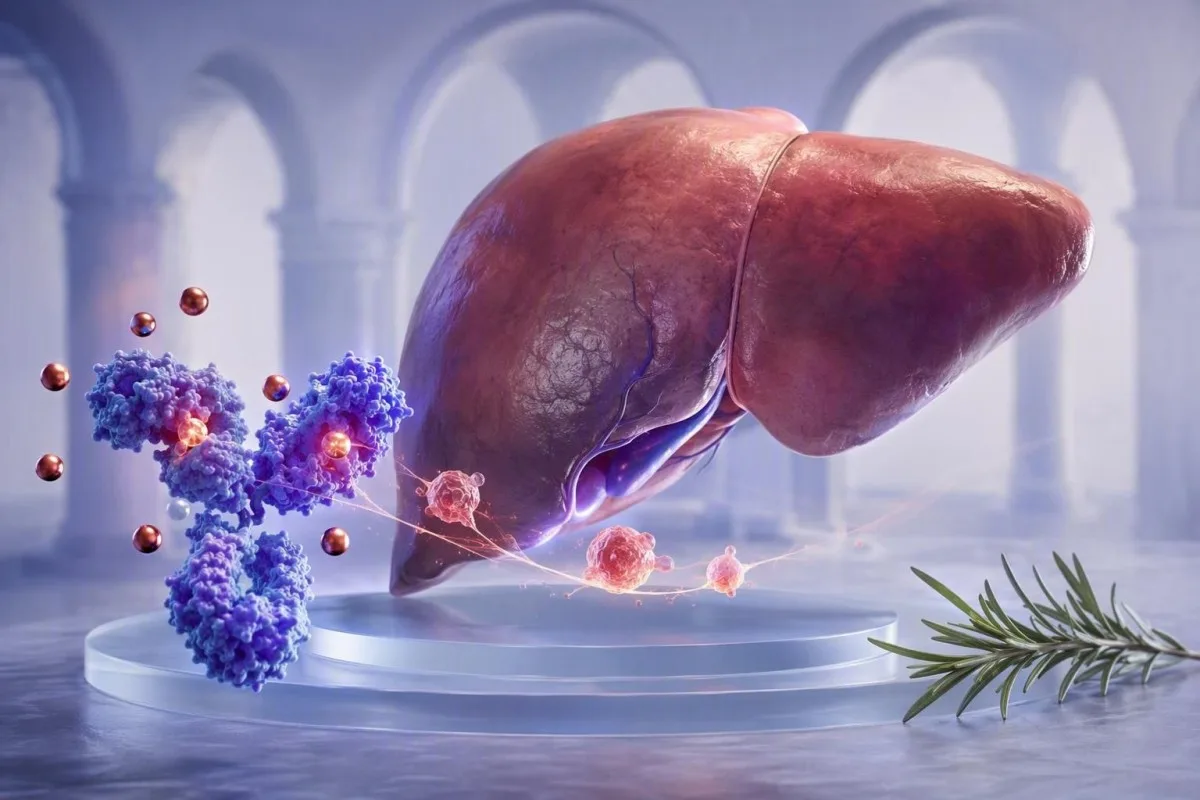
Lægur transferrín-niðurstaða getur endurspeglað viðbrögð lifrarinnar við bólgu frekar en tæmd járnbú. Lestur sermisjárns, ferritíns, CRP og transferríns saman kemur í veg fyrir algeng mistök.


- Neikvætt bráðafasa prótein: Transferrín lækkar algengt við sýkingar, sjálfsofnæmisvirkni, skurðaðgerðir og önnur bólguástand, jafnvel þegar líkamsjárnbú er fullnægjandi.
- Dæmigert viðmið fullorðinna: Flest rannsóknastofur nota transferrín viðmiðunarbilið nálægt 200-360 mg/dL (2,0-3,6 g/L), en staðbundin próf hafa forgang.
- TSAT útreikningur: Transferrínmettun jafngildir sermisjárni deilt með TIBC margfaldað með 100; lágt TIBC getur gert mettunina líta minna óeðlilega út en búist var við.
- Ferritín varúðarráðstöfun: Ferritín hækkar við bólgu, svo ferritín undir 30 ng/mL styður sterkt járnskort, en gildi 100 ng/mL útilokar það ekki áreiðanlega þegar CRP er hækkað.
- Gagnlegt meðfylgjandi próf: Leysanlegur transferrínviðtaki hefur venjulega minna áhrif á bólgu en ferritín eða transferrín og getur hjálpað til við að skýra blandað blóðleysi.
- Tímasetning endurmats: Fyrir ekki bráðar frávik, að endurtaka járnrannsóknir um 2-4 vikum eftir að skammvinn veikindi hafa lagast gefur oft betra fulltrúa grunnlína.
- Ekki meðhöndla sjálfan þig blindandi: Járnsuppbót getur verið gagnleg við staðfestan skort en getur verið óviðeigandi við járn ofhleðslu, virkan lifrarsjúkdóm eða blóðleysi sem aðallega stafar af bólgu.
Af hverju lækkar transferrínpróf við bólgu?
Bólgur lækka transferrín vegna þess að transferrín er neikvætt bráðafasa prótein. Sýtókín, sérstaklega interleukin-6, gefa lifrinni merki um að draga úr framleiðslu transferríns á meðan hepcidin takmarkar losun járns í plasma; þetta getur leitt til lágs transferríns og lágs sermisjárns án þess að sanna að járnbú séu tóm.
Stutta útgáfan er sú að líkaminn gerir járn tímabundið minna aðgengilegt við ónæmisvirkjun. Hepcidin dregur úr ferroportin-miðluðu járnútfæri frá þarmfrumum og átfrumum, svo sermisjárn lækkar oft innan 24-48 klukkustunda; lifrin getur samtímis dregið úr transferrín myndun. Þetta mynstur er svar líkamans til varnar, ekki bein mæling á inntöku járns úr fæðu.
Í klínísku starfi mínu getur einstaklingur með CRP upp á 68 mg/L eftir bakteríulungnabólgu haft sermisjárn 22 µg/dL og transferrín 165 mg/dL, samt sem áður ferritin 240 ng/mL. Að kalla þetta einangraða mynstur “járn ofhleðslu” eða útiloka með vissu járnskort væru bæði mistök. Járnrannsóknir leiðbeiningar útskýrir undirliggjandi útreikninga ítarlegar.
Kantesti er AI blóðprufugreiningartæki sem lesir transferrín við hlið CRP, ferritins, blóðkornavísitala, lifrargreina og fyrri niðurstöðu frekar en að meðhöndla eitt lágt gildi sem greiningu. Hagnýt regla Dr. Thomas Klein er einföld: transferrín niðurstaða sem fengin er við hita, blossa eða bata ætti að merkjast sem samhengisbundin áður en nokkur meðferðarákvörðun er tekin.
Bráðafasa stefnan skiptir máli
CRP, ferritin, fibrínógen og haptóglóbín hækka almennt sem jákvæð bráðafasa prótein, en transferrín og albumin geta lækkað. CRP yfir 10 mg/L gerir einstaka ferritíngildi verulega erfiðara að túlka, þó að enginn alhliða CRP þröskuldur geti leiðrétt hverja niðurstöðu sjúklings.
Hver eru eðlileg mörk transferríns og TIBC?
Transferrín fullorðinna er almennt á bilinu um 200-360 mg/dL, og TIBC er almennt á bilinu um 250-450 µg/dL. Þessi bil eru mismunandi eftir rannsóknaraðferð, kyni, meðgönguástandi og staðbundnum tilkynningareiningum, svo bil sem prentað er við hlið eigin niðurstöðu er það sem á að nota.
Transferrín er venjulega mælt beint í mg/dL eða g/L, en heildar járn bindandi geta, eða TIBC, áætlar hversu mikið járn lauslegt transferrín getur bundið. Margar rannsóknastofur leiða TIBC af transferrín frekar en að mæla það sérstaklega; gróft umreikningur er TIBC í µg/dL jafngildir transferrín í mg/dL margfaldað með 1.25. Þetta samband er gagnlegt til viðmiðunar, ekki til að hunsa eigin útreikning rannsóknarstofu.
Transferrín mettun, skammstafað TSAT, er reiknuð sem sermisjárn deilt með TIBC margfaldað með 100. TSAT nálægt 20-45% er dæmigert á mörgum rannsóknarstofum fyrir fullorðna; gildi undir 20% gefa til kynna takmarkað sirkulerandi járn, en þau greina ekki ein og sér algert járnskort frá bólgu-knúinni járn innilokun. Lág transferrínmettun fjallar um þann mun.
Sumar evrópskar rannsóknastofur tilkynna transferrín í g/L, þar sem 2.0-3.6 g/L samsvarar gróflega 200-360 mg/dL. Niðurstaða getur verið rétt fyrir neðan bil eftir veirusýkingu og normaliserast við endurtekningu, en viðvarandi gildi undir 150 mg/dL verðskulda markvissa leit að lifrar myndun vandamálum, prótein tapi, verulegri bólgu eða vannæringu.
Hvernig á að túlka járnrannsóknir sem heild?
Járnrannsóknir eru öruggastar þegar sermisjárn, transferrín eða TIBC, TSAT, ferritín og bólguviðmið eru túlkuð saman. Sermisjárn eins breytist verulega yfir daginn og getur fallið um meira en 30% milli sýna hjá sama einstaklingi.
Klassískt óbrotinn járnskortur veldur venjulega lágum sermisjárni, háu transferríni eða TIBC, TSAT undir 16%, og ferritíni undir 30 ng/mL. Hár TIBC kemur fram vegna þess að lifrin eykur framleiðslu transferríns þegar járn er lítið tiltækt. Hækkandi rauðkornadreifingarbreidd getur birst áður en blóðrauði fellur; sjá leiðbeiningar okkar um RDW breytingar eftir járnmeðferð fyrir tímaferilinn.
Blóðleysi vegna bólgu veldur oftar lágum sermisjárni, lágum eða eðlilegum transferríni, lágu TSAT, og ferritíni sem er eðlilegt eða hátt. Weiss og Goodnough lýstu þessu sem járntakmarkaðri rauðkornamyndun af völdum skertrar járnframboðs en ekki endilega fjarveru geymds járns (Weiss & Goodnough, 2005). Þessir tveir sjúkdómar koma oft saman fyrir, sérstaklega í bólgusjúkdómum í görnum, iktsýki, langvinnum nýrnasjúkdómum og krabbameini.
Eitt minna augljóst gildra er reikningsfræði: ef sermisjárn er 30 µg/dL og TIBC er lækkað í 180 µg/dL, er TSAT 17%. Ef TIBC væri 360 µg/dL, myndi sama sermisjárn gefa 8%. Lægri nefnarinn getur dulið hversu lítið járn er á sveimi, þess vegna lágt sermisjárn þarf samhengi fremur en sjálfvirka ávísun.
Hagnýt blönduð vísbending
Ferritín á milli 30 og 100 ng/mL með TSAT undir 16% og CRP yfir 10 mg/L er oft grásvæði, ekki fullvissa. Í þeim aðstæðum geta læknar notað leysanlegan transferrínviðtaka, hemóglóbíninnihald í nýmynduðum rauðum blóðkornum, tímaröðargögn, eða meðferðaráætlun sniðna að undirliggjandi sjúkdómi.
Af hverju ferritín getur litið eðlilegt út þegar járn er lítið
Ferritín er bæði járn-geymsluprótein og jákvætt bráðfasaefni, svo bólga getur hækkað það óháð geymdu járni. Ferritín undir 30 ng/mL styður sterklega járnskort hjá flestum fullorðnum, en hærra gildi getur ekki áreiðanlega útilokað skort þegar CRP eða ESR er hækkað.
Leiðbeiningar Alþjóðaheilbrigðismálastofnunarinnar frá 2020 um ferritín ráðleggja mælingu á bólguviðmiðum ásamt ferritíni þar sem sýking eða bólga er algeng. Hjá fullorðnum með bólgu, leggur WHO til að ferritín undir 70 µg/L geti bent til járnskorts; það er þjóðfélags upplýst viðmið, ekki staðgengill fyrir einstaklingsbundið klínískt mat (WHO, 2020).
Ég sé oft sjúklinga sem eru hræddir við ferritín upp á 180 ng/mL eftir bólguaukningu, og telja að járnbirgðir þeirra verði að vera miklar. Stundum eru þær það. Samt getur ferritín hækkað eftir erfiða líkamsrækt, lifrarskaða, efnaskiptasjúkdóm, útsetningu fyrir áfengi og ónæmisvirkni, svo talan er ekki einangruð birgðatalning. Ferritín og CRP er gagnleg samræðufélagi.
Kantesti er AI blóðrannsóknartúlkunarvettvangur sem bendir til ósamræmdrar samsetningar af lágum transferríni, lágum TSAT og hækkuðu CRP sem mögulegri bólgu-tengdri járntakmörkun. Gagnasett okkar frá 100.000 notendum er ekki staðgengill fyrir rannsóknarannsóknir, en það styrkir ítrekað gamlan kennslustund læknis: ferritín hegðar sér öðruvísi þegar sjúklingurinn er virkur veikur.
Ferritín gildi sem þurfa annað samtal
Ferritín yfir 1.000 ng/mL krefst tímanlegra læknisfræðilegra athugana vegna þess að alvarleg bólga, marktækur lifrarskaði, járn-ofhleðsluheilkenni og nokkrir aðrir sjúkdómar geta framkallað það svið. Bráðleiki fer eftir einkennum, lifrarensímum, transferrínmettun og hraða breytinga fremur en ferritíntölunni einni.
Hvaða próf skýra járnskort við bólgu?
Leysanlegt transferrínviðtaka og retíklóma hemóglóbíninnihald geta hjálpað til við að greina járn-takmarkaða rauðkornaframleiðslu þegar ferritín og transferrín eru ósammála. Leysanlegt transferrínviðtaka hækkar almennt með frumu-járnþörf og er minna raskað af bráðri bólgu en ferritín.
Leysanlegt transferrínviðtaka, eða sTfR, endurspeglar tjáningu transferrínviðtaka frá rauðkornaforsitum. Það hækkar venjulega við algjört járnskort og er oft eðlilegt við hreinan blóðleysi af völdum bólgu; þó eru viðmiðunarmörk mælinga ekki staðlað, og blóðskilun eða mikil rauðkornaframleiðsla getur hækkað það. Þetta er eitt af þeim svæðum þar sem nákvæm rannsóknaraðferð skiptir meira máli en viðmiðunarmörk á netinu.
Retíklóma hemóglóbíninnihald, tilkynnt sem CHr eða Ret-He eftir greiningartæki, endurspeglar járn sem er tiltækt nýframleiddum rauðkornum yfir um það bil síðustu 2-4 daga. Gildishlutföll undir um það bil 28-30 pg geta stutt járn-takmarkaða rauðkornaframleiðslu, þó staðbundin viðmiðunarmörk og siðareglur nýrnasjúkdóma séu mismunandi. Prófun á leysanlegu transferrínviðtaka útskýrir hvar það passar inn.
Járnlitað beinmerg er áfram söguleg viðmiðunaraðferð en er sjaldan nauðsynleg eingöngu til að leysa venjulegt útivistarlæknis járnspjald. Dr. Thomas Klein kýs almennt að endurtaka sýni eftir bata, skoða blæðingasögu og fæðusögu, og nota sTfR eða retíklóma mælingar þegar svarið mun sannarlega breyta meðferð; aukaprófanir án ákvörðunar til viðbótar hafa tilhneigingu til að skapa hávaða.
sTfR-ferritínvísitalan
Sumir sérfræðingar reikna sTfR/log ferritínvísitöluna, sem getur bætt aðgreiningu í bólguástandi. Viðmiðunarmörk eru mjög mismunandi eftir greiningartækjum, oft frá um það bil 1,0 til 3,2, svo niðurstöðu ætti að túlka með rannsóknarstofu-staðfestri aðferð frekar en með fengnu netviðmiðunarmörkum.
Hvenær á að endurtaka transferrínpróf?
Transferrínpróf er best að endurtaka eftir að skammlíf sýking, hiti eða meiriháttar bólguviðburður hefur lægt, venjulega eftir 2-4 vikur ef ástandið er ekki brátt. Morgunsöfnun og samfelldar aðstæður fyrir prófun draga úr óþarfa breytileika í sermisjárni og TSAT.
Sermisjárn hefur sólar-breytileika og getur verið lægra síðar um daginn, en nýlegt inntöku-járn getur tímabundið hækkað það. Margir læknar biðja um morgunsýni eftir 8-12 klst. föstu yfir nóttina þegar þeir þurfa áreiðanlegan samanburð, þó að fasta sé ekki nauðsynleg fyrir hverja járnprófun. Fylgdu fyrirmælum læknis frekar en að hætta ávísuðum lyfjum á eigin spýtur.
Transferrínniðurstöðu sem tekin er 48 klukkustundum eftir maraþon, tannlæknaaðgerð, bólusetningu eða bráða öndunarfærasjúkdóm getur endurspeglað bráða-áfanga svar frekar en varanlegt járnmynd. Hreyfing getur einnig breytt CK, plasma rúmmáli og ferritíni; íþróttamenn í þreki geta fundið leiðarvísir okkar um prófun á járni hjá hlaupurum gagnlegt.
TREND Greining Kantesti ber saman dagsetningar, ekki einfaldlega viðmiðunarmerki, og getur bent á lækkun úr 310 mg/dL í 180 mg/dL sem fellur saman við hækkun CRP úr 1 í 52 mg/L. Það er klínískt upplýsandi en hvor einasta niðurstaða. Endurskoða TIBC prófunar undirbúningur áður en skipulögð endurtekin prófun er gerð.
Hvenær á ekki að bíða eftir endurtekningu
Fresta ekki læknisúttekt fyrir endurtekna prófun ef alvarleg öndunarerfiðleiki, brjóstverkur, yfirlið, svartar hægðir, miklar áframhaldandi blæðingar, gulusótt eða hratt versnandi máttleysi er til staðar. Hemóglóbín undir 80 g/L (8 g/dL) er oft klínískt marktækt, en brýnt skiptir máli eftir einkennum, hraða lækkunar, meðgönguástandi og hjarta- og æðasjúkdómum.
Hvernig útskýra CRP, ESR og hepcidin lágt transferrín?
CRP og ESR mæla ekki járnbirgðir, en þau sýna hvort bólga gæti raskað transferrínprófi. CRP hækkar og lækkar á klukkustundum til dögum, á meðan ESR getur verið hækkað í vikur vegna þess að það hefur áhrif á fibrinogen, blóðleysi, aldur og ónæmisglóbúlín.
Interleukin-6 örvar lifrarframleiðslu hepcidins, og hepcidin binst ferroportin, sem veldur minni járnútflutningi frá átfrumum og þekjufrumum í þörmum. Endurskoðun Ganz og Nemeth lýsir þessari leið sem miðlægri fyrir blóðleysi vegna bólgu og járnþröngsins í rauðkornamyndun (Ganz & Nemeth, 2012). Afleiðingin getur verið lágt sermisjárn innan dags á meðan transferrin lækkar sem hluti af sama kerfisbundna viðbragðinu.
CRP undir 5 mg/L er oft talið innan viðmiðunarmarka, þó að rannsóknarstofur mismuni. CRP upp á 40 mg/L greinir ekki orsök bólgu, en það ætti að gera lækni varkárari við að greina járnskort út frá ferritíni eða transferríni einu sér. Orsakir hás ESR útskýrir hvers vegna ESR er hægari og óákveðnari.
Kantesti AI túlkar transferrin niðurstöður með því að bera saman stefnu CRP, ferritíns, albúmíns, hvítfrumnafjölda og nýlegra þróunar. Þetta er ekki greiningarvél fyrir sýkingar eða sjálfsofnæmissjúkdóma; þetta er skipulögð ábending til að spyrja hvort járnspjaldið hafi verið fengið á líffræðilega óstöðugu augnabliki.
Af hverju hepcidin er ekki reglulega mælt
Hepcidin próf eru enn takmörkuð vegna framboðs, stöðlunar og viðsnúningstíma í venjulegri læknisfræði. Hepcidin gildi getur verið upplýsandi í sérfræðirannsóknum eða óvenjulegum tilfellum blóðleysis, en sermis ferritín, TSAT, CRP, nýrnastarfsemi og blóðtalningarvísitölur eru enn hagnýt fyrsta línu verkfæri.
Hvernig er blóðleysi vegna bólgu frábrugðið járnskorti?
Fullkominn járnskortur þýðir að líkamsjárn er ófullnægjandi, en blóðleysi vegna bólgu þýðir að járn er til staðar en illa tiltækt fyrir framleiðslu rauðkorna. Báðir geta valdið þreytu, lágu TSAT, og lækkandi blóðrauða, og þeir koma oft saman.
Við óbrotinn járnskort er ferritín venjulega lágt og transferrin hækkar oft yfir 360 mg/dL þegar bindigetu eykst. Við blóðleysi vegna bólgu fellur transferrin algengt undir 200 mg/dL, ferritín er oft yfir 100 ng/mL og CRP getur verið hækkað. Enginn munstur er algildur; langvinnur nýrnasjúkdómur og lifrarsjúkdómur eru sérstaklega líklegir til að skarast.
Blóðrauði og MCV geta dregist eftir járnþröng. Einstaklingur getur haft ferritín 18 ng/mL, TSAT 14%, og eðlilegan blóðrauða 132 g/L, sérstaklega snemma í skorti; öfugt, bólga getur valdið blóðleysi með eðlilegu MCV frá 82-100 fL. Hvað blóðrauði þýðir hjálpar til við að setja CBC við hlið járnrannsókna.
Ástæðan fyrir því að við höfum áhyggjur af lágu transferríni ásamt lágu albúmíni er sú að saman benda þau annaðhvort á sterkara bólgubyggði, skerta lifrarstarfsemi eða próteintap, en lágt transferrin eitt eftir kvef er oft tímabundið. Læknir ætti að endurskoða nýrnastarfsemi, þvagsprótein, lifrarpróf, næringu, lyf, blæðingasögu og þróun yfir að minnsta kosti tvær sýnatökur.
Meðferð er ekki skiptanleg
Munnlegt járn bætir oft algildan skort, en það getur haft takmörkuð áhrif á meðan bólga heldur hepcidin háu. Í völdum sjúkdómum, eins og langvinnum nýrnasjúkdómum eða virkum bólgusjúkdómum í þörmum, geta læknar notað bláæðajárn eða meðhöndlað bólguna fyrst; nálgunin fer eftir greiningu, einkennum, blóðrauða og öryggissjónarmiðum.
Af hverju lágt transferrín getur brenglað transferrínmettun
Lágt transferrin getur gert transferrin mettun minna lágt vegna þess að TSAT notar TIBC sem nefnara. Eðlilegt eða lítið lágt TSAT þýðir ekki alltaf að járnflutningur sé fullnægjandi þegar TIBC er bælt af bólgu eða lifrarstarfsemi.
Hugleiddu sermisjárn 36 µg/dL. Með TIBC 360 µg/dL, TSAT er 10%; með TIBC 180 µg/dL, TSAT er 20%. Seinni niðurstaðan lítur minna áhyggjuefni út stærðfræðilega, en báðar sýnin innihalda sama lága járnmagn í blóði. Þess vegna ættu læknar að skoða hrágildin, ekki aðeins prósentuna.
Andstæða gildran kemur fram við langt gengið lifrarskaða eða bráða lifrarfrumuáverka: transferrin framleiðsla getur lækkað og sermisjárn getur hækkað vegna breyttrar meðhöndlunar, sem leiðir til hækkaðs TSAT. TSAT sem varir yfir 45% verðskuldar mat vegna járn ofhleðslu í réttu samhengi, en það ætti ekki að nota til að greina arfgenga blóðfrumuheilkenni við bráða lifraratvik. Lestu okkar lifrarhnútar í blóðprufum fyrir víðara lifrar samhengi.
Kantesti er AI-knúið blóðprófunargreiningartól notað í 127+ löndum, svo það staðalíðar einingar áður en mettun er reiknuð og merkir niðurstöður sem gætu verið stærðfræðilega óstöðugar vegna þess að TIBC er óvenju lágt. Útkomulagið ætti að styðja, ekki skipta út, lækninum sem veit hvort sjúklingur er með hita, lifrarbólgu, nýrnaheilkenni eða nýlegri járnameðferð.
Notið ekki TSAT sem rakastigsvísi
Vöknun getur þétt nokkrar sermismælingar en skapar ekki áreiðanlegt mynstur járn yfir mælingar. Ef albúmín, hematókrit, þvagefni og natríum benda til minni plasmavolúms, getur það verið skynsamlegt að endurtaka greininguna eftir eðlilegt rakastig áður en gefa merkingu takmörkuðu TSAT gildi.
Hvað annað veldur lágu transferríni fyrir utan bólgu?
Lágur transferrín kemur einnig fram við minnkaða lifrarframleiðslu, próteintap í gegnum nýru eða þörmum, ófullnægjandi prótein- og orkuinntöku og sjaldan meðfædda sjúkdóma. Bólga er algeng, en hún ætti aldrei að verða alhliða skýring án þess að athuga restina af greiningunni.
Lifrin framleiðir transferrín, svo lágt transferrín með aukinni bilirúbín, INR, AST, ALT, eða lágu albúmíni getur bent til lifrarsjúkdóms frekar en járnstaðu. Alvarleg lifrarbilun getur lækkað transferrín undir 150 mg/dL. Lifrarprófsgildi hjálpa til við að ákvarða hvort sú skýring sé líkleg.
Próteintap á nýrnaheilkennisviðmiðum getur fjarlægt transferrín ásamt albúmíni og öðrum próteinum. Albúmín-kreatínín hlutfall í þvagi yfir 300 mg/g, eða 30 mg/mmol, er alvarlegt aukinn albúmínúria og kallar á nýrna-miðaða mati; prótein stýripinnar einn er ekki nóg fyrir fullt svar. Sjá leiðbeiningar okkar um prótein í þvagi.
Lítil inntaka, vanfrásog, eða alvarleg katabólísk veikindi geta lækkað transferrín, þó að transferrín sé of næmis við bólgu til að þjóna sem næringarvísi einn sér. Meðfæddur ofurtransferrínæmi er afar sjaldgæft og kemur venjulega fram miklu fyrr á lífsleiðinni með alvarlegri blóðleysi og þversögn kerfisbundinni járnhleðslu. Þessi óvenjulega samsetning krefst sérfræðingameðferðar.
Lyfja- og hormónaáhrif
Estrogen útsetning og meðganga geta aukið transferrín, en andrógen geta lækkað það lítillega. Þessar breytingar eru venjulega minni en áhrif verulegrar bólgu eða lifrarsjúkdóms, en þær geta útskýrt takmörkuðu gildi þegar restin af járnprófinu er stöðug.
Hvernig breyta meðganga og blóðmissir frá tíðablæðingum transferríni?
Meðganga eykur oft transferrín og TIBC, á meðan járnþörf eykst mest á öðrum og þriðja þriðjungi meðgöngu. Því gefur lágt transferrín ástand á meðgöngu sérstaka athygli að bólgu, lifrarstarfsemi, próteintapi og rannsóknarstofu samhengi.
Plasma rúmmál eykst á meðgöngu, og estrógen eykur transferrín framleiðslu, svo TIBC eykst oft yfir það sem er í ófrjóum konum. Ferritín gengur einnig venjulega niður meðan á meðgöngu stendur; ferritín undir 30 µg/L er oft notað til að greina tæmda birgðir á meðgöngu, þó staðbundnar leiðbeiningar um mæðravernd geti verið mismunandi. Ferritín eftir þriðjungi meðgöngu veitir hagnýtar viðmiðanir.
Miklar tíðablæðingar eru algeng orsök algers járnskorts, sérstaklega þegar ferritín er undir 30 ng/mL og transferrín er aukið frekar en lækkað. Samt sem áður getur einstaklingur með sjálfsofnæmissjúkdóm og miklar blæðingar haft bæði blóðmissis skort og bólgu; “eðlilegt” ferritín upp á 75 ng/mL leysir ekki úr vafa þegar CRP er 24 mg/L.
Nýtt lágt transferrín með háan blóðþrýsting, bólgu, próteinúríu, höfuðverk, verk í efri hægri hluta kviðar, eða óeðlilegar lifrartölur á meðgöngu þarf tafarlaust fæðingar-mat. Það er ekki leið til að greina meðgöngueitrun, en víðara prótein og lifrar munstur getur skipt miklu meira máli en eingöngu járnáhrif.
Hvernig er transferrín túlkað við nýrnasjúkdóma og langvinna sjúkdóma?
Langvinnur nýrnasjúkdómur veldur oft virknilegum járnskorti þar sem bólga og minnkað ertrópóetín takmarka nýtanlegt járn fyrir framleiðslu rauðra blóðkorna. Í þessu samhengi styðja TSAT undir 20% og ferritín undir 100 ng/mL oft járnskort fyrir blóðskilun, þó meðferðarþröskuldar breytilegir eftir leiðbeiningum og klínísku samhengi.
KDIGO leiðbeiningar hafa sögulega notað tilraun með járni í mörgum fullorðnum með CKD þegar TSAT er á eða undir 30% og ferritín er á eða undir 500 ng/mL, að því tilskildu að klínískt markmið sé að auka blóðrauða eða draga úr ertrópóese-örvandi meðferð. Þetta eru meðferðaratriði, ekki alhliða skilgreiningar á eðlilegum járnbirgðum, og 500 ng/mL þak er oft misskilið.
Sjúklingur með eGFR 28 mL/min/1.73 m², blóðrauða 96 g/L, TSAT 16%, ferritín 220 ng/mL, og CRP 12 mg/L getur haft virknilegan skort þrátt fyrir ekki lágt ferritín. Oral járnupptaka getur verið skert, og læknar verða einnig að meta B12, fólínsýru, dulið tap, notkun ertrópóetíns, og nýrnaferil. Stigun CKD veitir gagnlegan bakgrunn.
Kantesti gervigreind getur sett transferrín í langtímasamhengi við nýrnaheilsu, en hún getur ekki ákvarðað hvort meðferð með járni í æð, meðferð sem örvar rauðkornamyndun eða tilvísun til sérfræðings sé viðeigandi. Sú ákvörðun krefst upplýsinga um einkenni, blóðþrýsting, sýkingarstöðu, yfirferðar á lyfjum og fullkominnar skráningar um nýrnastarfsemi.
Hvaða upplýsingar úr sjúkrasögu gera transferrín-niðurstöðu gagnlegri?
Gagnlegasta túlkun á transferríni tekur til einkenna, nýlegra veikinda, tíðablóðmissis eða blóðmissis úr meltingarvegi, mataræðis, áfengisneyslu, lyfja, hreyfingar og fyrri mæligilda. Eitt sýni er skyndimynd; tvö sambærileg sýni sýna oft hvort breytingin sé lífeðlisfræðileg, bólgutengd eða viðvarandi.
Spyrjið hvort hiti, bólusetning, meiðsli, sjúkrahúsinnlögn, tannlæknameðferð, mikil þjálfun eða þekkt versnun hafi átt sér stað á 14 dögum fyrir sýnatökuna. Skráið einnig járntöflur, fjölvítamín, prótónpumpuhemla, bólgueyðandi gigtarlyf, blóðþynningarlyf og nýlegar breytingar á mataræði. Þessar upplýsingar geta haft áhrif annaðhvort á upptöku járns eða líkur á duldu blóðtapi.
Breyting úr transferríni 290 í 205 mg/dL skiptir meira máli ef CRP hækkaði úr 2 í 35 mg/L og albúmín lækkaði úr 44 í 37 g/L. Aftur á móti getur 10 mg/dL breyting með stöðugu CRP og sama tímasetningu sýnatöku verið innan líffræðilegs og mælitæknilegs breytileika. Samanburður á blóðprufum útskýrir hvers vegna litlar breytingar eru ekki alltaf raunverulegar breytingar.
Fjöltyngd skráningartól Kantesti gera fólki kleift að vista samhengi við hlið niðurstöðu, þar á meðal dagsetningar veikinda og breytingar á fæðubótarefnum, á 75+ tungumálum. Þessi saga gerir yfirferð heilbrigðisstarfsmanns öruggari; okkar leiðbeiningar um blóðpróf yfir tíma telur upp þau hagnýtu atriði sem vert er að varðveita.
Einkenni eru ekki sértæk
Þreyta, skert úthald við áreynslu, hárlos, fótaóeirð og einbeitingarskortur geta komið fram við járnskort, bólgu, skjaldkirtilssjúkdóm, truflaðan svefn, þunglyndi og marga aðra sjúkdóma. Einkenni ættu að leiða til viðeigandi mats, en þau geta ekki sagt til um hvort lágt transferrín endurspegli tæmdar járnbirgðir.
Hvenær þarf skjótra yfirferðar á lágu transferrín-niðurstöðu?
Lágt transferrín krefst skjóts læknismats þegar það kemur fram samhliða verulegu blóðleysi, gulu, bjúg, miklum blæðingum, svörtum hægðum, óútskýrðu þyngdartapi eða vísbendingum um skerta nýrna- eða lifrarstarfsemi. Niðurstaðan sjálf er sjaldan neyðartilvik, en ástandið sem býr að baki getur stundum verið það.
Mat samdægurs er skynsamlegt við brjóstverk, yfirlið, verulega mæði, ringlun, svartar tjörukenndar hægðir, blóðuppköst eða hratt vaxandi bjúg. Blóðrauði undir 70 g/L (7 g/dL), bílirúbín yfir 50 µmol/L ásamt gulu eða óvænt hátt INR krefst einstaklingsbundins klínísks mats og gæti þurft brátt mat. Ekki reyna að leiðrétta þessi mynstur með járni sem fæst í lausasölu einu saman.
Við stöðugt, vægt lækkað transferrín, 185 mg/dL, með eðlilegum blóðrauða, eðlilegum lifrarprófum og CRP 18 mg/L meðan á skjalfestri öndunarfærasýkingu stendur, er oft skynsamlegt að endurtaka rannsóknir eftir bata. Ef það helst við í meira en 6–8 vikur víkka læknar oft matið til að taka til lifrarprófa, próteins í þvagi, næringarsögu, virkni bólgusjúkdóms og uppsprettna blóðmissis.
Klínískt efni okkar er yfirfarið með stuðningi Læknisfræðileg ráðgjafarnefnd, og túlkunarrökfræði Kantesti er skjalfest í gegnum læknisfræðilegt staðfestingar- og matsrammi. Frá og með 5. september 2026 stendur öruggasta skilaboðið óbreytt: lág niðurstaða fyrir transferrín er vísbending um flutning járns og altæka lífeðlisfræði, ekki greining ein og sér.
Algengar spurningar
Getur bólga valdið lágu transferríni án járnskorts?
Já. Bólgur geta lækkað transferrín, jafnvel þegar járnbirgðir eru fullnægjandi þar sem transferrín er neikvætt bráðfasa prótein sem lifrin framleiðir. Einstaklingur með CRP yfir 10 mg/L getur haft lágt transferrín, lágt sermisjárn og ferritín yfir 100 ng/mL vegna bólguvaktaðrar járnvöktunar fremur en hreinnar járnskorts. Járnskortur getur enn verið til staðar, sérstaklega þegar TSAT er undir 20% eða ferritín er undir 30 ng/mL. Læknir ætti að túlka heildarmynstrið og orsök bólgu.
Þýðir lágt transferrín lágt járn?
Lægt transferrín þýðir ekki sjálfkrafa lítið járn í allri líkamsgeymslu. Lægra transferrín kemur oft fram við bólgu, lifrarsjúkdóma, próteintap úr nýrum og ófullnægjandi próteininntöku, á meðan klassísk járnskortur hækkar oft transferrín yfir um það bil 360 mg/dL. Blóðjárn undir 50 µg/dL getur komið fram við annað hvort járnskort eða bólgu, svo þörf er á ferritíni, CRP, TIBC, TSAT og blóðtölu. Útkoma transferríns undir 200 mg/dL skal túlkuð í samhengi við viðmiðunarbil rannsóknastofunnar og sjúkrasögu.
Getur lágt transferrín látið transferrínmettun líta eðlilega út?
Já. Mettun transferrins er sermisjárn deilt með heildar járnbindgetu (TIBC) margfaldað með 100, svo lág TIBC vegna lágs transferrins skapar minni nefnara. Til dæmis gefur sermisjárn upp á 36 µg/dL TSAT upp á 10% við TIBC upp á 360 µg/dL en 20% við TIBC upp á 180 µg/dL. Þetta þýðir að takmörkuð TSAT getur vanmetið takmörkun á járni þegar transferrin er undir þrýstingi. Læknar ættu að skoða bæði prósentuna og hrá gildi sermisjárns og TIBC.
Hvað er ferritinmagn sem staðfestir járnskort við bólgu?
Ferritín undir 30 ng/mL eða µg/L styður sterklega járnskort hjá flestum fullorðnum, þar með talið mörgum með væga bólgu. Leiðbeiningar Alþjóðaheilbrigðismálastofnunarinnar frá 2020 benda til þess að ferritín undir 70 µg/L geti bent til járnskorts hjá fullorðnum með merki um bólgu, en þessi mörk eru ekki algild fyrir hvern einstakling. Ferritín getur hækkað með CRP hækkun, lifrarskaða og efnaskiptasjúkdómum, svo gildum milli 30 og 100 ng/mL er oft þörf á TSAT, CRP og stundum prófun á leysanlegum transferrínviðtaka. Ferritín yfir 100 ng/mL útilokar ekki alltaf járnskort þegar bólguvirkni er til staðar.
Hversu fljótt eftir veikindi á ég að endurtaka transferrínpróf?
Fyrir vægt, sjálf takmarkandi sjúkdómsástand er oft sanngjarnt að endurtaka transferrinpróf um 2-4 vikum eftir að einkenni og hiti hafa lægt, ef engin brýn einkenni eru til staðar. CRP getur lækkað innan nokkurra daga, en ESR, ferritín, albúmín og transferrín geta tekið lengri tíma að jafnast á við grunnlínugildi. Notaðu svipaðar aðstæður við endurtekna blóðsöfnun, helst á morgnana og sama rannsóknarstofu þegar mögulegt er. Hikaðu ekki við að endurskoða ef blóðrauði er að falla, blæðingar eru áframhaldandi eða lifrar- og nýrnasjúkdómar eru óeðlilegir.
Á ég að taka járninnskot vegna lágs tferssín?
Einungis lágt transferrín er ekki ástæða til að hefja járnuppbót. Talað er um að taka járn til inntöku þegar sannanir liggja fyrir um algert járnskort, svo sem ferritín undir 30 ng/mL, lágt TSAT, samhæf einkenni eða greinilega blæðingar; skammtur og tíðni ætti að vera einstaklingsbundin. Við blóðleysi vegna bólgu getur járn frásogast illa eða verið óaðgengilegt vegna hás hepcidins, og að meðhöndla undirliggjandi ástand getur skipt meira máli. Járn getur verið skaðlegt eða villandi við járn ofhleðslusjúkdóma og sum lifrarástand, svo staðfestu munstur með lækni.
Fáðu AI-knúna greiningu á blóðprufum í dag
Vertu með yfir 2 milljónir notenda um allan heim sem treysta Kantesti fyrir tafarlausa og nákvæma greiningu á blóðprufum. Hladdu upp niðurstöðum blóðrannsókna þinna og fáðu yfirgripsmikla túlkun á 15,000+ lífmerkjum á sekúndum.
📚 Tilvísuð rannsóknarútgáfa
Klein, T., Mitchell, S., & Weber, H. (2026). Forskráð, viðmiðabundin sjálfvirk tæknileg viðmiðun á Kantesti blóðprófatúlkunarvélina fyrir 100.000 gervi-prufutilvik. Kantesti AI Medical Research.
Klein, T., Mitchell, S., & Weber, H. (2026). Klínísk staðfestingarrammi v2.0 (Medical Validation Page). Kantesti AI Medical Research.
📖 Ytri læknisfræðilegar heimildir
Alþjóðaheilbrigðismálastofnunin (2020). Leiðbeiningar WHO um notkun á ferritínstyrk til að meta járnbúskap hjá einstaklingum og íbúum. Alþjóðaheilbrigðismálastofnunin.
⚕️ Fyrirvari vegna læknisfræðilegra mála
Þessi grein er eingöngu til fræðslu og felur ekki í sér læknisráðgjöf. Leitaðu alltaf til hæfs heilbrigðisstarfsmanns vegna ákvarðana um greiningu og meðferð.
E-E-A-T traustmerki
Reynsla
Læknastýrð klínísk yfirferð á vinnuferlum við túlkun rannsóknarniðurstaðna.
Sérþekking
Áhersla á rannsóknarstofulækningar: hvernig lífmarkarar hegða sér í klínísku samhengi.
Yfirvald
Skrifað af Dr. Thomas Klein með yfirferð Dr. Sarah Mitchell og próf. Dr. Hans Weber.
Traustleiki
Rökstudd túlkun byggð á gögnum með skýrum eftirfylgnileiðum til að draga úr ávörun.