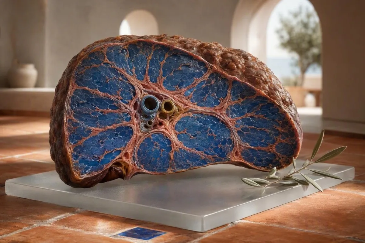
Ранні цыроз часта не выклікае відавочнага болю: стомленасць, змены сну, сінякі, сверб і зніжэнне колькасці трамбацытаў могуць з'явіцца да жаўтухі. Карысным падказкай звычайна з'яўляецца сукупнасць паказчыкаў аналізаў печані, аналізу крыві і балаў фіброзу, а не адзін адхілены фермент.


- Ціхае рубцаванне можа адбывацца пры нармальных ALT і AST, асабліва пры метабалічным захворванні печані, выкліканым тлушчам, і захворваннях печані, звязаных з алкаголем.
- Трамбацыты ніжэй за 150 × 10⁹/л можа быць раннім прыкметай партальнай гіпертэнзіі, калі яны паступова падаюць разам з фактарамі рызыкі захворвання печані.
- FIB-4 ніжэй за 1,3 звычайна сведчыць аб нізкай рызыцы развіцця фіброзу ў дарослых ва ўзросце да 65 гадоў; 2.67 або вышэй патрабуе неадкладнай кансультацыі спецыяліста.
- Альбумін ніжэй за 35 г/л або павышэнне INR можа сведчыць аб зніжэнні сінтэтычнай функцыі печані, нават калі ўзровень амінатрасфераз нязначны.
- AST:ALT вышэй за 1 не з'яўляецца дыягнастычным прыкметай цырозу, але выклікае занепакоенасць у спалучэнні з трамбацытапеніяй, павышэннем GGT або зменамі на выявах.
- Білірубін вышэй за 34 мкмоль/л з жаўтухай, цёмнай мочой, ліхаманкай, спутанностью свядомасці або азызлосцю жывата патрабуе тэрміновай клінічнай ацэнкі.
- Нармальныя ферменты азначаюць адсутнасць актыўнага вылучэння ферментаў у дадзены момант; яны не даказваюць, што архітэктура печані нармальная.
- Адмова ад алкаголю, лячэнне метабалічных фактараў рызыкі, лячэнне вірусных гепатытаў і перагляд медыкаментаў могуць запаволіць або часам часткова адмяніць фіброз на ранніх стагоддзях.
Тонкія раннія сімптомы цырозу, якія патрабуюць праверкі печані
Раннія сімптомы цырозу часта нязначныя і нерэгулярныя, прычым стомленасць, зніжэнне апетыту, парушэнне сну, сверб, лёгкае ўтварэнне сінякоў або зніжэнне пераноснасці фізічных нагрузак узнікаюць задоўга да жоўтай скуры ці затрымкі вадкасці. На мой клінічны досвед, трывожным сімптомам з'яўляецца не адзін стомлены дзень; гэта ціхі комплекс сімптомаў, які захоўваецца ад 6 да 12 тыдняў у чалавека з дыябетам, атлусценнем, рэгулярным ужываннем алкаголю, рызыкай гепатыту або сямейнай гісторыяй захворвання печані.
Чалавек можа працаваць, трэніравацца і выглядаць цалкам здаровым, пакуль прагрэсуе фіброз. Я бачыў 48-гадовага офіснага работніка, у якога адзінай зменай было абуджэнне ў 3 гадзіны ночы са свербам і стомленасцю; яго ALT складаў 31 МЕ/л, але колькасць трамбацытаў за чатыры гады знізілася з 214 да 136 × 10⁹/л. Гэтая тэндэнцыя прымусіла да эластаграфіі і выявіла запушчаны фіброз.
Цыроз звычайна не выклікае рэзкай болю ў печані. Плёнка печані можа расцягвацца на позніх стадыях працэсу, але ранні фіброз з'яўляецца біялагічна ціхім, паколькі фіброз развіваецца паміж мікраскапічнымі сасудзістымі каналамі. Новае адчуванне паўнаты ў жываце, ранняя насычанасць або азызласць лодыжак больш трывожныя, чым нявызначаны боль справа, асабліва калі вага павялічваецца больш чым на 2 кг за тыдзень.
Доктар Томас Кляйн раіць прыносіць папярэднія вынікі, а не толькі апошнюю панэль, на прыём да спецыяліста па печані. А базавы аналіз крыві становіцца значна больш карысным, калі яго можна параўнаць з вынікамі за 12, 24 і 48 месяцаў раней.
Сімптомы, якія не павінны чакаць руціннага прыёму
Спутаннасць свядомасці, новая дзённая дрымотнасць, ваніты крывёю, чорны крэсла, ліхаманка з азызласцю жывата або хуткае павелічэнне жаўтухі патрабуюць тэрміновай ацэнкі ў той жа дзень. Гэта можа адлюстроўваць дэкампенсаваны цыроз, страўнікава-кішачныя крывацёкі, інфекцыю або вострае пашкоджанне печані, а не бясшкодныя ваганні.
Чаму нармальныя ALT і AST не выключаюць цыроз
Нармальныя ALT і AST не выключаюць значны фіброз або цыроз. ALT і AST адлюстроўваюць бягучае вылучэнне ферментаў клеткамі, у той час як фіброз адлюстроўвае назапашанае архітэктурнае рубцаванне, якое можа прывесці да невялікага пастаяннага вылучэння пасля зніжэння масы клетак печані і запалення.
Многія лабараторыі выкарыстоўваюць верхнія межы ALT каля 35-45 МЕ/л для мужчын і 25-35 МЕ/л для жанчын, хоць рэферэнтныя інтэрвалы адрозніваюцца ў залежнасці ад аналізу і краіны. Вынік 28 МЕ/л можа быць нармальным на паперы, але ўсё яшчэ менш супакойвае ў чалавека з атлусценнем, цукровым дыябетам 2 тыпу, колькасцю трамбацытаў 128 × 10⁹/л і высокім балам FIB-4.
Кіраўніцтва AASLD 2023 гаворыць, што амінатрасферазы могуць быць нармальнымі ў пацыентаў з запушчанай неалкагольнай тлушчай печані; таму стратыфікацыя рызыкі павінна ўключаць неінвазіўнае тэсціраванне на фіброз, а не толькі ферменты (Rinella et al., 2023). Гэта адна з тых абласцей, дзе больш важны шаблон, чым сцяжок побач з адным значэннем.
Інтэнсіўныя трэніроўкі, пашкоджанне цягліц і некаторыя лекі могуць павысіць AST без рубцавання печані. 52-гадовы марафонца з AST 89 МЕ/л пасля гонкі можа мець мышачную крыніцу, асабліва калі креатінкіназа высокая, а білірубін, трамбацыты, INR і GGT нармальныя; наша кіраўніцтва па высокім AST тлумачыць гэтую розніцу.
Аналізы крыві на цыроз: сукупнасць паказчыкаў, якая выклікае занепакоенасць у клініцыстаў
Самая трывожная карціна аналізаў крыві на цыроз спалучае ў сабе зніжэнне колькасці трамбацытаў, перавышэнне AST над ALT, павышэнне білірубіна або GGT і парушэнне альбуміну або INR, а не ізаляванае павышэнне ферментаў. Кожны тэст адлюстроўвае розны аспект біялогіі печані: пашкоджанне, адток жоўці, парцылярны ціск або выпрацоўку бялку.
Трамбацыты ніжэй за 150 × 10⁹/л з'яўляюцца звычайна пры прасунутым фіброзе, паколькі партальная гіпертэнзія павялічвае селязёнку і спрыяе назапашванню трамбацытаў. Лік 142 × 10⁹/л не з'яўляецца доказам цырозу, аднак зніжэнне з 240 да 142 мае больш клінічнае значэнне, чым адзін ізаляваны вынік.
Суадносіны AST:ALT вышэй за 1,0 могуць узнікаць па меры прагрэсавання фіброзу, у той час як суадносіны вышэй за 2,0 класічна звязаны з алкагольным гепатытам, але не з'яўляюцца ні адчувальнымі, ні спецыфічнымі. ГГТ часта павышаецца пры ўжыванні алкаголю, метабалічным тлушчавым гепатозе, халестазе і ўплыве лекаў; чытайце больш пра што азначае ГГТ перад тым, як меркаваць, што прычынай з'яўляецца алкаголь.
Kantesti AI інтэрпрэтуе панэлі печані, узважваючы гэтыя звязаныя вынікі ў параўнанні з узростам, біялагічным полам, паведамленымі захворваннямі і папярэднімі значэннямі, а не прызначаючы дыягназ з аднаго паказчыка. Kantesti — гэта аналізатар крыві на базе AI прызначаны для вызначэння наступных шаблонаў, такіх як нізкі ўзровень трамбацытаў плюс павышанае суадносіны AST:ALT, якія патрабуюць агляду лекара.
Звычайная пастка ілжывага супакаення
Нармальны білірубін і нармальны альбумін могуць сустракацца разам з кампенсаваным цырозам. Гэтыя маркеры часта пагаршаюцца пазней, пасля таго, як печань страціла больш функцыянальнага рэзерву, таму яны з'яўляюцца дрэннымі самастойнымі скрынінгавымі тэстамі на ранняе рубцаванне.
FIB-4 і неінвазіўныя прыкметы клінічна значнага рубцавання
FIB-4 - гэта правераны першапачатковы паказчык фіброзу з выкарыстаннем узросту, AST, ALT і колькасці трамбацытаў. У дарослых ва ўзросце да 65 гадоў FIB-4 ніжэй за 1,3 звычайна паказвае на нізкі рызыку прасунутага фіброзу, у той час як вынік 2,67 або вышэй павінен паслужыць падставай для ацэнкі спецыялістам або эластаграфіі.
Формула: узрост × AST, падзелены на трамбацыты × квадратны корань з ALT, дзе AST і ALT у МЕ/л, а трамбацыты - у × 10⁹/л. Узрост істотна змяняе паказчык: EASL рэкамендуе больш высокі парог нізкага рызыкі 2,0 для людзей старэйшыя за 65 гадоў, паколькі звычайнае ўзроставае ацэньванне ў адваротным выпадку можа даць ілжывыя станоўчыя вынікі (European Association for the Study of the Liver, 2021).
Паказчык FIB-4 з'яўляецца інструментам сартавання, а не дыягназам. Востры гепатыт, нядаўняе моцнае ўжыванне алкаголю, хіміятэрапія або часовае зніжэнне колькасці трамбацытаў могуць павялічыць яго, таму я звычайна паўтараю нестабільныя тэсты праз 4-12 тыдняў перад інтэрпрэтацыяй памежнага выніку, калі ў чалавека няма жаўтухі або іншых трывожных сімптомаў.
Кантэсці — гэта сервісі інтерпретації тестів ШІ , якія могуць разлічваць і кантэкстуалізаваць FIB-4 пры наяўнасці неабходных вынікаў, але высокі паказчык патрабуе медыцынскага пацвярджэння з дапамогай транзіентнай эластаграфіі, ацэнкі спецыялістам або часам гісталагічнага даследавання. Наш агляд панэлі печані паказвае, якія тэсты звычайна даступныя.
Нізкі ўзровень трамбацытаў: ранні прыкмета партальнай гіпертэнзіі, а не прысуд
Устойлівая колькасць трамбацытаў ніжэй за 150 × 10⁹/л можа быць адным з самых ранніх паказчыкаў гіпертэнзіі порта па стандартных аналізах крыві, але яна мае шмат прычын, не звязаных з печанню. Сувязь з печанню ўзмацняецца, калі колькасць паступова зніжаецца і суправаджаецца павелічэннем селязёнкі, высокім FIB-4 або ненармальнымі візуалізацыйнымі даследаваннямі печані.
Гіпертэнзія порта змяняе кровазварот праз печань і селязёнку. Селязёнка можа ўтрымліваць больш трамбацытаў, так што колькасць ад 110 да 140 × 10⁹/л можа папярэднічаць асцыту або варыкознаму пашырэнню вен за гады пры кампенсаванай хваробе. Вось чаму тэндэнцыя трамбацытаў уключана ў FIB-4, а не разглядаецца як агульная паталёгія CBC.
Не кожны нізкі вынік адлюстроўвае сапраўднае зніжэнне. Згушчэнне трамбацытаў, звязанае з EDTA, можа памылкова знізіць колькасць, і лабараторыя можа пацвердзіць гэта аглядам мазка крыві або цытратным узорам; гл. наша тлумачэнне склейванне трамбацытаў EDTA. Вірусныя захворванні, імунная трамбацытапенія, дэфіцыт фалійнай кіслаты і захворванні касцявога мозку таксама застаюцца магчымымі.
Доктар Томас Кляйн выявіў, што зніжэнне колькасці на 20% за два гады часта больш карысна, чым пытанне, ці перавысіла яно ніжнюю мяжу лабараторыі. Злучыце яго з кампанентамі агульны аналіз крыві і папярэднімі справаздачамі УГД перад тым, як рабіць высновы.
Альбумін і INR: аналізы крыві, якія вымяраюць функцыянальны рэзерв печані
Нізкі ўзровень альбуміну і павышаны INR могуць сведчыць аб зніжэнні сінтэтычнай функцыі печані, асабліва калі яны з'яўляюцца разам. Альбумін ніжэй за 35 г/л або INR вышэй за 1.3 без антыкаагулянтнай тэрапіі патрабуе своечасовага клінічнага агляду, хоць ні адзін з вынікаў не з'яўляецца спецыфічным для цырозу.
Альбумін мае перыяд паўраспаду каля 20 дзён, таму ён змяняецца павольна і часта застаецца нармальным пры кампенсаваным цырозе. Недаяданне, страта бялку ныркамі, цяжкія сістэмныя захворванні, цяжарнасць і залішняе ўтрыманне вадкасці таксама могуць знізіць альбумін; альбумін і глабулін накіроўваюць дапамагае адрозніць гэтыя магчымасці.
INR адлюстроўвае актыўнасць фактараў згортвання, залежных ад вітаміна K, значная частка якіх выпрацоўваецца печанню. INR 1.5 у чалавека, які не прымае варфарын, выклікае больш занепакаення, чым мякка павышаны ALT, але дэфіцыт вітаміна K з-за парушэння ўсмоктвання, антыбіётыкаў або холестазу таксама можа працягнуць яго і можа быць зварачальным.
Kantesti AI — гэта Інструмент аналізу вынікаў аналізу крыві з падтрымкай AI , які падкрэслівае сукупнасць паказчыкаў альбуміну, білірубіну, INR і трамбацытаў, паколькі сінтэтычная функцыя не ахопліваецца толькі ALT. Для тэхнічнага кантэксту па аналізах згортвання азнаёмцеся з нашым даведнік па аналізах згусальнасці.
Жаўтуха, сверб, цёмная моча і светлы кал: што яны могуць азначаць
Жаўтуха, цёмная мача колеру гарбаты, светлы кал і агульны сверб могуць сведчыць аб парушэнні адтоку жоўці або парушэнні метабалізму білірубіну і павінны быць неадкладна ацэнены. Агульны білірубін вышэй за 34 мкмоль/л (прыкладна 2 мг/дл) часта становіцца бачным як жаўцізна на дзённым святле, хоць адценне скуры і асвятленне ўплываюць на вызначэнне.
Цёмная мача звычайна выклікана злучаным білірубінам, а не незлучаным білірубінам. Палачка для аналізу мачы, станоўчая на білірубін, такім чынам, накіроўвае клініцыстаў да захворвання печані і жоўцевых шляхоў або іх абструкцыі, а не простага разбурэння чырвоных крывяных цел; наш даведніку па білірубіне ў мачы тлумачыць наступныя праверкі.
Светлы або гліняны кал азначае, што менш пігменту трапляе ў кішачнік. Гэта можа адбыцца пры камянях у жоўцевым пузыры, холестазе, выкліканым лекамі, захворванні падстраўнікавай залозы або жоўцевых пратокаў, або пры цяжкай дысфункцыі печані, таму небяспечна называць гэта рэакцыяй на дэтоксікацыю; гл. прычынах бледнага кала.
Сверб ад холестазу можа быць моцным да таго, як білірубін стане прыкметна высокім. Ён звычайна дзівіць далоні і падэшвы і ўзмацняецца ноччу; клініцысты звычайна правяраюць ШФ, ГГТ, фракцыі білірубіну, уздзеянне лекаў, статус цяжарнасці, дзе гэта дастасавальна, і УГД жоўцевых пратокаў.
ALP, GGT і білірубін: распазнаванне халестатычнага шаблону
Холестатический тып печані характарызуецца павышэннем шчолачнай фасфатазы і ГГТ больш, чым АЛТ і АСТ, часта з павышэннем прамога білірубіну. Гэты тып павінен выклікаць пошук абструкцыі жоўцевых пратокаў, пашкоджанні лекамі, аутоіммуннага холестатического захворвання або інфільтратыўных захворванняў печані, а не аўтаматычнае меркаванне аб цырозе.
ШФ выпрацоўваецца тканінай жоўцевых пратокаў, а таксама косткамі, плацэнтай і кішкамі. Павышаная ШФ пры нармальнай ГГТ можа быць костнага паходжання, асабліва пасля пералому, падчас росту або пры дэфіцыце вітаміну D, у той час як высокая ШФ плюс высокая ГГТ сведчаць аб гепатабіліярнай крыніцы; наш тлумачэнне ШФ і ГГТ дае практычныя прыклады.
Каэфіцыент R дапамагае класіфікаваць вострыя ферментныя шаблоны: АЛТ, падзелены на яго верхнюю межу нормы, затым падзелены на ШФ, падзеленую на яго верхнюю межу. Каэфіцыент R вышэй за 5 сведчыць аб гепатацытарным пашкоджанні, ніжэй за 2 - аб холестазе, а ад 2 да 5 - змешаны; карысны для ацэнкі лекаў, але не з'яўляецца самастойнай дыягностыкай.
Kantesti AI выкарыстоўвае лабараторныя рэферэнтныя ліміты з загружанага справаздачы замест універсальнага парога ШФ, паколькі дыяпазоны адрозніваюцца. кіраўніцтва па аналізе крыві на холестаз з'яўляецца карыснай падрыхтоўкай да абмеркавання з клініцыстам.
Вызначэнне прычыны гэтак жа важна, як і вызначэнне рубцавання
Цыроз - гэта вынік, а не адно захворванне: алкаголь, метабалічнае тлушчавае захворванне печані, гепатыт B або C, аутоіммуннае захворванне, перагрузка жалезам і некаторыя лекі могуць выклікаць яго. Вызначэнне прычыны змяняе лячэнне, скрынінг сваякоў, назіранне за ракам і шанцы на запаволенне далейшага фіброзу.
Метабалічныя рызыкі заслугоўваюць прамой размовы. Цэнтральнае павелічэнне вагі, трыгліцерыды вышэй за 1,7 ммоль/л або 150 мг/дл, гіпертэнзія і дыябет 2-га тыпу павялічваюць верагоднасць тлушчавага захворвання печані, нават калі ўжыванне алкаголю нізкае. кантрольны спіс метабалічнага сіндрому можа ўдакладніць звязаныя рызыкі.
Феритин вышэй 300 нг/мл у мужчын або жанчын у постменопаузе недастатковы для дыягностыкі перагрузкі жалезам. Насычэнне трансферына стабільна вышэй 45% больш спецыфічнае для спадчыннай гемахроматозу і звычайна патрабуе генетычнага тэсціравання HFE у адпаведным клінічным кантэксце; гл. наша кіраўніцтва па сімптомах гемахроматозу.
Гісторыі прыёму лекаў і біялагічна актыўных дабавак руцінна недаацэньваюцца. Экстракт зялёнага гарбаты ў высокіх дозах, анабалічныя сродкі, шматкампанентныя прадукты для зніжэння вагі, аміядарон, метотрэксат і некаторыя антыбіётыкі могуць змяняць функцыю печані, у той час як так званая "ачыстка печані" можа затрымаць належную ацэнку; наш агляд бяспекі біялагічна актыўных дабавак усведамляе гэта.
Вірусны гепатыт павінен быць выяўлены, а не здагадвацца па сімптомах
Хранічны гепатыт C можа заставацца бессімптомным дзесяцігоддзямі, пакуль прагрэсуе фіброз. Тэсціраванне на антыцелы выяўляе ўздзеянне, у той час як тэст на РНК ВГС пацвярджае актыўную інфекцыю; наша кіраўніцтва па тэставанні на гепатыт C тлумачыць, чаму абедзве праверкі важныя.
Што адбываецца пасля адхіленага шаблону аналізаў крыві на цыроз
Анамальны паталагічны малюнак фіброзу звычайна прыводзіць да паўторных аналізаў крыві, УГД і эластаграфіі, а не да неадкладнай дыягностыкі цырозу. Эластаграфія ацэньвае цвёрдасць печані ў кілапаскалях і найбольш надзейная, калі ўлічваюцца вострае запаленне, застой і нядаўняе ўздзеянне алкаголю ў вялікіх дозах.
Клініцыст звычайна паўтарае аналізы AST, ALT, ALP, GGT, білірубіну, альбуміну, INR і агульнага аналізу крыві на працягу некалькіх тыдняў, калі вынікі нечаканыя. Яны могуць дадаць тэсты на гепатыт B і C, феритин з насычэннем трансферына, імунаглабуліны, аутоантыцелы, тэсты на целиакію і церулаплазмін у маладых дарослых, хоць дакладны спіс залежыць ад узросту і фактараў рызыкі.
Кіраўніцтва EASL 2021 па неінвазіўным тэсціраванні падтрымлівае двухступенчаты падыход: спачатку FIB-4, затым транзістарная эластаграфія або другі аналіз сыроваткі крыві для людзей з павышаным узроўнем рызыкі (European Association for the Study of the Liver, 2021). Цвёрдасць печані не ідэнтычная фіброзу; застой з-за сардэчнай недастатковасці, халестаз і востры гепатыт могуць часова павысіць яе.
Кантэсці — гэта платформа інтэрпрэтацыі біямаркераў AI якія могуць арганізаваць загружаную панэль у пытанні для наступнага прызначэння, у тым ліку, якія значэнні змяніліся і якія патэнцыйныя прычыны неабходна выключыць. Наша клінічная метадалогія апісана ў агляд медыцынскай валідацыі, але яна не замяняе агляд або прызначэнне візуалізацыйных даследаванняў.
Алкоголь, метабалічная рызыка і калі паўтарыць аналізы печані
Зніжэнне ўжывання алкаголю і лячэнне метабалічных фактараў рызыкі могуць палепшыць функцыю печані на працягу некалькіх тыдняў, але рызыку фіброзу нельга ацэньваць па адным палепшаным выніку ALT. GGT часта зніжаецца на працягу 2-6 тыдняў пасля адмовы ад алкаголю, у той час як змены ў колькасці трамбацытаў і паказчыках фіброзу могуць заняць больш часу і залежаць ад стадыі асноўнага захворвання.
Не існуе універсальна бяспечнага парога алкаголю для чалавека з усталяваным цырозам; адмова ад алкаголю з'яўляецца стандартнай рэкамендацыяй, паколькі нават невялікія колькасці могуць пагоршыць парэнхіматозную гіпертэнзію або перашкаджаць аднаўленню. Для людзей без цырозу справаздачнае спажыванне ўсё роўна патрабуе кантэксту: эпізадычнае ўжыванне, маса цела, дыябет і прыём лекаў значна змяняюць рызыку.
Пры метабалічных захворваннях печані ўстойлівае зніжэнне вагі на 7% да 10% звязана з паляпшэннем стеатогепатыту і можа палепшыць фіброз у некаторых пацыентаў, хоць індывідуальныя вынікі адрозніваюцца. Хуткія дыеты могуць часова павысіць ALT, таму аналізуйце панэль разам з нядаўнімі зменамі вагі; наша артыкул пра enzimas hepáticas após perda de peso тлумачыць, чаму.
Аналіз тэндэнцый AI Kantesti найбольш інфарматыўны, калі аналізы праводзяцца ў параўнальныя моманты часу, у ідэале пазбягаючы інтэнсіўных фізічных нагрузак на працягу 48 гадзін і адзначаючы ўжыванне алкаголю, хваробы і новыя біялагічна актыўныя дабаўкі. графік біямаркераў адмовы ад алкаголю апісвае, што можа нармалізавацца першым.
Міфы аб харчаванні і дадатках, якія могуць адтэрмінаваць лячэнне печані
Ніводная дэтоксікацыйная дыета, травяны прадукт або дадатак не могуць надзейна выдаліць рубцовую тканіну пры цырозе. Міжземнаморская дыета, дастатковы прыём бялку, лячэнне дыябету і пазбяганне гепататаксічных дадаткаў могуць падтрымліваць здароўе печані, але яны не з'яўляюцца заменай для вызначэння прычыны фіброзу.
Людзям з цырозам часам раяць пазбягаць усякага бялку, што можа пагоршыць страту мышачнай масы і слабасць. Калі няма спецыяльных часовых інструкцый падчас цяжкай печаначнай энцэфалапатыі, многім пацыентам трэба прыкладна ад 1,2 да 1,5 г бялку на кг масы цела штодня, падзеленых на прыёмы ежы, пад кіраўніцтвам дыетолага.
Будзьце асцярожныя з прадуктамі, якія прадаюцца як дэтоксікатары. Канцэнтраваная куркума, авакада, чорны кагош і шматкампанентныя дабаўкі для бодзібілдынгу або пахудання звязваліся з пашкоджаннем печані ў серыях выпадкаў, і прадукты могуць не ўтрымліваць таго, што пазначана на іх этыкетках. Наша доказна-базіраванае кіраўніцтва па харчаванні для печані аддзяляе падтрымліваючыя харчовыя звычкі ад маркетынгавых заяваў.
Калі развіваецца затрымка вадкасці, абмежаванне натрыю часта мае больш важнае значэнне, чым абмежаванне вадкасці. Агульная мэта складае менш за 2 г натрыю штодня, але дыўрэтыкі, функцыя нырак, крывяны ціск і харчовая дастатковасць патрабуюць індывідуальнага нагляду, а не інтэрнэт-плану харчавання.
Калі сімптомы цырозу становяцца неадкладнымі
Ваніты крывёю, чорны дёгцепадобны крэсла, спутанность свядомасці, ліхаманка з ацёкам жывата, моцнае недастатковае дыханне або хуткае пагаршэнне жаўтухі з'яўляюцца тэрміновымі сімптомамі ў чалавека з вядомым або падазраваным цырозам. Гэтыя прыкметы могуць сведчыць аб крывацёку з варыкозна пашыраных вен, печаначнай энцэфалапатыі, спантанным бактэрыяльным перытаніце, пашкоджанні нырак або вострай-на-хранічнай пячоначнай недастатковасці.
Новая спутанность свядомасці або парушэнне цыклу сну-няспання - гэта не проста стомленасць пры цырозе. Гэта можа сведчыць аб печаначнай энцэфалапатыі, асабліва калі яна суправаджаецца завалом, абязводжваннем, інфекцыяй, страўнікава-кішачным крывацёкам, ужываннем седатыўных сродкаў або высокім узроўнем аміяку; аднак сам па сабе аміяк не можа надзейна дыягнаставаць або ацаніць энцэфалапатыю.
Раздуты жывот можа адлюстроўваць асцыт, але ён таксама можа быць выкліканы атлусценнем, уздуццем кішачніка, захворваннямі яечнікаў або сардэчнай недастатковасцю. Ліхаманка, хваравітасць, нізкі крывяны ціск або пагаршэнне функцыі нырак у чалавека з асцытам павінны быць неадкладна ацэнены, бо інфекцыя можа быць няўлоўнай і небяспечнай.
У выпадку неадкладнай нявызначанасці запішыце сімптомы, лекі, спажыванне алкаголю, дадаткі і вагу са штэмпелямі дат перад званком лекару. А Checkliste für Bluttestergebnisse можа зрабіць гэтую размову больш эфектыўнай.
Практычны кантрольны спіс прыёму ў спецыяліста па захворваннях печані, які змяняе рашэнні
Лепшы прыём у гепатолага ўключае папярэднія лабараторныя справаздачы, рэалістычную гісторыю спажывання алкаголю і лекаў, метабалічную гісторыю і любыя справаздачы аб візуалізацыі. Гэтыя дэталі дазваляюць лекарам адрозніць часовы вынік ферментаў ад шматгадовай тэндэнцыі, якая сведчыць аб фіброзе.
Прынясіце ўсе вынікі, якія вы можаце знайсці, уключаючы CBC, панэль печані, INR, феритин, тэсты на гепатыт, справаздачы аб ультрагукавым даследаванні і значэнні эластаграфіі ў кПа. Адзін вынік можа быць няпэўным; тры вынікі за 18 месяцаў могуць паказаць, падае ці колькасць трамбацытаў ці білірубін стабільны. Наш кіраўніцтва па параўнанні вынікаў побач тлумачыць, якія змены звычайна значныя.
Задайце чатыры прамыя пытанні: Якая верагодная прычына? Якая стадыя фіброзу падазраецца? Ці патрэбна мне эластаграфія або накіраванне да гепатолага? Якія сімптомы павінны прывесці мяне ў аддзяленне хуткай дапамогі? Калі цыроз пацверджаны, спытайце пра паўгадавы ультрагукавы скрынінг на рак печані і ці патрэбна эндаскапія для ацэнкі варыкознага пашырэння вен.
Па стане на 4 верасня 2026 года нашы лекары па-ранейшаму рэкамендуюць разглядаць тлумачэнні, згенераваныя штучным інтэлектам, як падрыхтоўку, а не дыягназ. клінічная праца ажыццяўляецца пад наглядам Медыцынская кансультатыўная рада, і канчатковае рашэнне адносна цырозу належыць які лечыць лекару, які можа вас агледзець і прызначыць пацвярджаючыя аналізы.
Часта задаваныя пытанні
Ці можа быць цыроз пры нармальных узроўнях пячоначных ферментаў?
Так. Нармальныя АЛТ і АСТ не выключаюць цырозу, паколькі гэтыя ферменты адлюстроўваюць бягучае пашкоджанне клеткаў печані, а не колькасць устаноўленай рубцовай тканіны. Кампансаваны цыроз можа праяўляцца АЛТ і АСТ у межах лабараторнага рэферэнснага інтэрвалу, асабліва пры метабалічных захворваннях печані. Лік трамбацытаў ніжэй за 150 × 10⁹/л, FIB-4 2,67 або больш, анамальная эластаграфія, нізкі альбумін або павышэнне INR могуць даць больш карысных падказак, чым толькі ферменты.
Якія першыя прыкметы цырозу?
Раннія сімптомы цырозу могуць уключаць пастаянную стомленасць, дрэнны апетыт, сверб, лёгкія сінякі, зніжэнне пераноснасці фізічных нагрузак, змены сну і адчуванне перапаўнення жывата, але ў многіх людзей сімптомы адсутнічаюць. Жаўтуха, цёмная мача, светлы кал, ацёк лодыжак, вадкасць у жываце, спутанность свядомасці, ваніты крывёю і чорны кал звычайна з'яўляюцца больш познімі або тэрміновымі прыкметамі. Толькі сімптомаў недастаткова для дыягностыкі цырозу, таму звычайна неабходны аналіз печані, CBC with platelets, INR, альбумін і ацэнка фіброзу.
Якая колькасць трамбацытаў паказвае на цыроз?
Паказчык колькасці трамбацытаў ніжэй за 150 × 10⁹/л можа выклікаць падазрэнне на прагрэсіўны фіброз печані або партальную гіпертэнзію, калі ён захоўваецца і мае тэндэнцыю да зніжэння, але не з'яўляецца дыягназам цырозу. Зніжэнне колькасці таксама можа быць выклікана агрэгацыяй трамбацытаў, віруснымі захворваннямі, імуннай трамбацытапеніяй, прыёмам лекаў, дэфіцытам пажыўных рэчываў і захворваннямі касцявога мозгу. Прагрэсіўнае зніжэнне, напрыклад, з 220 да 135 × 10⁹/л на працягу двух гадоў разам з павышаным AST і высокім FIB-4, з'яўляецца больш інфарматыўным, чым адзін нязначна нізкі паказчык.
Што азначае ацэнка FIB-4 пры цырозе?
FIB-4 не ставіць дыягназ цырозу, але ацэньвае рызыку развіцця фіброзу па ўзросце, AST, ALT і колькасці трамбацытаў. У дарослых маладзейшых за 65 гадоў паказчык ніжэй за 1,3 звычайна сведчыць аб нізкай рызыцы, паказчык ад 1,3 да 2,66 патрабуе далейшай неінвазіўнай ацэнкі, а паказчык 2,67 або вышэй патрабуе кансультацыі спецыяліста. У дарослых старэйшых за 65 гадоў часта выкарыстоўваецца мяжа вышэй за 2,0 для зніжэння колькасці ілжывых станоўчых вынікаў, звязаных з узростам.
Ці можа цыроз палепшыцца, калі яго выявіць на ранніх стадыях?
Раннія стадыі фіброзу могуць палепшыцца пры выдаленні або кантролі асноўнай прычыны, уключаючы працяглую алкагольную абстыненцыю, паспяховае лячэнне віруснага гепатыту, зніжэнне вагі пры метабалічным захворванні печані або лячэнне імунамедыяваных захворванняў. Усталяваны цыроз можа не цалкам знікнуць, але ўскладненні і далейшае рубцаванне часта можна паменшыць пры спецыялізаваным лячэнні. Устойлівае зніжэнне вагі на 7%-10% можа палепшыць стеатагепатыт у многіх людзей з метабалічным захворваннем печані, хаця вынікі адрозніваюцца ў залежнасці ад пацыентаў.
Калі трэба звярнуцца ў бальніцу з падазрэннем на цыроз?
Звярніцеся па неадкладную дапамогу пры ванітах крывёй, чорным крэслападобным кале, новай блытаніне, страце прытомнасці, ліхаманцы з ацёкам жывата, моцнай жаўтусе або хуткім пагаршэнні дыхавіцы. Гэтыя сімптомы могуць сведчыць аб варыказным крывацёку, інфекцыі ў асцытычнай вадкасці, пячоначнай энцэфалапатыі або вострай пячоначнай недастатковасці і не павінны чакаць паўторных амбулаторных аналізаў крыві. Калі ў вас ёсць лёгкія сімптомы, але білірубін перавышае 34 мкмоль/л, INR перавышае 1.3 без антыкаагулянтаў, або колькасць трамбацытаў зніжаецца, неадкладна звярніцеся да ўрача за парадай.
Атрымаць аналіз крыві з AI сёння
Далучайцеся больш чым да 2 мільёнаў карыстальнікаў па ўсім свеце, якія давяраюць Kantesti для імгненнага, дакладнага аналізу лабараторных тэстаў. Загрузіце вынікі аналізу крыві і атрымайце поўную інтэрпрэтацыю біямаркераў 15,000+ за лічаныя секунды.
📚 Публікацыі даследаванняў са спасылкамі
Кляйн, Т., Мітчэл, С., і Вебер, Г. (2026). Kantesti Research Team. (2026). aPTT Normal Range: D-Dimer, Protein C Blood Clotting Guide. Zenodo. https://doi.org/10.5281/zenodo.18262555. Спасылкі на даступнасць ResearchGate і Academia.edu захоўваюцца праз запіс аб публікацыі.. Kantesti AI Medical Research.
Кляйн, Т., Мітчэл, С., і Вебер, Г. (2026). Kantesti Research Team. (2026). Serum Proteins Guide: Globulins, Albumin & A/G Ratio Blood Test. Zenodo. https://doi.org/10.5281/zenodo.18316300. Спасылкі на даступнасць ResearchGate і Academia.edu захоўваюцца праз запіс аб публікацыі.. Kantesti AI Medical Research.
📖 Знешнія медыцынскія спасылкі
⚕️ Медыцынская адмова ад адказнасці
Гэты артыкул прызначаны толькі для адукацыйных мэт і не з’яўляецца медычнай парадай. Заўсёды кансультуйцеся з кваліфікаваным спецыялістам аховы здароўя для прыняцця рашэнняў па дыягностыцы і лячэнні.
Сігналы даверу E-E-A-T
Вопыт
Клінічны агляд, які вядзе лекар, працэсаў інтэрпрэтацыі лабараторных паказчыкаў.
Экспертыза
Фокус лабараторнай медыцыны на тым, як біямаркеры паводзяць сябе ў клінічным кантэксце.
Аўтарытэтнасць
Напісана доктарам Томасам Кляйнам, з аглядам доктара Сара Мітчэл і праф. доктара Ганса Вебера.
Надзейнасць
Інтэрпрэтацыя на аснове доказаў з выразнымі шляхамі далейшых дзеянняў, каб паменшыць трывогу.