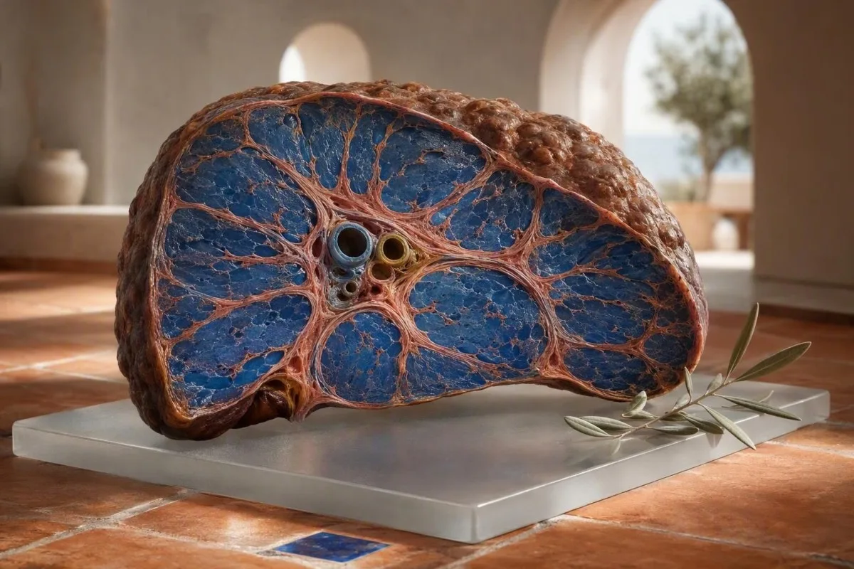
ภาวะตับแข็งระยะแรกมักไม่ทำให้เกิดอาการเจ็บปวดที่ชัดเจน: ความเหนื่อยล้า การเปลี่ยนแปลงของการนอนหลับ การฟกช้ำ อาการคัน และจำนวนเกล็ดเลือดที่ลดลง อาจเกิดขึ้นก่อนดีซ่าน สัญญาณที่เป็นประโยชน์มักเป็นรูปแบบที่ปรากฏในผลการตรวจตับ ความสมบูรณ์ของเม็ดเลือด และคะแนนพังผืด ไม่ใช่เอนไซม์ที่ผิดปกติเพียงตัวเดียว.


- การเกิดแผลเป็นที่ไม่มีอาการ สามารถเกิดขึ้นได้กับ ALT และ AST ปกติ โดยเฉพาะอย่างยิ่งในโรคไขมันพอกตับจากเมตาบอลิซึม และโรคตับที่เกี่ยวข้องกับแอลกอฮอล์.
- កម្រិតផ្លាកែតទាបជាង 150 × 10⁹/L สามารถเป็นสัญญาณบ่งชี้ระยะแรกของความดันในหลอดเลือดดำพอร์ทัล เมื่อลดลงอย่างต่อเนื่องพร้อมกับปัจจัยเสี่ยงต่อตับ.
- FIB-4 ຕໍ່າກວ່າ 1.3 มักบ่งชี้ถึงความเสี่ยงต่ำของพังผืดขั้นสูงในผู้ใหญ่ที่มีอายุต่ำกว่า 65 ปี; 2.67 หรือสูงกว่า ควรได้รับการประเมินจากผู้เชี่ยวชาญอย่างเร่งด่วน.
- Albumin ក្រោម 35 g/L หรือ INR ที่เพิ่มขึ้น อาจบ่งชี้ถึงการทำงานในการสังเคราะห์ของตับที่ลดลง แม้ว่าระดับอะมิโนทรานสเฟอเรสจะอยู่ในระดับปานกลางก็ตาม.
- AST:ALT สูงกว่า 1 ไม่ได้เป็นการวินิจฉัยภาวะตับแข็ง แต่ทำให้เกิดความกังวลเมื่อคู่กับภาวะเกล็ดเลือดต่ำ, GGT สูง, หรือการเปลี่ยนแปลงจากการถ่ายภาพ.
- บิลิรูบินสูงกว่า 34 µmol/L ຜູ້ທີ່ມີອາການເຫຼືອງ, ปัสสาวะเข้ม, ໄຂ້, ສັບສົນ, ຫລືທ້ອງບວມຕ້ອງໄດ້ຮັບການປະເມີນທາງການແພດຢ່າງຮີບດ່ວນ.
- ເອນໄຊມ์ປົກກະຕິ ໝາຍເຖິງບໍ່ມີການຮົ່ວໄຫລຂອງເອນໄຊມ์ໃນເວລານັ້ນ; ພວກມັນບໍ່ໄດ້ພິສູດວ່າໂຄງສ້າງຕັບປົກກະຕິ.
- ການຢຸດເຫຼົ້າ, ການປິ່ນປົວຄວາມສ່ຽງດ້ານການເຜົາຜລານ, ການດູແລຕັບອັກເສບຈາກໄວຣັສ, ແລະການທົບທວນຢາ ສາມາດຊະລໍຕົວລົງຫລືບາງຄັ້ງກໍປີ້ນກັບຂັ້ນຕອນທີ່ຜ່ານມາ.
อาการระยะแรกของภาวะตับแข็งที่ละเอียดอ่อนซึ่งควรได้รับการตรวจตับ
ອາການເລີ່ມຕົ້ນຂອງພະຍາດຕັບແຂງມັກຈະບໍ່ຊັດເຈນແລະເປັນບາງຄັ້ງຄາວ, ໂດຍມີອາການເມື່ອຍລ້າ, ຄວາມຢາກອາຫານຫຼຸດລົງ, ການນອນຫລັບຜິດປົກກະຕິ, ຄັນ, ເລືອດອອກງ່າຍ, ຫລືຄວາມທົນທານຕໍ່ການອອກກໍາລັງກາຍຫຼຸດລົງເກີດຂຶ້ນດົນກ່ອນຜິວຫນັງເຫຼືອງຫລືການຄັ່ງນໍ້າ. ຈາກປະສົບການທາງຄລີນິກຂອງຂ້າພະເຈົ້າ, ຄຸນນະສົມບັດທີ່ໜ້າເປັນຫ່ວງບໍ່ແມ່ນຕອນບ່າຍທີ່ເມື່ອຍລ້າ; ມັນເປັນການລວມຕົວທີ່ງຽບສະຫງົບທີ່ຍັງຄົງຢູ່ເປັນເວລາ 6 ຫາ 12 ອາທິດໃນຄົນທີ່ເປັນໂລກເບົາຫວານ, ໂລກອ້ວນ, ການສໍາຜັດເຫຼົ້າເປັນປະຈໍາ, ຄວາມສ່ຽງຕໍ່ການເປັນພະຍາດຕັບ, ຫລືປະຫວັດຄອບຄົວເປັນພະຍາດຕັບ.
ບຸກຄົນຍັງສາມາດເຮັດວຽກ, ຝຶກອົບຮົມ, ແລະເບິ່ງດີທັງຫມົດໃນຂະນະທີ່ການກໍ່ຕົວເພີ່ມຂຶ້ນ. ຂ້າພະເຈົ້າໄດ້ເຫັນພະນັກງານຫ້ອງການອາຍຸ 48 ປີ ຜູ້ທີ່ມີການປ່ຽນແປງພຽງແຕ່ຕື່ນນອນເວລາ 3 ໂມງເຊົ້າ ດ້ວຍອາການຄັນແລະເມື່ອຍລ້າ; ALT ຂອງລາວແມ່ນ 31 IU/L, ແຕ່ເມັດເລືອດໄດ້ຫຼຸດລົງຈາກ 214 ຫາ 136 × 10⁹/L ໃນໄລຍະສີ່ປີ. ແນວໂນ້ມນັ້ນໄດ້ກະຕຸ້ນການກວດຫາຄື້ນສຽງແລະກວດພົບການກໍ່ຕົວຂັ້ນສູງ.
ພະຍາດຕັບແຂງປົກກະຕິແລ້ວບໍ່ເຮັດໃຫ້ເກີດອາການເຈັບຕັບແຫຼມ. ເຍື່ອຫຸ້ມຕັບສາມາດຍືດອອກໃນຕອນທ້າຍຂອງຂະบวนການ, ແຕ່ການເປັນແຜໃນໄລຍະເລີ່ມຕົ້ນແມ່ນງຽບສະຫງົບທາງຊີວະສາດເນື່ອງຈາກການເປັນພະຍາດກໍ່ຕົວຂຶ້ນລະຫວ່າງຊ່ອງທາງການໄຫຼຂອງເສັ້ນເລືອດຂະຫນາດນ້ອຍ. ຄວາມຮູ້ສຶກແໜ້ນທ້ອງໃຫມ່, ຄວາມອີ່ມໄວ, ຫລືອາການບວມຂໍ້ຕີນເປັນສິ່ງທີ່ໜ້າເປັນຫ່ວງກວ່າອາການເຈັບເລັກນ້ອຍຢູ່ເບື້ອງຂວາ, ໂດຍສະເພາະເມື່ອນໍ້າໜັກເພີ່ມຂຶ້ນຫຼາຍກວ່າ 2 ກິໂລໃນອາທິດຫນຶ່ງ.
ດຣ. Thomas Klein ແນະນຳໃຫ້ເອົາຜົນການກວດກ່ອນຫນ້ານີ້ມານໍາ, ບໍ່ແມ່ນພຽງແຕ່ການກວດລ່າສຸດເທົ່ານັ້ນ, ເພື່ອກວດຕັບ. A ການກວດເລືອດຄັ້ງຕົ້ນ ຈະມີປະໂຫຍດຫລາຍຂຶ້ນເມື່ອສາມາດປຽບທຽບກັບຜົນການກວດຂອງ 12, 24, ແລະ 48 ເດືອນກ່ອນຫນ້ານີ້.
ອາການທີ່ບໍ່ຄວນລໍຖ້ານັດຫມາຍປົກກະຕິ
ອາການສັບສົນ, ງ່ວງເຫຼືອໃນເວລາກາງເວັນໃຫມ່, ຮາກມີເລືອດອອກ, ຖອກດໍາ, ໄຂ້ກັບອາການທ້ອງບວມ, ຫລືອາການເຫຼືອງທີ່ເພີ່ມຂຶ້ນຢ່າງໄວວາຮຽກຮ້ອງໃຫ້ມີການປະເມີນສຸກເສີນໃນມື້ດຽວ. ສິ່ງເຫລົ່ານີ້ສາມາດສະທ້ອນເຖິງການເປັນພະຍາດຕັບແຂງທີ່ບໍ່ດີ, ການມີເລືອດອອກໃນລະບົບຍ່ອຍອາຫານ, ການຕິດເຊື້ອ, ຫລືການບາດເຈັບຕັບສ້ຽວແທນທີ່ຈະເປັນການປ່ຽນແປງທີ່ບໍ່ເປັນອັນຕະລາຍ.
เหตุใด ALT และ AST ปกติจึงไม่สามารถตัดภาวะตับแข็งออกไปได้
ALT ແລະ AST ປົກກະຕິບໍ່ໄດ້ຍົກເວັ້ນການເປັນພະຍາດທີ່ມີນัยສໍາຄັນຫລືຕັບແຂງ. ALT ແລະ AST ສະທ້ອນໃຫ້ເຫັນການຮົ່ວໄຫລຂອງເອນໄຊມ์ໃນຈຸລັງໃນປະຈຸບັນ, ໃນຂະນະທີ່ການເປັນພະຍາດສະທ້ອນເຖິງການເປັນແຜໃນໂຄງສ້າງທີ່ສະສົມທີ່ອາດຈະເຮັດໃຫ້ມີການຮົ່ວໄຫລເລັກນ້ອຍເມື່ອມวลຈຸລັງຕັບແລະການອັກເສບຫຼຸດລົງ.
ຫ້ອງທົດລອງຈໍານວນຫຼາຍໃຊ້ຂີດຈໍາກັດເທິງຂອງ ALT ປະມານ 35 ຫາ 45 IU/L ສໍາລັບຜູ້ຊາຍ ແລະ 25 ຫາ 35 IU/L ສໍາລັບຜູ້ຍິງ, ເຖິງແມ່ນວ່າໄລຍະອ້າງອີງຈະແຕກຕ່າງກັນໄປຕາມການວິເຄາະແລະປະເທດ. ຜົນການກວດ 28 IU/L ອາດຈະເປັນເລື່ອງປົກກະຕິຢູ່ເທິງເຈ້ຍໃນຂະນະທີ່ຍັງຄົງບໍ່ໜ້າໄວ້ວາງໃຈໃນຄົນທີ່ເປັນໂລກອ້ວນ, ໂລກເບົາຫວານຊະນິດ 2, ຈໍານວນເມັດເລືອດ 128 × 10⁹/L, ແລະຄະແນນ FIB-4 ສູງ.
ຄໍາແນະນໍາ AASLD ປີ 2023 ລະບຸວ່າອາມິໂນທຣານສະເຟເຣສອາດຈະເປັນເລື່ອງປົກກະຕິໃນຄົນເຈັບທີ່ເປັນພະຍາດໄຂມັນເກາະຕັບທີ່ບໍ່ໄດ້ມາຈາກເຫຼົ້າຂັ້ນສູງ; ດັ່ງນັ້ນ, ການຈັດລໍາດັບຄວາມສ່ຽງຄວນລວມເອົາການທົດສອບການເປັນພະຍາດທີ່ບໍ່ມີການບຸກລຸກແທນທີ່ຈະເປັນພຽງແຕ່ເອນໄຊມ์ (Rinella et al., 2023). ນີ້ແມ່ນໜຶ່ງໃນບັນດາຂົງເຂດທີ່ຮູບແບບມີຄວາມສໍາຄັນຫຼາຍກວ່າທຸງຢູ່ຂ້າງຄຽງຂອງຄ່າດຽວ.
ການຝຶກອົບຮົມຢ່າງຫນັກແຫນ້ນ, ການບາດເຈັບກ້າມເນື້ອ, ແລະຢາບາງຊະນິດສາມາດເພີ່ມ AST ໂດຍບໍ່ມີການເປັນແຜໃນຕັບ. ນັກແລ່ນມາຣາທອນອາຍຸ 52 ປີ ທີ່ມີ AST 89 IU/L ຫລັງຈາກແຂ່ງຂັນອາດມີແຫຼ່ງກໍາເນີດຈາກກ້າມເນື້ອ, ໂດຍສະເພາະຖ້າ creatine kinase ສູງແລະ bilirubin, platelets, INR, ແລະ GGT ປົກກະຕິ; ຂອງພວກເຮົາ ຄູ່ມື AST ສູງ ອະທິບາຍຄວາມແຕກຕ່າງນັ້ນ.
การตรวจเลือดเพื่อวินิจฉัยภาวะตับแข็ง: รูปแบบที่แพทย์ให้ความกังวลจริงๆ
ຮູບແບບການກວດເລືອດພະຍາດຕັບແຂງທີ່ໜ້າເປັນຫ່ວງທີ່ສຸດລວມມີການຫຼຸດລົງຂອງເມັດເລືອດ, AST ທີ່ສູງກວ່າ ALT, bilirubin ຫລື GGT ທີ່ເພີ່ມຂຶ້ນ, ແລະ albumin ຫລື INR ທີ່ອ່ອນແອແທນທີ່ຈະເປັນການເພີ່ມຂຶ້ນຂອງເອນໄຊມ์ທີ່ແຍກອອກມາ. ແຕ່ລະການທົດສອບຈັບເອົາແງ່ມຸມທີ່ແຕກຕ່າງກັນຂອງຊີວະສາດຕັບ: ການບາດເຈັບ, ການໄຫຼຂອງນໍ້າດີ, ຄວາມດັນໃນເສັ້ນເລືອດດໍາໃນຕັບ, ຫລືການຜະລິດໂປຼຕີນ.
កម្រិតផ្លាកែតទាបជាង 150 × 10⁹/L ພົບເລື້ອຍໃນຂັ້ນຕອນສຸດທ້າຍຂອງການເປັນພະຍາດຕັບແຂງເພາະວ່າຄວາມດັນເລືອດໃນເສັ້ນເລືອດດໍາໃນຕັບເພີ່ມຂຶ້ນ, ເຮັດໃຫ້ກະເພາະຂະຫຍາຍໃຫຍ່ຂຶ້ນ ແລະ ເພີ່ມການສະສົມຂອງ platelet. ການນັບ 142 × 10⁹/L ບໍ່ແມ່ນຫຼັກຖານຂອງພະຍາດຕັບແຂງ, ແຕ່ການຫຼຸດລົງຈາກ 240 ຫາ 142 ມີຄວາມໝາຍທາງດ້ານຄລີນິກຫຼາຍກວ່າຜົນລັບດຽວ.
ອັດຕາສ່ວນ AST:ALT ສູງກວ່າ 1.0 ສາມາດເກີດຂຶ້ນໄດ້ເມື່ອພະຍາດຕັບແຂງຮ້າຍແຮງຂຶ້ນ, ໃນຂະນະທີ່ອັດຕາສ່ວນສູງກວ່າ 2.0 ຕາມປົກກະຕິແມ່ນກ່ຽວຂ້ອງກັບພະຍາດຕັບອັກເສບທີ່ກ່ຽວຂ້ອງກັບເຫຼົ້າ, ແຕ່ບໍ່ມີຄວາມອ່ອນໄຫວຫຼືສະເພາະ. GGT ມັກຈະເພີ່ມຂຶ້ນກັບເຫຼົ້າ, ໄຂມັນໃນຕັບທີ່ກ່ຽວຂ້ອງກັບການເຜົາຜານ, ภาวะดีซ่าน, ແລະ ຜົນກະທົບຂອງຢາ; ອ່ານເພີ່ມເຕີມກ່ຽວກັບ ຄວາມໝາຍຂອງ GGT ກ່ອນທີ່ຈະຖືວ່າເຫຼົ້າເປັນສາເຫດ.
Kantesti AI ຕີຄວາມໝາຍການກວດເລືອດຂອງຕັບໂດຍການຊັ່ງນໍ້າໜັກຜົນລັບທີ່ເຊື່ອມໂຍງເຫຼົ່ານີ້ກັບອາຍຸ, ເພດ, ສະພາບທີ່ລາຍງານ, ແລະ ຄ່າກ່ອນໜ້າ ແທນທີ່ຈະກໍານົດການວິນິດໄສຈາກທຸງດຽວ. Kantesti ແມ່ນເຄື່ອງວິເຄາະກວດເລືອດດ້ວຍ AI ອອກແບບມາເພື່ອລະບຸຮູບແບບການຕິດຕາມເຊັ່ນ: platelet ຕ່ໍາ ບວກກັບອັດຕາສ່ວນ AST:ALT ທີ່ເພີ່ມຂຶ້ນ, ເຊິ່ງສົມຄວນໄດ້ຮັບການກວດສອບຈາກທ່ານໝໍ.
ກັບດັກທີ່ເຮັດໃຫ້ສະບາຍໃຈຜິດທໍາມະດາ
ລະດັບ bilirubin ແລະ albumin ປົກກະຕິສາມາດຢູ່ຮ່ວມກັບພະຍາດຕັບແຂງທີ່ຊົດເຊີຍໄດ້. ຕົວຊີ້ວັດເຫຼົ່ານັ້ນມັກຈະຊຸດໂຊມລົງໃນພາຍຫຼັງ, ຫຼັງຈາກທີ່ຕັບໄດ້ສູນເສຍສຳຮອງການເຮັດວຽກໄປຫຼາຍຂຶ້ນ, ດັ່ງນັ້ນພວກມັນຈຶ່ງເປັນການກວດຄັດກອງທີ່ບໍ່ດີສໍາລັບການເປັນຮອຍແຜໃນໄລຍະເລີ່ມຕົ້ນ.
FIB-4 และสัญญาณที่ไม่รุกรานที่บ่งชี้ถึงแผลเป็นที่มีนัยสำคัญทางคลินิก
FIB-4 ເປັນຄະແນນການເປັນພະຍາດຕັບແຂງຄັ້ງທໍາອິດທີ່ໄດ້ຮັບການຢືນຢັນ, ໃຊ້ອາຍຸ, AST, ALT, ແລະ ຈໍານວນ platelet. ໃນຜູ້ໃຫຍ່ທີ່ອາຍຸຕໍ່າກວ່າ 65 ປີ, FIB-4 ຕໍ່າກວ່າ 1.3 ໂດຍທົ່ວໄປແລ້ວຊີ້ໃຫ້ເຫັນຄວາມສ່ຽງຕ່ໍາຂອງການເປັນພະຍາດຕັບແຂງຂັ້ນສູງ, ໃນຂະນະທີ່ຜົນລັບ 2.67 ຫຼື ສູງກວ່າຄວນຈະກະຕຸ້ນການປະເມີນຜົນໂດຍຜູ້ຊ່ຽວຊານຫຼື elastography.
ສູດແມ່ນອາຍຸ × AST ຫານດ້ວຍ platelets × ຮາກທີສອງຂອງ ALT, ໂດຍ AST ແລະ ALT ຢູ່ໃນ IU/L ແລະ platelets ຢູ່ໃນ × 10⁹/L. ອາຍຸມີການປ່ຽນແປງຄະແນນຢ່າງຫຼວງຫຼາຍ: EASL ແນະນໍາຄ່າ threshold ຄວາມສ່ຽງຕ່ໍາທີ່ສູງກວ່າ 2.0 ສໍາລັບຄົນອາຍຸຫຼາຍກວ່າ 65 ປີເພາະວ່າຄະແນນທີ່ກ່ຽວຂ້ອງກັບອາຍຸທໍາມະດາອາດຈະເຮັດໃຫ້ເກີດຜົນບວກທີ່ຜິດພາດ (European Association for the Study of the Liver, 2021).
ຄະແນນ FIB-4 ເປັນເຄື່ອງມືຄັດແຍກ, ບໍ່ແມ່ນການວິນິດໄສ. ພະຍາດຕັບອັກເສບສ້ວຍ, ການດື່ມເຫຼົ້າໜັກເມື່ອບໍ່ດົນມານີ້, ການປິ່ນປົວດ້ວຍຄີໂມເທຣປີ, ຫຼື ການຫຼຸດລົງຂອງ platelet ທີ່ເປັນການຊົ່ວຄາວສາມາດເພີ່ມຄະແນນໄດ້., ດັ່ງນັ້ນ ຂ້ອຍມັກຈະກວດຊ້ຳການທົດສອບທີ່ບໍ່ໝັ້ນຄົງໃນ 4 ຫາ 12 ອາທິດ ກ່ອນທີ່ຈະຕີຄວາມໝາຍຜົນລັບທີ່ບໍ່ຊັດເຈນ ເວັ້ນເສຍແຕ່ຄົນນັ້ນມີອາການເຫຼືອງ ຫຼື ສັນຍານອັນຕະລາຍອື່ນໆ.
ແຄນເທສຕີ ເປັນ ບໍລິການຕີຄວາມຜົນການກວດຂອງ AI ທີ່ສາມາດຄຳນວນ ແລະ ຈັດວາງຄຳອະທິບາຍຂອງ FIB-4 ເມື່ອມີຜົນລັບທີ່ຈຳເປັນ, ແຕ່ຄະແນນສູງຕ້ອງການການຢືນຢັນທາງການແພດດ້ວຍ transient elastography, ການປະເມີນຜົນໂດຍຜູ້ຊ່ຽວຊານ, ຫຼື ບາງຄັ້ງການກວດເນື້ອເຍື່ອ. ຂອງພວກເຮົາ overview ຂອງຕັບ ສະແດງໃຫ້ເຫັນວ່າການທົດສອບໃດມີຢູ່ທົ່ວໄປ.
เกล็ดเลือดต่ำ: สัญญาณบ่งชี้ระยะแรกของความดันในหลอดเลือดดำพอร์ทัล ไม่ใช่ข้อสรุป
ຈຳນວນເກັດເລືອດທີ່ຫຼຸດລົງຕ່ຳກວ່າ 150 × 10⁹/L ຢ່າງຕໍ່ເນື່ອງ ສາມາດເປັນໜຶ່ງໃນອາການທຳອິດຂອງການກວດເລືອດເປັນປະຈຳທີ່ຊີ້ໃຫ້ເຫັນເຖິງຄວາມດັນເລືອດສູງໃນເສັ້ນເລືອດดำ, ແຕ່ມັນກໍ່ມີສາເຫດອື່ນທີ່ບໍ່ແມ່ນຕັບຫຼາຍ. ການເຊື່ອມຕໍ່ກັບຕັບຈະແຂງແຮງຂຶ້ນເມື່ອຈຳນວນຫຼຸດລົງເທື່ອລະໜ້ອຍ ແລະ ເກີດຂື້ນກັບການຂະຫຍາຍຕົວຂອງ ໝາກໄຂ່ຫຼັງ, FIB-4 ສູງ, ຫລື ການສະແດງຮູບພາບຕັບທີ່ຜິດປົກກະຕິ.
ຄວາມດັນເລືອດສູງໃນເສັ້ນເລືອດดำ ປ່ຽນແປງການໄຫຼວຽນຜ່ານຕັບ ແລະ ໝາກໄຂ່ຫຼັງ. ໝາກໄຂ່ຫຼັງອາດຈະເກັບຮັກສາເກັດເລືອດຫຼາຍຂຶ້ນ, ດັ່ງນັ້ນ ຈຳນວນ 110 ຫາ 140 × 10⁹/L ສາມາດເກີດຂື້ນກ່ອນ ອາການນ້ຳໃນທ້ອງ ຫລືเส้นเลือดขยาย ຫຼາຍປີໃນພະຍາດທີ່ຊົດເຊີຍໄດ້. ນີ້ແມ່ນເຫດຜົນທີ່ແນວໂນ້ມຂອງເກັດເລືອດຖືກລວມຢູ່ໃນ FIB-4 ແທນທີ່ຈະຖືກຈັດການເປັນພຽງແຕ່ໝາຍເຫດ CBC ທົ່ວໄປ.
ບໍ່ແມ່ນທຸກຜົນການຊອກຫາທີ່ຕ່ຳ ສະທ້ອນເຖິງການຫຼຸດລົງທີ່ແທ້ຈິງ. ການຈັບກຸ່ມຂອງເກັດເລືອດທີ່ກ່ຽວຂ້ອງກັບ EDTA ສາມາດເຮັດໃຫ້ຈຳນວນຫຼຸດລົງຢ່າງຜິດພາດ, ແລະຫ້ອງທົດລອງສາມາດຢືນຢັນເລື່ອງນີ້ໄດ້ດ້ວຍການກວດຮູບເງົາເລືອດຫລືຕົວຢ່າງ citrate; ເບິ່ງຄຳອະທິບາຍຂອງພວກເຮົາກ່ຽວກັບ ການຈັບກຸ່ມຂອງເກັດເລືອດ EDTA. ພະຍາດໄວຣັດ, ພູມຕ້ານທານຕໍ່เกล็ดเลือด, ການຂາດວິຕາມິນໂຟລິກ, ແລະສະພາບໄຂກະດູກ ກໍ່ຍັງເປັນໄປໄດ້.
ດຣ. Thomas Klein ໄດ້ພົບວ່າ ຈຳນວນທີ່ຫຼຸດລົງ 20% ໃນໄລຍະສອງປີ ມັກຈະມີປະໂຫຍດຫຼາຍກວ່າການຖາມວ່າໄດ້ຂ້າມຂອບເຂດຕ່ຳສຸດຂອງຫ້ອງທົດລອງແລ້ວຫລືບໍ່. ຄູ່ມັນກັບ ພາກສ່ວນຂອງການກວດເລືອດຄົບຖ້ວນ ແລະ ລາຍງານ ultrasound ກ່ອນໜ້ານີ້ກ່ອນທີ່ຈະສະຫລຸບ.
อัลบูมิน และ INR: การตรวจเลือดที่วัดการสำรองของตับ
ລະດັບ albumin ຕ່ຳ ແລະ INR ທີ່ຍືດເຍື້ອ ສາມາດຊີ້ບອກເຖິງການເຮັດວຽກຂອງຕັບທີ່ຫຼຸດລົງ, ໂດຍສະເພາະເມື່ອມັນພັດທະນາຮ່ວມກັນ. Albumin ຕ່ຳກວ່າ 35 g/L ຫລື INR ສູງກວ່າ 1.3 ໂດຍບໍ່ໄດ້ຮັບການປິ່ນປົວດ້ວຍຢາຕ້ານການແຂງຕົວ ຕ້ອງການການກວດສອບທາງຄລີນິກທີ່ທັນເວລາ, ເຖິງແມ່ນວ່າບໍ່ມີຜົນການຊອກຫາໃດທີ່ສະເພາະເຈາະຈົງກັບ cirrhosis.
Albumin ມີເຄິ່ງຊີວິດປະມານ 20 ວັນ, ດັ່ງນັ້ນມັນຈຶ່ງປ່ຽນແປງຊ້າໆ ແລະ ມັກຈະເປັນປົກກະຕິໃນ cirrhosis ທີ່ຊົດເຊີຍໄດ້. ການຂາດສານອາຫານ, ການສູນເສຍທາດໂປຼຕີນຂອງไต, ພະຍາດລະບົບທີ່ຮ້າຍແຮງ, ການຖືພາ, ແລະການໄຫຼວຽນຂອງນ້ຳຫຼາຍເກີນໄປ ກໍ່ສາມາດເຮັດໃຫ້ albumin ຫຼຸດລົງ; មគ្គុទេសក៍អាល់ប៊ុមីន និងโกลប៊ូលីន ຊ່ວຍແຍກແຍະຄວາມເປັນໄປໄດ້ເຫຼົ່ານີ້.
INR ສະທ້ອນເຖິງກິດຈະກຳຂອງປັດໄຈການແຂງຕົວທີ່ຂຶ້ນກັບວິຕາມິນ K, ສ່ວນໃຫຍ່ແມ່ນຜະລິດໂດຍຕັບ. INR 1.5 ໃນບຸກຄົນທີ່ບໍ່ໄດ້ກິນ warfarin ຈະໜ້າເປັນຫ່ວງຫຼາຍກວ່າ ALT ທີ່ເພີ່ມຂຶ້ນເລັກນ້ອຍ, ແຕ່ການຂາດວິຕາມິນ K ຈາກການດູດຊຶມ, ຢາຕ້ານເຊື້ອ, ຫລື ภาวะดีซ่าน ຍັງສາມາດເຮັດໃຫ້ມັນຍືດເຍື້ອ ແລະ ອາດຈະສາມາດຟື້ນຟູໄດ້.
Kantesti AI គឺជាអ្វីមួយដែល ឧបករណ៍វិភាគតេស្តឈាមដែលដំណើរការដោយ AI ທີ່ເນັ້ນສີ albumin, bilirubin, INR, ແລະຮູບແບບ platelet ຮ່ວມກັນ ເພາະວ່າການທໍາງານຂອງການສັງເຄາະບໍ່ໄດ້ຖືກບັນທຶກໂດຍ ALT ຢ່າງດຽວ. ສໍາລັບບໍລິບົດດ້ານເຕັກນິກກ່ຽວກັບການທົດສອບການແຂງຕົວ, ກວດສອບ ຄູ່ມືການກວດການກໍ່ຕົວຂອງເລືອດ.
ดีซ่าน อาการคัน ปัสสาวะสีเข้ม และอุจจาระสีซีด: สิ่งที่อาจบ่งบอกได้
ອາການເຫຼືອງ, ປັດສະວະສີຊາເຂັ້ມ, ອາຈົມສີຈາງ, ແລະອາການຄັນຕາມຮ່າງກາຍທົ່ວໄປອາດຊີ້ບອກເຖິງການໄຫຼຂອງນໍ້າດີທີ່ຜິດປົກກະຕິຫຼືການປຸງແຕ່ງບິລິລູບິນທີ່ຜິດປົກກະຕິ ແລະຄວນໄດ້ຮັບການປະເມີນຢ່າງຮີບດ່ວນ. ລະດັບບິລິລູບິນທັງໝົດສູງກວ່າ 34 µmol/L, ປະມານ 2 mg/dL, ມັກຈະເຫັນເປັນສີເຫຼືອງໃນແສງແດດ, ເຖິງແມ່ນວ່າສີຜິວແລະແສງສະຫວ່າງຈະສົ່ງຜົນຕໍ່ການກວດຫາ.
ປັດສະວະສີເຂັ້ມສ່ວນໃຫຍ່ມັກຈະເກີດຈາກບິລິລູບິນທີ່ຖືກລວມ, ບໍ່ແມ່ນຈາກບິລິລູບິນທີ່ບໍ່ໄດ້ລວມ. ການກວດປັດສະວະດ້ວຍແຖບທົດສອບທີ່ໃຫ້ຜົນບວກຕໍ່ບິລິລູບິນ, ດັ່ງນັ້ນ, ຈຶ່ງເຮັດໃຫ້ແພດໝໍສົງໄສວ່າເປັນພະຍາດຕັບແລະຖົງນໍ້າດີ ຫຼືການອຸດຕັນ ຫຼາຍກວ່າການແຕກຂອງເມັດເລືອດແດງແບບງ່າຍໆ; មគ្គុទេសក៍ bilirubin ក្នុងទឹកនោម ອະທິບາຍການກວດຕໍ່ໄປ.
ອາຈົມສີຈາງຫຼືສີດິນຈີ່ໝາຍຄວາມວ່າມີສີໜ້ອຍລົງໃນລຳໄສ້. ມັນອາດເກີດຂຶ້ນກັບນໍ້າດີ, ຖົງນໍ້າດີ, ພະຍາດຕັບອ່ອນ, ພະຍາດທໍ່ນໍ້າດີ, ຫຼືການເຮັດວຽກຂອງຕັບທີ່ລົ້ມເຫຼວໃນໄລຍະກ້າວໜ້າ, ດັ່ງນັ້ນ, ມັນຈຶ່ງບໍ່ປອດໄພທີ່ຈະຕິດປ້າຍວ່າເປັນການຕອບສະໜອງການຂັບລ້າງສານພິດ; ເບິ່ງ สาเหตุของอุจจาระสีซีด.
ອາການຄັນຈາກການເປັນພິດໃນນໍ້າດີອາດຮຸນແຮງກ່ອນທີ່ບິລິລູບິນຈະສູງຂຶ້ນຢ່າງເຫັນໄດ້ຊັດ. ມັນມັກຈະສົ່ງຜົນກະທົບຕໍ່ຝາມືແລະຝາຕີນ ແລະຮ້າຍແຮງຂຶ້ນໃນຕອນກາງຄືນ; ໂດຍທົ່ວໄປແລ້ວ, ແພດໝໍຈະກວດ ALP, GGT, ຕ່ອນບິລິລູບິນ, ການໄດ້ຮັບຢາ, ສະຖານະການຖືພາ (ໃນກໍລະນີທີ່ກ່ຽວຂ້ອງ), ແລະການເຂົ້າເຖິງອัลตราซาวนด์ຂອງທໍ່ນໍ້າດີ.
ALP, GGT, และบิลิรูบิน: การรับรู้รูปแบบของการตีบตันของน้ำดี
ຮູບແບບການກວດເລືອດຂອງຕັບທີ່ເປັນພິດໝາຍຄວາມວ່າອາຊິດຟอสຟໍຣິກ ແລະ GGT ເພີ່ມຂຶ້ນຫຼາຍກວ່າ ALT ແລະ AST, ມັກຈະມີລະດັບບິລິລູບິນໂດຍກົງເພີ່ມຂຶ້ນ. ຮູບແບບນີ້ຄວນກະຕຸ້ນໃຫ້ຄົ້ນຫາການອຸດຕັນຂອງທໍ່ນໍ້າດີ, ຄວາມເສຍຫາຍຈາກຢາ, ພະຍາດຕັບອັກເສບທີ່ເກີດຈາກພູມຕ້ານທານ, ຫຼືສະພາບຕັບທີ່ແຊກຊຶມ ເພື່ອແທນທີ່ຈະສົມມຸດຕິຖານວ່າເປັນຕັບແຂງໂດຍອັດຕະໂນມັດ.
ALP ຜະລິດໂດຍເນື້ອເຍື່ອຂອງທໍ່ນໍ້າດີ, ແຕ່ຍັງໂດຍກະດູກ,รก, ແລະ ລຳໄສ້. ALP ທີ່ເພີ່ມຂຶ້ນໂດຍມີ GGT ປົກກະຕິອາດຈະມາຈາກກະດູກ, ໂດຍສະເພາະຫຼັງຈາກກະດູກຫັກ, ໃນລະຫວ່າງການຈະເລີນເຕີບໂຕ, ຫຼືມີຂາດວິຕາມິນດີ, ໃນຂະນະທີ່ ALP ສູງບວກກັບ GGT ສູງສະໜັບສະໜູນແຫຼ່ງກໍາເນີດຈາກຕັບແລະຖົງນໍ້າດີ; ຄຳອະທິບາຍ ALP ແລະ GGT ໃຫ້ຕົວຢ່າງທີ່ເປັນປະໂຫຍດ.
ອັດຕາສ່ວນ R ຊ່ວຍຈັດປະເພດຮູບແບບเอนไซม์ທີ່ເປັນສ້ວຍແຫຼມ: ALT ຫານດ້ວຍຂີດຈຳກັດເທິງຂອງມັນ, ຈາກນັ້ນຫານດ້ວຍ ALP ຫານດ້ວຍຂີດຈຳກັດເທິງຂອງມັນ. ອັດຕາສ່ວນ R ສູງກວ່າ 5 ຊີ້ບອກເຖິງການບາດເຈັບຂອງຈຸລັງຕັບ, ຕ່ຳກວ່າ 2 ຊີ້ບອກເຖິງການເປັນພິດໃນນໍ້າດີ, ແລະ 2 ຫາ 5 ແມ່ນປະສົມ - ມີປະໂຫຍດສຳລັບການປະເມີນຢາ, ແຕ່ບໍ່ແມ່ນການວິນິດໄສແບບດຽວ.
Kantesti AI ໃຊ້ຂີດຈຳກັດອ້າງອີງຫ້ອງທົດລອງຈາກລາຍງານທີ່ອັບໂຫຼດ, ແທນທີ່ຈະບັງຄັບໃຊ້ຂີດຈຳກັດ ALP ສາກົນ, ເພາະວ່າຊ່ວງຕ່າງໆແຕກຕ່າງກັນ. ຄູ່ມືການກວດເລືອດເປັນພິດໃນນໍ້າດີ ເປັນການກະກຽມທີ່ເປັນປະໂຫຍດສໍາລັບການປຶກສາຫາລືກັບແພດ.
การหาสาเหตุมีความสำคัญพอๆ กับการหาแผลเป็น
ຕັບແຂງແມ່ນຈຸດສຸດທ້າຍ, ບໍ່ແມ່ນພະຍາດດຽວ: ເຫຼົ້າ, ພະຍາດໄຂມັນໃນຕັບຈາກການເຜົາຜານ, ຕັບອັກເສບ B ຫຼື C, ພະຍາດພູມຕ້ານທານ, ທາດເຫຼັກເກີນ, ແລະຢາບາງຊະນິດສາມາດເຮັດໃຫ້ເກີດຂຶ້ນໄດ້. ການລະບຸສາເຫດປ່ຽນການປິ່ນປົວ, ການຄັດເລືອກຄອບຄົວ, ການກວດຫາມະເຮັງ, ແລະໂອກາດທີ່ຈະຊ້າລົງ fibrosis ຕື່ມອີກ.
ຄວາມສ່ຽງດ້ານການເຜົາຜານອາຫານສົມຄວນໄດ້ຮັບການສົນທະນາໂດຍກົງ. ການເພີ່ມນໍ້າໜັກໃນສ່ວນກາງ, ໄຂມັນໄຕຣກລີເຊີດສູງກວ່າ 1.7 mmol/L ຫຼື 150 mg/dL, ຄວາມດັນເລືອດສູງ, ແລະພະຍາດເບົາຫວານຊະນິດທີ 2 ເພີ່ມຄວາມເປັນໄປໄດ້ຂອງພະຍາດຕັບ steatotic, ເຖິງແມ່ນວ່າເມື່ອການບໍລິໂພກເຫຼົ້າຕ່ຳ. ລາຍການກວດໂຣກ metabolic syndrome ສາມາດຊີ້ແຈງຄວາມສ່ຽງທີ່ກ່ຽວຂ້ອງໄດ້.
Ferritin ທີ່ສູງກວ່າ 300 ng/mL ໃນເພດຊາຍ ຫຼື ແມ່ຍິງຫຼັງໝົດປະຈຳເດືອນ ບໍ່ພຽງພໍໃນການວິນິດໄສການສະສົມທາດເຫຼັກເກີນ. Transferrin saturation ທີ່ສູງກວ່າ 45% ຢ່າງຕໍ່ເນື່ອງແມ່ນສະເພາະເຈາະຈົງຫຼາຍກວ່າສຳລັບພະຍາດ hereditary haemochromatosis ແລະ ໂດຍປົກກະຕິແລ້ວແມ່ນຊຸກຍູ້ການກວດຫາເຊື້ອ HFE ທາງພັນທຸກຳໃນສະພາບທາງຄລີນິກທີ່ເໝາະສົມ; ເບິ່ງຄູ່ມືອາການພະຍາດ haemochromatosis ຂອງພວກເຮົາ ອາການຂອງພະຍາດ haemochromatosis.
ປະຫວັດການໃຊ້ຢາ ແລະ ອາຫານເສີມ ມັກຈະຖືກປະເມີນຕໍ່າກວ່າຄວາມເປັນຈິງ. ອາຫານເສີມຊາຂຽວຂະໜາດສູງ, ສານສະເຕີຣອຍສ໌, ຜະລິດຕະພັນຫຼຸດນ້ຳໜັກຫຼາຍຊະນິດ, amiodarone, methotrexate, ແລະ ຢາຕ້ານເຊື້ອບາງຊະນິດ ສາມາດປ່ຽນແປງການທົດສອບຕັບໄດ້, ໃນຂະນະທີ່ການລ້າງຕັບທີ່ຖືກກ່າວອ້າງ ອາດຊັກຊ້າການປະເມີນຜົນທີ່ເໝາະສົມ; ບົດຄວາມທົບທວນຄວາມປອດໄພຂອງອາຫານເສີມຂອງພວກເຮົາ ການທົບທວນຄວາມປອດໄພຂອງອາຫານເສີມ ລະມັດລະວັງຢ່າງຈົງໃຈກ່ຽວກັບເລື່ອງນີ້.
ຄວນທົດສອບການອັກເສບຕັບອັກເສບຈາກໄວຣັດ, ບໍ່ແມ່ນການຄາດເດົາຈາກອາການ
ພະຍາດຕັບອັກເສບ ຊະນິດ ຊໍາເຮື້ອ ສາມາດບໍ່ສະແດງອາການໄດ້ເປັນເວລາຫຼາຍທົດສະວັດ ໃນຂະນະທີ່ພະຍາດ fibrosis ກ້າວໜ້າ. ການທົດສອບພູມຕ້ານທານ ລະບຸການສໍາຜັດ, ໃນຂະນະທີ່ການທົດສອບ HCV RNA ຢືນຢັນການຕິດເຊື້ອທີ່ກໍາລັງດໍາເນີນຢູ່; ບົດຄວາມຂອງພວກເຮົາ មគ្គុទេសក៍ការធ្វើតេស្ត hepatitis C ອະທິບາຍວ່າເປັນຫຍັງການທົດສອບທັງສອງຈຶ່ງສຳຄັນ.
จะเกิดอะไรขึ้นหลังจากการตรวจเลือดภาวะตับแข็งที่ผิดปกติ
ຮູບແບບ fibrosis ທີ່ຜິດປົກກະຕິ ໂດຍທົ່ວໄປແລ້ວ ຈະນຳໄປສູ່ການກວດເລືອດຄືນ, ອัลตราซาวด์, ແລະ elastography ແທນທີ່ຈະເປັນການວິນິດໄສ cirrhosis ຢ່າງທັນທີ. Elastography ປະເມີນຄວາມແຂງຂອງຕັບເປັນ kilopascals ແລະ ມີຄວາມໜ້າເຊື່ອຖືຫຼາຍທີ່ສຸດເມື່ອພິຈາລະນາການອັກເສບສ້ວຍຄົມ, ການຄັ່ງ, ແລະການສໍາຜັດເຫຼົ້າໜັກບໍ່ດົນມານີ້.
ໂດຍທົ່ວໄປແລ້ວ ທ່ານໝໍຈະກວດ AST, ALT, ALP, GGT, bilirubin, albumin, INR, ແລະ ຈຳນວນເມັດເລືອດທັງໝົດຄືນພາຍໃນສອງສາມອາທິດ ຖ້າຜົນການກວດບໍ່ຄາດຄິດ. ພວກເຂົາອາດເພີ່ມການທົດສອບຕັບອັກເສບ B ແລະ C, ferritin ພ້ອມ transferrin saturation, immunoglobulins, autoantibodies, ການທົດສອບ coeliac, ແລະ ceruloplasmin ໃນຜູ້ໃຫຍ່ທີ່ອາຍຸນ້ອຍກວ່າ—ເຖິງແມ່ນວ່າລາຍການທີ່ແນ່ນອນຈະເປັນໄປຕາມອາຍຸ ແລະ ປະຫວັດຄວາມສ່ຽງ.
ຄຳແນະນຳ EASL's 2021 ກ່ຽວກັບການທົດສອບແບບບໍ່ຮຸກຮານ ສະໜັບສະໜູນວິທີການສອງຂັ້ນຕອນ: ທໍາອິດ FIB-4, ຈາກນັ້ນ transient elastography ຫຼື ການທົດສອບ serum ຄັ້ງທີສອງສຳລັບຄົນທີ່ຢູ່ເໜືອລະດັບຄວາມສ່ຽງຕໍ່າ (European Association for the Study of the Liver, 2021). ຄວາມແຂງຂອງຕັບບໍ່ແມ່ນສິ່ງດຽວກັບ fibrosis; ການຄັ່ງຂອງຫົວໃຈລົ້ມເຫຼວ, cholestasis, ແລະ ຕັບອັກເສບສ້ວຍຄົມ ສາມາດເພີ່ມຂຶ້ນໄດ້ຊົ່ວຄາວ.
ແຄນເທສຕີ ເປັນ ແພລດຟອມການຕີຄວາມໝາຍ biomarker ຂອງ AI ທີ່ສາມາດຈັດລຽງແຜງທີ່ອັບໂຫຼດເປັນຄຳຖາມສຳລັບການນັດໝາຍຄັ້ງຕໍ່ໄປ, ລວມທັງຄ່າໃດທີ່ປ່ຽນແປງ ແລະ ສາເຫດທີ່ອາດເປັນໄປໄດ້ທີ່ຕ້ອງໄດ້ຍົກເວັ້ນ. ວິທີການທາງຄລີນິກຂອງພວກເຮົາໄດ້ຖືກອະທິບາຍໄວ້ໃນ ພາບລວມການຢືນຢັນທາງການແພດ, ແຕ່ມັນບໍ່ສາມາດແທນທີ່ການກວດຮ່າງກາຍ ຫຼື ຄຳສັ່ງຮູບພາບໄດ້.
แอลกอฮอล์ ความเสี่ยงต่อเมตาบอลิซึม และเมื่อใดควรทำการตรวจตับซ้ำ
ການຫຼຸດຜ່ອນເຫຼົ້າ ແລະ ການປິ່ນປົວຄວາມສ່ຽງດ້ານການເຜົາຜານອາຫານ ສາມາດປັບປຸງເອນໄຊມ์ຕັບໄດ້ພາຍໃນສອງສາມອາທິດ, ແຕ່ຄວາມສ່ຽງຂອງ fibrosis ບໍ່ຄວນຖືກຕັດສິນຈາກຜົນ ALT ທີ່ດີຂຶ້ນພຽງຄັ້ງດຽວ. GGT ມັກຈະຫຼຸດລົງໃນໄລຍະ 2 ຫາ 6 ອາທິດ ຫຼັງຈາກການຢຸດດື່ມເຫຼົ້າ, ໃນຂະນະທີ່ການປ່ຽນແປງຂອງລະດັບ platelet ແລະ ຕົວຊີ້ວັດ fibrosis ອາດໃຊ້ເວລາດົນກວ່າ ແລະ ຂຶ້ນກັບຂັ້ນຕອນຂອງພະຍາດພື້ນຖານ.
ບໍ່ມີຂອບເຂດເຫຼົ້າທີ່ປອດໄພສຳລັບຄົນທີ່ມີ cirrhosis ທີ່ໄດ້ຮັບການຢືນຢັນແລ້ວ; ການງົດເວັ້ນແມ່ນຄຳແນະນຳມາດຕະຖານ ເພາະວ່າເຖິງແມ່ນວ່າໃນປະລິมาณໜ້ອຍກໍ່ອາດເຮັດໃຫ້ຄວາມດັນ portal ເພີ່ມຂຶ້ນ ຫຼື ຂັດຂວາງການຟື້ນຟູ. ສຳລັບຄົນທີ່ບໍ່ມີ cirrhosis, ການບໍລິໂພກທີ່ລາຍງານຍັງຕ້ອງການບໍລິບົດ: ຮູບແບບການດື່ມໜັກ, ນ້ຳໜັກໂຕ, ພະຍາດເບົາຫວານ, ແລະ ການໃຊ້ຢາ ປ່ຽນແປງຄວາມສ່ຽງຢ່າງຫຼວງຫຼາຍ.
ໃນພະຍາດຕັບເມື່ອທຽບກັບການເຜົາຜານອາຫານ, ການຫຼຸດນ້ຳໜັກ 7% ຫາ 10% ຢ່າງຕໍ່ເນື່ອງ ໄດ້ເຊື່ອມໂຍງກັບການປັບປຸງ steatohepatitis ແລະ ສາມາດປັບປຸງ fibrosis ໃນຄົນເຈັບບາງຄົນ, ເຖິງແມ່ນວ່າຜົນໄດ້ຮັບຂອງແຕ່ລະຄົນຈະແຕກຕ່າງກັນ. ການກິນອາຫານທີ່ໜັກໜ່ວງສາມາດເພີ່ມ ALT ໄດ້ຊົ່ວຄາວ, ດັ່ງນັ້ນ ຄວນຕີຄວາມໝາຍແຜງ ຮ່ວມກັບການປ່ຽນແປງນ້ຳໜັກບໍ່ດົນມານີ້; ບົດຄວາມຂອງພວກເຮົາກ່ຽວກັບ ລະດັບເອນໄຊມ໌ໃນຕັບ ຫຼັງຈາກຫຼຸດນ້ຳໜັກ ການກວດເລືອດທີ່ສາມາດຊີ້ໄປຫາມະເຮັງ.
ການວິເຄາະແນວໂນ້ມ AI Kantesti ມີຂໍ້ມູນຫຼາຍທີ່ສຸດເມື່ອການທົດສອບຖືກປະຕິບັດໃນຈຸດທີ່ສາມາດປຽບທຽບກັນໄດ້—ໂດຍທີ່ດີທີ່ສຸດແມ່ນຫຼີກລ້ຽງການອອກກຳລັງກາຍໜັກເປັນເວລາ 48 ຊົ່ວໂມງ ແລະ ບັນທຶກການດື່ມເຫຼົ້າ, ການເຈັບປ່ວຍ, ແລະ ອາຫານເສີມໃໝ່. Timeline ຂອງສິ່ງທີ່ຊ່ວຍໃນການຢຸດເຫຼົ້າ ອະທິບາຍວ່າອັນໃດອາດຈະກັບຄືນສູ່ສະພາບປົກກະຕິ ກ່ອນ.
ความเชื่อผิดๆ เกี่ยวกับอาหารและอาหารเสริมที่อาจทำให้การดูแลตับล่าช้า
ບໍ່ມີການລ້າງສານພິດດ້ວຍນ້ຳໝາກໄມ້, ຜະລິດຕະພັນສະໝຸນໄພ, ຫລື ອາຫານເສີມໃດໆ ທີ່ສາມາດລົບລ້າງຮອຍແປ້ວຂອງພະຍາດຕັບແຂງໄດ້ຢ່າງໜ້າເຊື່ອຖື. ອາຫານຮູບແບບເມດີເຕີເຣນ, ໂປຣຕີນທີ່ພຽງພໍ, ການຈັດການພະຍາດເບົາຫວານ, ແລະການຫລີກລ້ຽງອາຫານເສີມທີ່ເປັນພິດຕໍ່ຕັບສາມາດສະໜັບສະໜູນສຸຂະພາບຕັບໄດ້, ແຕ່ສິ່ງເຫລົ່ານີ້ບໍ່ແມ່ນສິ່ງທົດແທນສໍາລັບການກໍານົດສາເຫດຂອງພະຍາດເສັ້ນໃຍໃນຕັບ.
ບາງຄັ້ງຄົນເຈັບທີ່ເປັນພະຍາດຕັບແຂງຈະຖືກບອກໃຫ້ຫລີກລ້ຽງໂປຣຕີນທຸກຊະນິດ, ເຊິ່ງສາມາດເຮັດໃຫ້ກ້າມເນື້ອອ່ອນເພຍ ແລະ ຄວາມເມື່ອຍລ້າຮ້າຍແຮງຂຶ້ນ. ນອກເຫນືອໄປຈາກຄໍາແນະນໍາສະເພາະຊົ່ວຄາວໃນລະຫວ່າງສະພາບສະໝອງເປັນພິດຈາກຕັບທີ່ຮ້າຍແຮງ, ຄົນເຈັບຈໍານວນຫຼາຍຕ້ອງການປະມານ 1.2 ຫາ 1.5 g ຂອງໂປຣຕີນຕໍ່ກິໂລກຣາມຂອງນ້ຳໜັກໂຕ ຕໍ່ມື້, ແບ່ງອອກເປັນແຕ່ລະຄາບ, ພາຍໃຕ້ການຊີ້ນໍາດ້ານອາຫານ.
ລະວັງຜະລິດຕະພັນທີ່ໂຄສະນາວ່າເປັນສານລ້າງພິດ. ຂີ້ໝິ້ນເຂັ້ມຂຸ້ນ, ກະລໍ້າ, ດອກແດງ, ແລະ ຜະລິດຕະພັນເສີມຫຼາຍສ່ວນປະກອບສຳລັບການເພີ່ມກ້າມເນື້ອຫລືຫຼຸດນ້ຳໜັກ ໄດ້ເຊື່ອມໂຍງກັບຄວາມເສຍຫາຍຕໍ່ຕັບໃນກໍລະນີສຶກສາ, ແລະ ຜະລິດຕະພັນອາດບໍ່ມີສິ່ງທີ່ປ້າຍກ່າວອ້າງ. ຂໍ້ມູນທີ່ອີງຕາມຫຼັກຖານຂອງພວກເຮົາ ຄູ່ມືອາຫານສໍາລັບຕັບ ແຍກນິໄສການກິນອາຫານທີ່ສະໜັບສະໜູນອອກຈາກຄໍາຮຽກຮ້ອງທາງການຕະຫຼາດ.
ຖ້າມີອາການບວມນ້ຳເກີດຂຶ້ນ, ການຈຳກັດໂຊດຽມມັກຈະສຳຄັນກວ່າການຈຳກັດນ້ຳ. ເປົ້າໝາຍທົ່ວໄປແມ່ນໜ້ອຍກວ່າ 2 g ໂຊດຽມຕໍ່ມື້, ແຕ່ຢາຂັບປັດສະວະ, ການເຮັດວຽກຂອງไต, ຄວາມດັນເລືອດ, ແລະ ຄວາມພຽງພໍທາງດ້ານໂພຊະນາການຮຽກຮ້ອງໃຫ້ມີການຕິດຕາມສ່ວນບຸກຄົນແທນທີ່ຈະເປັນແຜນອາຫານໃນອິນເຕີເນັດ.
เมื่อใดที่อาการของภาวะตับแข็งจะกลายเป็นเรื่องเร่งด่วน
ການຮາກເລືອດ, ອາຈົມສີດຳເໝືອນຢາງ, ອາການສັບສົນ, ໄຂ້ດ້ວຍທ້ອງອືດ, ຫາຍໃຈສັ້ນຢ່າງຮ້າຍແຮງ, ຫລື ອາການຕົວເຫຼືອງທີ່ຮ້າຍແຮງຂຶ້ນຢ່າງໄວວາແມ່ນອາການສຸກເສີນໃນຜູ້ທີ່ມີພະຍາດຕັບແຂງທີ່ຮູ້ຈັກຫຼືສົງสัย. ສັນຍານເຫລົ່ານີ້ອາດເປັນຕົວແທນຂອງເລືອດອອກເສັ້ນເລືອດຂອດ, ສະໝອງເປັນພິດຈາກຕັບ, ການຕິດເຊື້ອແບັກທີເຣຍໃນທ້ອງ, ໝາກໄຂ່ຫລັງອັກເສບ, ຫລື ຄວາມລົ້ມເຫຼວຂອງຕັບຊໍາເຮື້ອສ້ວຍພ້ອມກັບເຫດການສ້ວຍ.
ອາການສັບສົນໃຫມ່ ຫລື ການນອນ-ຕື່ນ ທີ່ປີ້ນກັບກັນ ບໍ່ແມ່ນພຽງແຕ່ຄວາມເມື່ອຍລ້າໃນພະຍາດຕັບແຂງ. ມັນອາດຊີ້ບອກເຖິງສະໝອງເປັນພິດຈາກຕັບ, ໂດຍສະເພາະຖ້າປະກອບກັບອາການທ້ອງຜູກ, ຂາດນ້ໍາ, ການຕິດເຊື້ອ, ເລືອດອອກໃນລະບົບຍ່ອຍອາຫານ, ການໃຊ້ຢາສະຫລົບ, ຫລື ຜົນຂອງແອມໂມເນຍສູງ; ແຕ່ແອມໂມເນຍພຽງຢ່າງດຽວ ບໍ່ສາມາດວິນິດໄສ ຫລື ຈັດລຳດັບຄວາມຮຸນແຮງຂອງສະໝອງເປັນພິດໄດ້ຢ່າງໜ້າເຊື່ອຖື.
ທ້ອງອືດອາດສະທ້ອນເຖິງອາການທ້ອງບວມ, ແຕ່ມັນກໍອາດເກີດຈາກໂລກອ້ວນ, ລຳໄສ້ອືດ, ພະຍາດຮັງໄຂ່, ຫລື ຫົວໃຈລົ້ມເຫຼວ. ໄຂ້, ເຈັບ, ຄວາມດັນເລືອດຕໍ່າ, ຫລື ການເຮັດວຽກຂອງไตທີ່ຮ້າຍແຮງຂຶ້ນໃນຜູ້ທີ່ມີອາການທ້ອງບວມຄວນໄດ້ຮັບການປະເມີນທັນທີ ເພາະການຕິດເຊື້ອອາດຈະເບົາບາງແຕ່ເປັນອັນຕະລາຍ.
ສໍາລັບຄວາມບໍ່ແນ່ນອນທີ່ບໍ່ແມ່ນສຸກເສີນ, ໃຫ້ຂຽນອາການ, ຢາ, ການດື່ມເຫຼົ້າ, ອາຫານເສີມ, ແລະ ນ້ຳໜັກທີ່ຕິດວັນທີ ກ່ອນໂທຫາທ່ານໝໍ. ຫນຶ່ງ ບັນຊີລາຍຊື່ສະຫຼຸບການກວດເລືອດ ສາມາດເຮັດໃຫ້ການສົນທະນານັ້ນມີປະສິດທິພາບຫຼາຍຂຶ້ນ.
รายการตรวจสอบการนัดหมายตับที่นำไปปฏิบัติได้จริงซึ่งเปลี่ยนแปลงการตัดสินใจ
ການນັດພົບທ່ານໝໍທີ່ດີທີ່ສຸດສຳລັບຕັບ ລວມມີລາຍງານຜົນກວດຫ້ອງທົດລອງກ່ອນໜ້າ, ປະຫວັດການດື່ມເຫຼົ້າ ແລະ ການໃຊ້ຢາທີ່ເປັນຈິງ, ປະຫວັດການເຜົາຜານອາຫານ, ແລະ ລາຍງານຮູບພາບໃດໆ. ລາຍລະອຽດເຫລົ່ານີ້ ຊ່ວຍໃຫ້ທ່ານໝໍສາມາດຈຳແນກຜົນ enzyme ທີ່ຜັນຜວນອອກຈາກຮູບແບບຫຼາຍປີທີ່ຊີ້ບອກເຖິງພະຍາດເສັ້ນໃຍໃນຕັບ.
ນຳຜົນກວດທຸກຢ່າງທີ່ທ່ານຊອກຫາໄດ້, ລວມທັງ CBC, ຜົນກວດຕັບ, INR, ferritin, ການກວດຫາພະຍາດຕັບອັກເສບ, ລາຍງານ ultrasound, ແລະ ຄ່າ elastography ໃນ kPa. ຜົນດຽວອາດຈະມີຄວາມຜັນຜວນ; ຜົນສາມຄັ້ງ ທີ່ກວມໄລຍະ 18 ເດືອນສາມາດສະແດງໃຫ້ເຫັນວ່າຈໍານວນเกล็ดเลือด ກໍາລັງຫຼຸດລົງຫລື bilirubin ຄົງທີ່. ຂອງພວກເຮົາ ຄູ່ມືການເຂົ້າກັນຜົນແບບຂ້າງຂ້າງ อธิบายว่าการเปลี่ยนแปลงใดมีความสำคัญโดยทั่วไป.
ถามคำถามโดยตรงสี่ข้อ: สาเหตุที่เป็นไปได้คืออะไร? สงสัยว่ามีระยะพังผืดเท่าใด? ฉันต้องการการตรวจวัดความยืดหยุ่นของตับ หรือส่งต่อผู้เชี่ยวชาญด้านโรคตับหรือไม่? อาการใดที่ควรพาไปห้องฉุกเฉิน? หากได้รับการยืนยันว่าเป็นโรคตับแข็ง ให้สอบถามเกี่ยวกับการตรวจอัลตราซาวด์ทุกหกเดือนเพื่อคัดกรองมะเร็งตับ และจำเป็นต้องส่องกล้องเพื่อประเมินเส้นเลือดขอดหรือไม่.
ณ วันที่ 4 กันยายน 2026 แพทย์ของเรายังคงแนะนำให้ถือว่าคำอธิบายที่สร้างโดย AI เป็นการเตรียมความพร้อม ไม่ใช่การวินิจฉัย การทำงานทางคลินิกของ Kantesti จะได้รับการตรวจสอบภายใต้การดูแลของ ຄະນະທີ່ປຶກສາທາງການແພດ, และการตัดสินใจขั้นสุดท้ายเกี่ยวกับโรคตับแข็งขึ้นอยู่กับแพทย์ผู้ทำการรักษาที่สามารถตรวจคุณและสั่งการทดสอบเพื่อยืนยันได้.
ຄໍາຖາມທີ່ຖາມເລື້ອຍໆ
ທ່ານສາມາດເປັນໂຣກຕັບແຂງໂດຍມີເອນໄຊມ์ຕັບປົກກະຕິໄດ້ບໍ?
ແມ່ນແລ້ວ. ALT ແລະ AST ທີ່ເປັນປົກກະຕິບໍ່ໄດ້ຕັດຄວາມເປັນໄປໄດ້ຂອງພະຍາດຕັບແຂງອອກ ເພາະເອນໄຊມ์ເຫຼົ່ານີ້ສະທ້ອນເຖິງຄວາມເສຍຫາຍຂອງຈຸລັງຕັບໃນປັດຈຸບັນ ແທນທີ່ຈະເປັນປະລິມານຂອງເນື້ອເຍື່ອແຜເປັນແລ້ວ. ພະຍາດຕັບແຂງທີ່ຊົດເຊີຍແລ້ວອາດສະແດງອອກດ້ວຍ ALT ແລະ AST ຢູ່ໃນຊ່ວງອ້າງອີງຂອງຫ້ອງທົດລອງ, ໂດຍສະເພາະໃນພະຍາດຕັບທີ່ກ່ຽວຂ້ອງກັບການເຜົາຜະຫລັກ. ການນັບຈໍານວນเกล็ดเลือดຕ່ໍາກວ່າ 150 × 10⁹/L, FIB-4 2.67 ຫຼືຫຼາຍກວ່ານັ້ນ, elastography ຜິດປົກກະຕິ, albumin ຕ່ໍາ, ຫຼື INR ທີ່ເພີ່ມຂຶ້ນ ສາມາດໃຫ້ຄໍາແນະນໍາທີ່ເປັນປະໂຫຍດຫຼາຍກວ່າເອນໄຊມ์ພຽງຢ່າງດຽວ.
ສັນຍານເຕືອນທຳອິດຂອງໂລກຕັບແຂງແມ່ນຫຍັງ?
ອາການເລີ່ມຕົ້ນຂອງພະຍາດຕັບແຂງອາດລວມມີ: ຄວາມເມື່ອຍລ້າຢ່າງຕໍ່ເນື່ອງ, ຄວາມຢາກອາຫານຫຼຸດລົງ, ອາການຄັນ, ມີຮອຍຟົກຊ້ຳໄດ້ງ່າຍ, ຄວາມທົນທານຕໍ່ການອອກກຳລັງກາຍຫຼຸດລົງ, ການປ່ຽນແປງການນອນ, ແລະອາການທ້ອງເຕັມ, ແຕ່ຫຼາຍຄົນບໍ່ມີອາການ. ອາການຕົວເຫຼືອງ, ປັດສະວະສີເຂັ້ມ, ອາຈົມສີຈາງ, ຂໍ້ຕີນບວມ, ນ້ຳໃນທ້ອງ, ອາການສັບສົນ, ຮາກເປັນເລືອດ, ແລະອາຈົມສີດຳແນວໂນ້ມທີ່ຈະເປັນອາການທີ່ເກີດຂຶ້ນຊ້າໆ ຫຼືຮີບດ່ວນ. ອາການພຽງຢ່າງດຽວບໍ່ສາມາດວິນິດໄສພະຍາດຕັບແຂງໄດ້, ສະນັ້ນໂດຍທົ່ວໄປແລ້ວຈຶ່ງຕ້ອງການການກວດເລືອດ, CBC with platelets, INR, albumin, ແລະ ການປະເມີນພະຍາດເຍື່ອຫຸ້ມປອດ.
ຈະນັບເລືອດຄົບຖ້ວນ ທີ່ຊີ້ບອກວ່າເປັນພະຍາດຕັບແຂງ?
ຈໍານວນเกล็ดเลือดທີ່ຕໍ່າກວ່າ 150 × 10⁹/L ສາມາດເຮັດໃຫ້ເກີດຄວາມສົງໄສກ່ຽວກັບການເປັນພະຍາດເຍື່ອຫຸ້ມຕັບຂັ້ນສູງຫຼືຄວາມດັນໃນເສັ້ນເລືອດດໍາໃນຕັບສູງເມື່ອມັນຍັງຄົງຢູ່ແລະມີແນວໂນ້ມຫຼຸດລົງ, ແຕ່ມັນບໍ່ສາມາດບົ່ງມະຕິໂຣກຕັບແຂງໄດ້. ການລວມຕົວຂອງเกล็ดเลือด, ພະຍາດໄວຣັສ, ພູມຄຸ້ມກັນຕ່ອມໄທຣອຍ, ຢາ, ການຂາດສານອາຫານ, ແລະຄວາມຜິດປົກກະຕິຂອງໄຂກະດູກກໍ່ສາມາດເຮັດໃຫ້ຈໍານວນຫຼຸດລົງໄດ້. ການຫຼຸດລົງຢ່າງຕໍ່ເນື່ອງ, ເຊັ່ນ 220 ຫາ 135 × 10⁹/L ໃນໄລຍະສອງປີພ້ອມກັບ AST ທີ່ສູງຂຶ້ນແລະ FIB-4 ທີ່ສູງ, ໃຫ້ຂໍ້ມູນຫຼາຍກ່ວາຄ່າທີ່ຕໍ່າເລັກນ້ອຍຫນຶ່ງ.
ຄະແນນ FIB-4 ໝາຍເຖິງໂລກຕັບແຂງບໍ?
FIB-4 ບໍ່ໄດ້ تشخيص ภาวะตับแข็ง, ແຕ່ມັນປະເມີນຄວາມສ່ຽງຂອງ fibrosis ຂັ້ນສູງຈາກອາຍຸ, AST, ALT, ແລະ platelets. ໃນຜູ້ໃຫຍ່ອາຍຸຕ່ຳກວ່າ 65 ປີ, ຄະແນນຕ່ຳກວ່າ 1.3 ໂດຍທົ່ວໄປຊີ້ບອກເຖິງຄວາມສ່ຽງຕ່ຳ, 1.3 ຫາ 2.66 ຕ້ອງການການປະເມີນແບບບໍ່ຮຸກຮານເພີ່ມເຕີມ, ແລະ 2.67 ຫຼື ສູງກວ່າແມ່ນຮຽກຮ້ອງໃຫ້ມີການປະເມີນໂດຍຜູ້ຊ່ຽວຊານ. ໃນຜູ້ໃຫຍ່ອາຍຸຫຼາຍກວ່າ 65 ປີ, ເກນສູງກວ່າ 2.0 ຖືກນຳໃຊ້ທົ່ວໄປເພື່ອຫຼຸດຜ່ອນ false positives ທີ່ກ່ຽວຂ້ອງກັບອາຍຸ.
ภาวะตับแข็งสามารถดีขึ้นได้หรือไม่หากตรวจพบตั้งแต่เนิ่นๆ?
ภาวะพังผืดในระยะเริ่มต้นสามารถดีขึ้นได้เมื่อสาเหตุที่ก่อให้เกิดโรคได้รับการแก้ไขหรือควบคุมแล้ว ซึ่งรวมถึงการงดดื่มแอลกอฮอล์อย่างต่อเนื่อง การรักษาไวรัสตับอักเสบให้หายขาด การลดน้ำหนักในผู้ป่วยโรคตับจากเมตาบอลิซึม หรือการรักษาโรคที่เกิดจากภูมิคุ้มกันตนเอง ภาวะตับแข็งที่ตั้งหลักแล้วอาจไม่สามารถกลับคืนสู่สภาพปกติได้ทั้งหมด แต่ภาวะแทรกซ้อนและแผลเป็นที่เพิ่มขึ้นมักจะสามารถลดลงได้ด้วยการดูแลโดยผู้เชี่ยวชาญ การลดน้ำหนักอย่างต่อเนื่อง 7% ถึง 10% สามารถปรับปรุงภาวะไขมันเกาะตับชนิดอักเสบในผู้ป่วยโรคตับจากเมตาบอลิซึมหลายรายได้ แม้ว่าผลลัพธ์จะแตกต่างกันไปในผู้ป่วยแต่ละราย.
ຂ້ອຍຄວນໄປໂຮງໝໍເມື່ອໃດ ຖ້າສົງໃສວ່າເປັນໂຣກຕັບແຂງ?
ຕິດຕໍ່您就診的診所尋求協助,以處理吐血、柏油狀黑便、新出現的意識模糊、昏厥、腹脹發燒、嚴重黃疸或呼吸困難迅速惡化等情況。這些症狀可能表示食道靜脈瘤出血、腹水感染、肝性腦病變或急性肝衰竭,不應等待門診回診的血液檢查。如果您有輕微症狀,但膽紅素超過 34 µmol/L,或 INR 超过 1.3 且未使用抗凝血劑,或血小板計數下降,請儘快聯絡臨床醫師尋求建議。.
ຮັບການວິເຄາະຜົນກວດເລືອດດ້ວຍ AI ທັນທີ
ເຂົ້າຮ່ວມຜູ້ໃຊ້ຫຼາຍກວ່າ 2 ລ້ານຄົນທົ່ວໂລກ ທີ່ໄວ້ໃຈ Kantesti ສຳລັບການວິເຄາະການກວດເລືອດທີ່ທັນທີ ແລະຖືກຕ້ອງ. ອັບໂຫຼດຜົນກວດເລືອດຂອງທ່ານ ແລະຮັບການຕີຄວາມໝາຍຢ່າງຄົບຖ້ວນຂອງ biomarker 15,000+ ໃນວິນາທີ.
📚 ບົດຄວາມວິຈັຍທີ່ອ້າງອີງ
Klein, T., Mitchell, S., & Weber, H. (2026). Kantesti Research Team. (2026). aPTT Normal Range: D-Dimer, Protein C Blood Clotting Guide. Zenodo. https://doi.org/10.5281/zenodo.18262555. ลิงก์ความพร้อมใช้งานของ ResearchGate และ Academia.edu จะถูกดูแลผ่านบันทึกการตีพิมพ์.. ການຄົ້ນຄວ້າທາງການແພດຂອງ AI Kantesti.
Klein, T., Mitchell, S., & Weber, H. (2026). Kantesti Research Team. (2026). Serum Proteins Guide: Globulins, Albumin & A/G Ratio Blood Test. Zenodo. https://doi.org/10.5281/zenodo.18316300. ลิงก์ความพร้อมใช้งานของ ResearchGate และ Academia.edu จะถูกดูแลผ่านบันทึกการตีพิมพ์.. ການຄົ້ນຄວ້າທາງການແພດຂອງ AI Kantesti.
📖 ເອກະສານອ້າງອີງທາງການແພດພາຍນອກ
⚕️ ຂໍ້ສັງເກດທາງການແພດ
ບົດຄວາມນີ້ມີຈຸດປະສົງເພື່ອການສຶກສາເທົ່ານັ້ນ ແລະບໍ່ແມ່ນຄຳແນະນຳທາງການແພດ. ຄວນປຶກສາຜູ້ໃຫ້ບໍລິການດ້ານສຸຂະພາບທີ່ມີຄຸນວຸດທິສະເໝີ ສຳລັບການວິນິດໄຊ ແລະ ການຕັດສິນໃຈດ້ານການຮັກສາ.
ສັນຍານຄວາມໄວ້ໃຈ E-E-A-T
ປະສົບການ
ການທົບທວນຄລີນິກຂອງແພດຜູ້ນຳພາ ກ່ຽວກັບຂັ້ນຕອນການຕີຄວາມໝາຍຜົນການກວດໃນຫ້ອງທົດລອງ.
ຄວາມຊ່ຽວຊານ
ວິຊາການແພດທົດລອງ (ການແພດທາງຫ້ອງທົດລອງ) ເນັ້ນໃສ່ວ່າຕົວຊີ້ວັດ (biomarkers) ມີພຶດຕິກຳແນວໃດໃນບັນບົດທາງຄລີນິກ.
ຄວາມເປັນອຳນາດ
ຂຽນໂດຍທ່ານດຣ. Thomas Klein ໂດຍມີການກວດທານໂດຍທ່ານດຣ. Sarah Mitchell ແລະ ສາດສະດາຈານດຣ. Hans Weber.
ຄວາມໜ້າເຊື່ອຖື
ການຕີຄວາມໝາຍອີງຕາມຫຼັກຖານດ້ວຍເສັ້ນທາງຕິດຕາມທີ່ຊັດເຈນ ເພື່ອຫຼຸດການຕົກໃຈ.