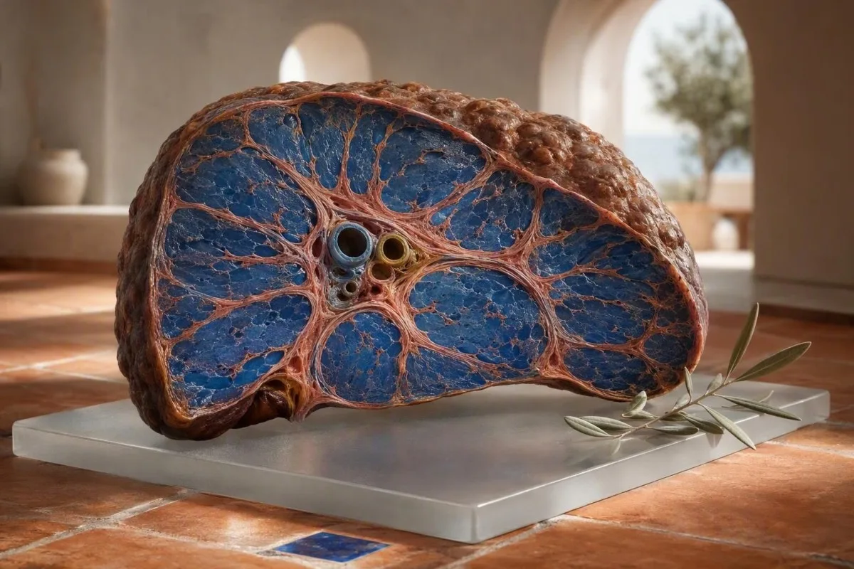
肝硬変の初期には、はっきりとした痛みがないことがよくあります。黄疸が現れる前に、疲労感、睡眠の変化、あざができやすい、かゆみ、血小板数の減少などが現れることがあります。肝臓の検査、血液算定、線維症スコアにおけるパターンが、異常な酵素一つではなく、通常は有用な手がかりとなります。.


- サイレントスカーリング は、特に代謝性脂肪肝疾患およびアルコール性肝疾患において、正常なALTおよびASTでも起こり得ます。.
- 血小板数が150 × 10⁹/L未満 は、肝臓のリスク因子と並行して徐々に低下する場合、門脈圧亢進症の初期の手がかりとなり得ます。.
- FIB-4が1.3未満 は、通常、65歳未満の成人における進行した線維症のリスクが低いことを示しますが;; 2.67以上 は、迅速な専門医による評価を保証します。.
- アルブミンが35 g/L未満 またはINRの上昇は、アミノトランスフェラーゼが軽度であっても、肝臓の合成機能の低下を示している可能性があります。.
- AST:ALTが1超 は肝硬変の診断ではありませんが、血小板減少症、GGTの上昇、または画像検査の変化と組み合わされると懸念が高まります。.
- ビリルビンが34 µmol/L超 黄疸、尿の色が濃い、発熱、錯乱、または腹部膨満がある場合は、緊急の臨床評価が必要です。.
- 酵素値が正常 は、その時点では活発な酵素漏出がないことを意味しますが、肝臓の構造が正常であることを証明するものではありません。.
- 飲酒中止、代謝リスクの管理、ウイルス性肝炎のケア、および薬剤の見直し は、早期の線維化を遅らせたり、場合によっては部分的に逆転させたりすることができます。.
肝臓の検査を検討すべき、初期の肝硬変の微妙な症状
肝硬変の初期症状はしばしば不明瞭で断続的であり、, 、疲労、食欲不振、睡眠障害、かゆみ、あざができやすい、または運動耐容能の低下などが、皮膚の黄色化や体液貯留よりもはるか前に現れます。私の臨床経験では、懸念すべき特徴は、ある午後の倦怠感ではなく、糖尿病、肥満、定期的なアルコール摂取、肝炎のリスク、または肝疾患の家族歴がある人で、6~12週間続く静かな症状の集まりです。.
線維症が進行していても、人はまだ仕事をし、運動をし、完全に健康に見えることがあります。私は、48歳の会社員で、唯一の変化は、かゆみと疲労で午前3時に目が覚めることでした。ALTは31 IU/Lでしたが、血小板は4年間で214から136 × 10⁹/Lに減少していました。この傾向がエラストグラフィーを促し、進行した線維症を特定しました。.
肝硬変は通常、鋭い肝臓の痛み does not cause。. 肝臓の被膜は、過程の後半で伸びることがありますが、初期の瘢痕形成は、微細な血管チャネルの間に線維症が発達するため、生物学的に静かです。腹部膨満感、早期の満腹感、または足首の腫れといった新しい感覚は、特に1週間で体重が2 kg以上増加した場合、右側のはっきりしない痛みよりも懸念されます。.
トーマス・クライン医師は、最新の検査結果だけでなく、以前の検査結果も持参して肝臓の診察を受けることを勧めています。 ベースライン血液検査 は、12、24、48か月前の結果と比較できる場合に、はるかに有用になります。.
定期的な診察を待つべきではない症状
錯乱、新たな日中の眠気、吐血、黒色便、腹部膨満を伴う発熱、または急速に進行する黄疸は、即日の緊急評価が必要です。. これらは、無害な変動ではなく、肝硬変の代償不全、消化管出血、感染症、または急性の肝障害を反映している可能性があります。.
なぜ正常なALTおよびASTでは肝硬変を除外できないのか
ALTおよびASTが正常であっても、有意な線維症または肝硬変を除外することはできません。. ALTおよびASTは現在の細胞酵素漏出を反映しますが、線維症は、肝細胞量と炎症が低下するとほとんど継続的な漏出をほとんど生じない可能性のある、蓄積された構造的瘢痕を反映します。.
多くの検査室では、男性でALT上限を約35~45 IU/L、女性で25~35 IU/Lとしていますが、基準範囲はアッセイや国によって異なります。28 IU/Lという結果は、紙面上では正常ですが、肥満、2型糖尿病、血小板数128 × 10⁹/L、および高いFIB-4スコアを持つ人にとっては、それほど安心できるものではありません。.
2023年のAASLDガイドラインでは、非アルコール性脂肪性肝疾患の進行した患者ではアミノトランスフェラーゼが正常である可能性があると述べられています。したがって、リスク層別化は、酵素のみではなく、非侵襲的線維症検査を含むべきです(Rinella et al.、2023)。これは、単一の値の横にあるフラグよりもパターンが重要である領域の1つです。.
ハードトレーニング、筋肉の損傷、および一部の薬剤は、肝臓の瘢痕なしにASTを上昇させることがあります。レース後にAST 89 IU/Lの52歳のマラソンランナーは、特にクレアチンキナーゼが高く、ビリルビン、血小板、INR、およびGGTが正常であれば、筋肉が原因である可能性があります。 AST高値ガイド が、その違いを説明しています。.
肝硬変の血液検査:臨床医が実際に懸念するパターン
最も懸念される肝硬変の血液検査パターンは、血小板の減少、ALTを超えるAST、ビリルビンまたはGGTの上昇、およびアルブミンまたはINRの低下を組み合わせたものであり、孤立した酵素の上昇ではありません。. 各検査は、肝臓の生物学の異なる側面、つまり損傷、胆汁の流れ、門脈圧、またはタンパク質産生を捉えています。.
血小板数が150 × 10⁹/L未満 肝硬変でよく見られます。門脈圧亢進により脾臓が腫大し、血小板の貯留が増加するためです。142 × 10⁹/L という値は肝硬変の証明ではありませんが、240 から 142 への減少傾向は、単一の結果よりも臨床的に意味のあるものです。.
AST:ALT 比が 1.0 を超える 線維化が進むにつれて起こり得ますが、2.0 を超える比率は古典的にアルコール関連肝炎と関連付けられますが、感度も特異度も高くありません。GGT は、アルコール、代謝性脂肪肝、胆汁うっ滞、薬剤の影響で上昇することがよくあります。 GGT の意味 アルコールが原因であると仮定する前に、詳細をご覧ください。.
Kantesti AI は、単一の所見から診断を下すのではなく、これらの関連する結果を年齢、性別、報告された病状、および過去の値と比較して肝機能パネルを解釈します。. KantestiはAI血液検査アナライザーです 低血小板数と AST:ALT 比の上昇のような、臨床医によるレビューに値するフォローアップパターンを特定するように設計されています。.
よくある誤った安心感の落とし穴
正常なビリルビン値と正常なアルブミン値は、代償性肝硬変と併存することがあります。. これらのマーカーは、肝臓がより多くの機能予備能力を失った後に悪化することが多いため、初期の瘢痕化のスクリーニング検査としては不十分です。.
FIB-4および非侵襲的な臨床的に有意な瘢痕の手がかり
FIB-4 は、年齢、AST、ALT、および血小板数を使用した、検証済みの第一選択線維化スコアです。. 65 歳未満の成人では、FIB-4 が 1.3 未満であれば通常、進行性線維化のリスクは低いことを示しますが、2.67 以上であれば専門医の評価またはエラストグラフィーをトリガーすべきです。.
計算式は、年齢 × AST ÷ (血小板 × √ALT) で、AST と ALT は IU/L、血小板は × 10⁹/L です。年齢はスコアに大きく影響します。EASL は、65 歳以上の人では、通常の年齢関連スコアリングが偽陽性を生み出す可能性があるため、より高い低リスク閾値 2.0 を推奨しています (European Association for the Study of the Liver, 2021)。.
FIB-4 スコアはトリアージツールであり、診断ではありません。. 急性肝炎、最近の大量飲酒エピソード、化学療法、または一時的な血小板減少は、スコアを増加させる可能性があります, 、そのため、黄疸やその他の危険信号がない限り、境界線上の結果を解釈する前に、通常 4 ~ 12 週間後に不安定な検査を再検査します。.
カンテスティは AIラボ検査解釈サービス 必要な結果が存在する場合に FIB-4 を計算および文脈化できるものですが、高スコアは、経皮的肝エラストグラフィー、専門医の評価、または場合によっては組織検査による医療確認が必要です。私たちの 肝機能パネルの概要 には、一般的に利用可能な検査が示されています。.
血小板減少:門脈圧亢進症の初期の手がかりであり、最終診断ではない
持続的な血小板数が150 × 10⁹/L未満であることは、門脈圧亢進症の最も初期のルーチン血液検査の兆候の1つになる可能性がありますが、多くの非肝臓性原因があります。. 血小板数の減少が徐々に進行し、脾腫、FIB-4の上昇、または肝臓の画像検査の異常を伴う場合、肝臓との関連性が強くなります。.
門脈圧亢進症は、肝臓と脾臓への血流を変化させます。脾臓はより多くの血小板を保持する可能性があるため、110〜140 × 10⁹/Lの血小板数は、代償性疾患では腹水または静脈瘤よりも数年前に先行する可能性があります。これが、血小板の傾向がFIB-4に含まれる理由であり、一般的なCBCの脚注として扱われない理由です。.
すべての低い結果が真の低下を反映しているわけではありません。EDTA関連の血小板凝集は血小板数を偽って低下させる可能性があり、検査室は血液塗抹標本またはクエン酸サンプルでこれを確認できます。当社の説明を参照してください。 EDTAによる血小板の凝集. ウイルス性疾患、免疫性血小板減少症、葉酸欠乏症、および骨髄の状態も依然として可能性として残ります。.
Thomas Klein医師は、2年間で20%低下する血小板数は、検査室の下限を超えたかどうかを尋ねるよりも有用であることが多いと発見しました。これをCBCの構成要素と以前の超音波検査レポートと組み合わせて、結論を導き出してください。 血球計算 の項目 および以前の超音波検査レポートと組み合わせて、結論を導き出してください。.
アルブミンおよびINR:肝臓の予備能力を測定する血液検査
低アルブミン血症とINRの延長は、特にそれらが同時に発生した場合、肝臓の合成機能の低下を示す可能性があります。. アルブミンが35 g/L未満またはINRが1.3を超えている(抗凝固療法を受けていない場合)は、適時の臨床的レビューを必要としますが、どちらの結果も肝硬変に特異的ではありません。.
アルブミンは半減期が約20日であるため、ゆっくりと変化し、代償性肝硬変では正常であることがよくあります。栄養失調、腎臓のタンパク質喪失、重度の全身疾患、妊娠、および体液過剰もアルブミンを低下させる可能性があります。 アルブミンとグロブリンのガイド は、これらの可能性を分離するのに役立ちます。.
INRは、ビタミンK依存性凝固因子の活性を反映しており、その大部分は肝臓によって産生されます。. よりも懸念されますが、吸収不良、抗生物質、または胆汁うっ滞によるビタミンK欠乏症もそれを延長させる可能性があり、可逆的である可能性があります。, 、しかし、吸収不良、抗生物質、または胆汁うっ滞によるビタミンK欠乏症もそれを延長させる可能性があり、可逆的である可能性があります。.
Kantesti AIは、 AI搭載の血液検査解析ツール は、合成機能はALTだけでは捕捉されないため、アルブミン、ビリルビン、INR、および血小板のパターンをまとめて強調します。凝固アッセイに関する技術的な文脈については、当社の 凝固検査のガイド.
黄疸、かゆみ、尿の色が濃い、便の色が薄い:それらが意味するもの
閉塞性黄疸、濃いお茶のような尿、白色便、全身のかゆみは、胆汁の流れやビリルビン処理の障害を示唆しており、速やかに評価されるべきです。. 総ビリルビン値が34 µmol/L(約2 mg/dL)を超えると、日中の光で黄色くなることがありますが、肌の色や照明によって見え方が変わります。.
濃い尿は通常、抱合型ビリルビンによって引き起こされ、未抱合型ビリルビンによるものではありません。. したがって、尿中ビリルビン陽性の尿検査スティックは、単なる赤血球の破壊ではなく、肝胆道系疾患や閉塞を疑う医師の判断につながります。 尿ビリルビンガイド は、次の検査を説明します。.
淡いまたは粘土色の便は、腸に到達する色素が少ないことを意味します。胆石、薬剤誘発性胆汁うっ滞、膵臓または胆管疾患、または進行した肝機能障害で起こる可能性があり、解毒反応とラベル付けするのは安全ではありません。 淡い便の原因.
胆汁うっ滞によるかゆみは、ビリルビン値が著しく高くなる前に重度になることがあります。手のひらや足の裏によく現れ、夜間に悪化します。医師は通常、ALP、GGT、ビリルビン分画、薬剤曝露、関連する場合の妊娠状況、および胆管への超音波アクセスを確認します。.
ALP、GGT、およびビリルビン:胆汁うっ滞パターンを認識する
胆汁うっ滞性肝機能検査パターンは、アルカリホスファターゼとGGTがALTとASTよりも上昇し、しばしば直接ビリルビン値の上昇を伴うことを意味します。. このパターンは、肝硬変の自動的な仮定ではなく、胆管閉塞、薬剤性肝障害、自己免疫性胆汁うっ滞性疾患、または浸潤性肝疾患の検索を促すべきです。.
ALPは胆管組織によって産生されますが、骨、胎盤、腸によっても産生されます。. ALPの上昇とGGTの正常は、骨由来である可能性があります。, 、特に骨折後、成長期、またはビタミンD欠乏症の場合に起こりますが、ALPとGGTの両方が高い場合は肝胆道系が原因であることを支持します。 ALPおよびGGTの説明 は実用的な例を示します。.
R比は、急性酵素パターンを分類するのに役立ちます。ALTを正常上限で割り、それをALPを正常上限で割った値で割ります。R比が5を超える場合は肝細胞傷害を示唆し、2未満の場合は胆汁うっ滞を示唆し、2から5の場合は混合型です。これは薬剤評価に役立ちますが、単独の診断ではありません。.
Kantesti AIは、範囲が異なるため、普遍的なALPカットオフを課すのではなく、アップロードされたレポートの実験室参照上限を使用します。 胆汁うっ滞血液検査ガイド は、医師との議論のための有用な準備です。.
原因の特定は、瘢痕の特定と同じくらい重要です
肝硬変は終末期であり、単一の疾患ではありません。アルコール、代謝性脂肪肝疾患、B型またはC型肝炎、自己免疫疾患、鉄過剰症、および一部の薬剤が原因となる可能性があります。. 原因を特定することで、治療法、家族のスクリーニング、がんサーベイランス、およびさらなる線維化を遅らせる可能性が変わります。.
代謝リスクは直接的な会話に値します。中心性肥満、トリグリセリド値1.7 mmol/Lまたは150 mg/dL超、高血圧、2型糖尿病は、アルコール摂取量が少ない場合でも、脂肪肝疾患の可能性を高めます。 代謝症候群チェックリスト は、関連するリスクを明確にすることができます。.
男性または閉経後の女性でフェリチン値が300 ng/mLを超える場合は、鉄過剰症の診断には不十分です。. トランスフェリン飽和度が持続的に45%を超えることは、遺伝性ヘモクロマトーシスにより特異的であり、適切な臨床的状況下でHFE遺伝子検査を通常促します。当社の ヘモクロマトーシス症状ガイド.
薬物およびサプリメントの病歴は日常的に過小評価されています。高用量の緑茶エキス、アナボリック剤、複数の成分を含む減量製品、アミオダロン、メトトレキサート、一部の抗生物質は肝臓の検査値を変化させる可能性があり、肝臓の浄化を謳うものは適切な評価を遅らせる可能性があります。当社の サプリメント安全性レビュー は、この点について意図的に慎重です。.
ウイルス性肝炎は、症状から推測するのではなく、検査されるべきです。
慢性C型肝炎は、線維化が進行する間、数十年無症状のまま経過することがあります。. 抗体検査は曝露を特定し、HCV RNA検査は活動性感染を確認します。当社の C型肝炎検査ガイド は、両方の検査が重要である理由を説明しています。.
肝硬変の血液検査で異常なパターンが見られた後の対応
異常な線維化パターンは通常、すぐに肝硬変と診断するのではなく、血液検査の再検、超音波検査、およびエラストグラフィーにつながります。. エラストグラフィーはキロパスカル単位で肝臓の硬さを推定し、急性炎症、うっ血、最近の大量のアルコール曝露が考慮される場合に最も信頼性があります。.
臨床医は、結果が予期せぬものであった場合、通常、数週間以内にAST、ALT、ALP、GGT、ビリルビン、アルブミン、INR、および全血球計算を繰り返します。若年成人では、B型およびC型肝炎検査、トランスフェリン飽和度を伴うフェリチン、免疫グロブリン、自己抗体、セリアック病検査、セルロプラスミンを追加する場合がありますが、正確なリストは年齢とリスク歴に従います。.
EASLの2021年非侵襲的検査ガイドラインは、2段階アプローチを支持しています。まずFIB-4、次に低リスクしきい値を超える人々に対する一過性エラストグラフィーまたは2回目の血清検査(欧州肝臓病学会、2021年)。肝臓の硬さは線維化と同一ではありません。心不全によるうっ血、胆汁うっ滞、急性肝炎は一時的に上昇させることがあります。.
カンテスティは AIバイオマーカー解釈プラットフォーム アップロードされたパネルを次の予約のための質問に整理でき、どの値が変化し、どのような潜在的な原因を除外する必要があるかを含みます。当社の臨床方法論は 医学的検証の概要, に記載されていますが、診察や画像検査の注文に代わるものではありません。.
アルコール、代謝リスク、および肝臓の検査を再検査する時期
アルコール摂取量の削減と代謝リスクの治療は、数週間以内に肝臓酵素を改善させることができますが、線維化リスクは、ALTの結果が1つ改善しただけでは判断すべきではありません。. GGTは、断酒後2〜6週間で低下することがよくありますが、血小板数と線維化スコアの変化には時間がかかる場合があり、根本的な疾患の段階に依存します。.
確立された肝硬変のある人に対して、普遍的に安全なアルコールのしきい値はありません。少量の摂取でも門脈圧亢進を悪化させたり回復を妨げたりする可能性があるため、禁酒が標準的な推奨事項です。肝硬変のない人については、報告された摂取量であっても文脈が必要です。 bingeパターン、体重、糖尿病、薬の使用はリスクを大幅に変化させます。.
代謝性肝疾患では、持続的な 体重の7〜10%の減少 は、脂肪肝炎の改善と関連しており、一部の患者では線維化を改善する可能性がありますが、個々の結果は異なります。急激なダイエットは一時的にALTを上昇させる可能性があるため、最近の体重変化と合わせてパネルを解釈してください。当社の 減量後の肝酵素 が、その理由を説明しています。.
に関する記事は、検査が比較可能な時点で実施された場合に最も有益です。理想的には、激しい運動を48時間避け、アルコール、病気、新しいサプリメントを記録してください。 禁酒バイオマーカーのタイムライン 何が最初に正常化する可能性があるかを説明しています。.
肝臓のケアを遅らせる可能性のある食品やサプリメントの誤解
ジュースクレンズ、ハーブ製品、サプリメントで肝硬変の瘢痕組織を確実に除去することはできません。. 地中海食、適切なタンパク質摂取、糖尿病管理、肝毒性のあるサプリメントの回避は肝臓の健康をサポートできますが、線維症の原因を特定する代わりにはなりません。.
肝硬変の患者は、筋肉の低下と衰弱を悪化させる可能性があるため、タンパク質をすべて避けるように指示されることがありますが、重度の肝性脳症の期間中の特別な一時的な指示がない限り、多くの患者はほぼ 体重1kgあたり1.2〜1.5gのタンパク質を毎日, 、食事ごとに分け、栄養士の指導のもとで摂取する必要があります。.
解毒剤を謳う製品には注意してください。濃縮ターメリック、カバ、ブラックコホッシュ、および複数の成分を含むボディビルディングまたは痩身サプリメントは、症例シリーズで肝臓の損傷と関連付けられており、製品にはラベルに記載されている内容が含まれていない場合があります。私たちのエビデンスに基づいた 肝臓のための食事ガイド は、サポート的な食事習慣とマーケティングの主張を区別します。.
体液貯留が発生した場合、水分制限よりもナトリウム制限が重要になることがよくあります。一般的な目標は 1日あたり2g未満のナトリウム, ですが、利尿薬、腎機能、血圧、栄養の充足度は、インターネットの食事プランではなく、個別化された監視が必要です。.
肝硬変の症状が緊急性を帯びる場合
吐血、黒色タール状便、錯乱、腹部膨満を伴う発熱、重度の息切れ、または急速に悪化する黄疸は、既知または疑われる肝硬変のある人における緊急症状です。. これらの兆候は、静脈瘤出血、肝性脳症、自然細菌性腹膜炎、腎障害、または急性-慢性肝不全を表す可能性があります。.
新たな錯乱や逆転した睡眠-覚醒サイクルは、肝硬変における単なる疲労ではありません。. 特に便秘、脱水、感染、消化管出血、鎮静剤の使用、または高アンモニアの結果を伴う場合、肝性脳症を示している可能性があります。ただし、アンモニア単独では脳症を確実に診断または等級付けすることはできません。.
腹部膨満は腹水を示している可能性がありますが、肥満、腸の膨満、卵巣疾患、または心不全から生じることもあります。腹水のある人で発熱、圧痛、低血圧、または腎機能の悪化は、感染症が微妙で危険な場合があるため、直ちに評価する必要があります。.
緊急ではない不確実な場合は、医師に電話する前に、症状、薬、アルコール摂取量、サプリメント、および日付スタンプ付きの体重を書き留めてください。 血液検査の概要チェックリスト は、その会話をより効率的にすることができます。.
診断を変える実用的な肝臓受診チェックリスト
最良の肝臓の診察には、事前の検査結果、現実的なアルコールと薬剤の病歴、代謝の病歴、および画像診断のレポートが含まれます。. これらの詳細は、臨床医が一時的な酵素の結果と線維症を示唆する数年間のパターンを区別するのに役立ちます。.
CBC、肝機能パネル、INR、フェリチン、肝炎検査、超音波検査レポート、およびkPa単位の弾性測定値を含む、見つけられるすべての結果を持参してください。単一の結果はノイズが多い可能性があります。18ヶ月にわたる3つの結果は、血小板が減少しているか、ビリルビンが安定しているかを示すことができます。私たちの 並列結果比較ガイド 変化が通常意味のあるものであるかを説明します。.
4つの直接的な質問をしてください:原因は何である可能性が高いですか? どの線維症の段階が疑われますか? エラストグラフィまたは肝臓専門医の紹介は必要ですか? どのような症状で救急外来を受診すべきですか? 肝硬変が確認された場合は、肝臓がんの6ヶ月ごとの超音波スクリーニングと、食道静脈瘤の評価に必要な内視鏡検査の有無について尋ねてください。.
2026年9月4日現在、当院の医師はAIによって生成された説明を診断ではなく準備として扱うことを引き続き推奨しています。 Kantestiの臨床業務は、監督のもとでレビューされます。 医療諮問委員会, 、そして肝硬変に関する最終的な決定は、診察を行い、確定検査を指示できる担当医に委ねられます。.
よくある質問
肝酵素値が正常でも肝硬変になることはありますか?
はい。正常な ALT および AST は、肝硬変が確立された瘢痕組織の量ではなく、現在の肝細胞障害を反映するため、肝硬変を否定するものではありません。代償性肝硬変では、特に代謝性肝疾患において、ALT および AST が検査室の基準範囲内にあることがあります。血小板数 150 × 10⁹/L 未満、FIB-4 が 2.67 以上、異常なエラストグラフィ、低アルブミン血症、または INR の上昇は、酵素単独よりも有用な手がかりとなる可能性があります。.
肝硬変の最初の兆候は何ですか?
肝硬変の初期症状には、持続的な倦怠感、食欲不振、かゆみ、あざができやすい、運動耐容能の低下、睡眠の変化、腹部膨満感などが含まれる場合がありますが、多くの人は症状がありません。黄疸、尿の黒色化、便の白色化、足首のむくみ、腹水、錯乱、吐血、黒色便などは、後期または緊急の兆候となる傾向があります。症状だけでは肝硬変を診断することはできないため、通常は肝機能パネル、血小板を含むCBC、INR、アルブミン、線維化評価が必要です。.
肝硬変を示唆する血小板数はいくつですか?
150 × 10⁹/L未満の血小板数は、持続的かつ低下傾向にある場合、進行した肝線維症や門脈圧亢進症を疑う根拠となり得ますが、肝硬変を診断するものではありません。血小板凝集、ウイルス性疾患、免疫性血小板減少症、薬剤、栄養不足、骨髄疾患なども血小板数を低下させる可能性があります。2年間で220から135 × 10⁹/Lへの進行性の低下は、ASTの上昇やFIB-4の高値を伴う場合、軽度に低い1回の測定値よりも情報量が多くなります。.
FIB-4スコアは肝硬変を意味しますか?
FIB-4は肝硬変を診断するものではありませんが、年齢、AST、ALT、血小板から進行した線維化のリスクを推定します。65歳未満の成人では、一般的に1.3未満のスコアは低リスクを示し、1.3から2.66はさらなる非侵襲的評価が必要であり、2.67以上は専門医の評価を必要とします。65歳を超える成人では、年齢に関連する偽陽性を減らすために、一般的に2.0を超える閾値が使用されます。.
早期発見で肝硬変は改善しますか?
기저 원인을 제거하거나 조절하면 진행 초기 섬유증이 개선될 수 있는데, 여기에는 지속적인 금주, 바이러스성 간염의 성공적인 치료, 대사성 간 질환에서의 체중 감량, 면역 매개 질환의 치료 등이 포함됩니다. 확립된 간경변증은 완전히 역전되지 않을 수 있지만, 전문적인 관리로 합병증과 추가적인 흉터를 줄이는 경우가 많습니다. 대사성 간 질환이 있는 많은 사람들에게서 지속적인 7% ~ 10% 체중 감량은 지방간염을 개선할 수 있지만, 환자마다 결과가 다릅니다.
肝硬変の疑いがある場合、いつ病院に行くべきですか?
吐血、黒色タール状便、新たな錯乱、失神、腹部膨満を伴う発熱、重度の黄疸、または急速に悪化する息切れがある場合は、救急医療を求めてください。これらの症状は、食道静脈瘤出血、腹水感染、肝性脳症、または急性肝不全を示している可能性があり、外来での再度の血液検査を待つべきではありません。症状は軽度でも、ビリルビンが34 µmol/Lを超える場合、抗凝固薬なしでINRが1.3を超える場合、または血小板が減少している場合は、速やかに医師に連絡してアドバイスを受けてください。.
今日、AIによる血液検査分析を
いますぐ利用しませんか。即時で正確な検査分析を提供するKantestiを信頼する、世界中の200万人以上のユーザーに参加してください。血液検査結果をアップロードすると、15,000+のバイオマーカーについて数秒で包括的な解釈が得られます。.
📚 参考文献
Klein, T., Mitchell, S., & Weber, H. (2026). Kantesti Research Team. (2026). aPTT Normal Range: D-Dimer, Protein C Blood Clotting Guide. Zenodo. https://doi.org/10.5281/zenodo.18262555. ResearchGateおよびAcademia.eduの利用可能性リンクは、公開記録を通じて維持されています。..。 Kantesti AI Medical Research.
Klein, T., Mitchell, S., & Weber, H. (2026). Kantesti Research Team. (2026). Serum Proteins Guide: Globulins, Albumin & A/G Ratio Blood Test. Zenodo. https://doi.org/10.5281/zenodo.18316300. ResearchGateおよびAcademia.eduの利用可能性リンクは、公開記録を通じて維持されています。..。 Kantesti AI Medical Research.
📖 外部の医学的参考文献
⚕️ 医療免責事項
この記事は教育目的のみを対象としており、医療助言を構成するものではありません。診断や治療の判断を行う際は、必ず有資格の医療提供者にご相談ください。.
E-E-A-T 信頼性シグナル
経験
医師主導による、検査結果解釈ワークフローの臨床レビュー。.
専門知識
臨床的な文脈においてバイオマーカーがどのように振る舞うかに焦点を当てた検査医学。.
権威
トーマス・クライン博士が執筆し、サラ・ミッチェル博士およびハンス・ヴェーバー教授によるレビュー。.
信頼性
アラームを減らすための明確なフォローアップ経路を備えた、エビデンスに基づく解釈。.