肝硬化症状:早期体征和血液检查线索

类别
文章
肝脏健康 实验室解读 2026年更新 面向患者的说明

早期肝硬化通常不引起明显疼痛:在出现黄疸之前,可能会出现疲劳、睡眠改变、瘀伤、瘙痒和血小板计数下降。有用的线索通常是肝脏检查、血常规和纤维化评分的模式,而不是单一的异常酶。.

📖 ~11分钟 📅
📝 发表: 🩺 医学审阅: ✅ 基于证据
⚡ 快速概要 v1.0 —
  1. 隐匿性瘢痕 可能会在 ALT 和 AST 正常的情况下发生,尤其是在代谢性脂肪肝和酒精相关性肝病中。.
  2. 血小板低于 150 × 10⁹/L 当血小板计数与肝脏风险因素一起逐渐下降时,可能是早期门静脉高压的线索。.
  3. FIB-4低于1.3 通常表明 65 岁以下成人患晚期纤维化风险较低;; 2.67 或更高 需要及时的专科评估。.
  4. 白蛋白低于35 g/L 或 INR 升高可能表明肝脏合成功能下降,即使转氨酶水平适中。.
  5. AST:ALT ratio above 1 并非肝硬化的诊断依据,但当与血小板减少、GGT 升高或影像学改变并存时,会引起担忧。.
  6. 胆红素高于 34 µmol/L 出现黄疸、尿色加深、发热、意识模糊或腹胀者,需要紧急临床评估。.
  7. 酶正常 意味着在那一刻没有活跃的酶泄漏;它们不能证明肝脏结构正常。.
  8. 戒酒、代谢风险治疗、病毒性肝炎护理和药物审查 可以减缓或有时部分逆转早期纤维化。.

值得进行肝脏检查的早期肝硬化隐匿性症状

早期肝硬化症状通常模糊且间歇性, ,如疲劳、食欲减退、睡眠障碍、瘙痒、容易瘀伤或运动耐力下降,远早于皮肤发黄或体液潴留。根据我的临床经验,令人担忧的不是某个下午的疲劳;而是在患有糖尿病、肥胖、经常饮酒、有肝炎风险或有肝病家族史的人群中,持续 6 至 12 周的“安静”迹象。.

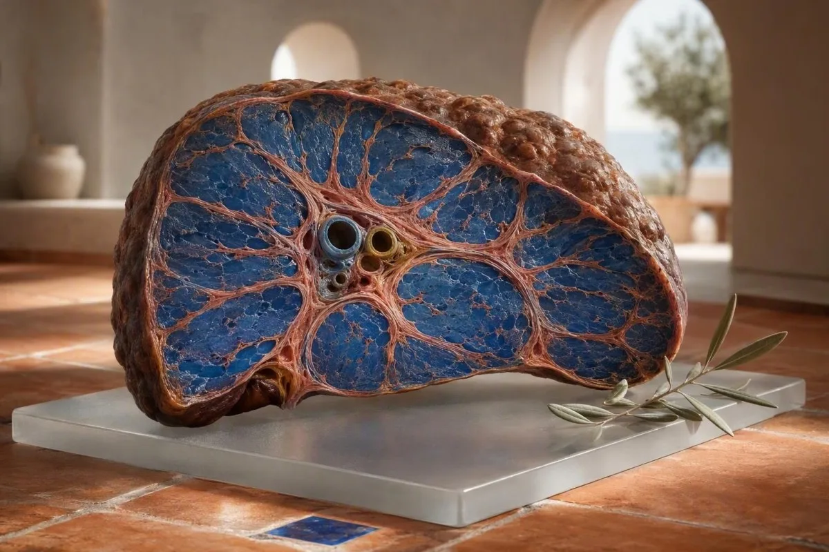
Cirrhosis symptoms shown through an anatomically accurate liver cross-section with subtle fibrous bands
图1: 肝脏横截面图显示,在出现明显外部症状之前,会逐渐形成瘢痕带。.

一个人可能仍然可以工作、训练,并且看起来完全健康,而纤维化仍在进展。我曾见过一名 48 岁的办公室职员,他唯一的改变是凌晨 3 点因瘙痒和疲劳醒来;他的 ALT 为 31 IU/L,但血小板在四年内从 214 降至 136 × 10⁹/L。这一趋势促使进行了弹性成像,并发现了晚期纤维化。.

肝硬化通常不会引起剧烈的肝痛。. 肝包膜可能在后期被拉伸,但早期的瘢痕形成在生物学上是“安静”的,因为纤维化是在微小的血管通道之间发生的。新的腹胀感、早饱感或脚踝肿胀比模糊的右侧腹痛更令人担忧,特别是当体重在一周内增加超过 2 公斤时。.

Thomas Klein 医生建议在进行肝脏检查时,携带之前的检查结果,而不仅仅是最近一次的检查。 基线血液检查 当可以与 12、24 和 48 个月前的结果进行比较时,就会变得更有价值。.

不应推迟至常规预约的症状

意识模糊、白天嗜睡加重、呕血、黑便、发热伴腹胀或黄疸迅速加重,需要当日急诊评估。. 这些可能反映了肝硬化失代偿、胃肠道出血、感染或急性肝损伤,而不是无害的波动。.

为什么正常的 ALT 和 AST 不能排除肝硬化

ALT 和 AST 正常并不能排除明显的纤维化或肝硬化。. ALT 和 AST 反映了当前的细胞酶泄漏,而纤维化反映了累积的结构性瘢痕,一旦肝细胞质量和炎症消退,可能不会产生多少持续的泄漏。.

Cirrhosis symptoms context with paired liver tissue views showing active injury and established scarring
图2: 确立的瘢痕组织可能与中度或正常的氨基转移酶结果并存。.

许多实验室对男性的 ALT 上限约为 35 至 45 IU/L,对女性约为 25 至 35 IU/L,尽管参考区间因检测方法和国家/地区而异。在肥胖、2 型糖尿病、血小板计数为 128 × 10⁹/L 且 FIB-4 评分高的患者中,28 IU/L 的结果可能在纸面上正常,但仍不那么令人放心。.

2023 年 AASLD 指南指出,非酒精性脂肪性肝病晚期患者的氨基转移酶可能正常;因此,风险分层应包括无创纤维化检测,而不仅仅依赖于酶水平(Rinella 等人,2023)。这是模式比单个数值的标志物更重要的领域之一。.

剧烈训练、肌肉损伤和某些药物会升高 AST 而没有肝脏瘢痕。一名 52 岁的马拉松运动员在比赛后 AST 为 89 IU/L,可能源于肌肉,特别是如果肌酸激酶升高而胆红素、血小板、INR 和 GGT 正常;我们的 AST 升高指南 解释了这种差别。.

肝硬化血液检查:医生实际担心的模式

最令人担忧的肝硬化血液检查模式是血小板进行性下降、AST 超过 ALT、胆红素或 GGT 升高以及白蛋白或 INR 受损,而不是孤立的酶升高。. 每项检查都反映了肝脏生物学的不同方面:损伤、胆汁流动、门脉高压或蛋白质生成。.

Cirrhosis symptoms laboratory pattern with liver panel, platelet count, and coagulation sample materials
图 3: 联合肝功能检查和全血细胞计数显示不同程度的肝功能障碍。.

血小板低于 150 × 10⁹/L 在晚期纤维化中很常见,因为门静脉高压会使脾脏肿大并增加血小板聚集。血小板计数为 142 × 10⁹/L 并非肝硬化的证据,但从 240 下降到 142 的趋势比单一的孤立结果在临床上更有意义。.

AST:ALT 比率高于 1.0 随着纤维化的进展而发生,而比率高于 2.0 通常与酒精相关性肝炎有关,但它既不敏感也不特异。GGT 常常随着酒精、代谢性脂肪肝、胆汁淤积和药物影响而升高;了解更多关于 GGT 的意义 再假定酒精是原因。.

Kantesti AI 通过权衡这些关联结果与年龄、性别、报告的病症和先前的值来解释肝功能检查,而不是根据单一标志物分配诊断。. Kantesti是一种AI血液检测分析仪 旨在识别随访模式,例如低血小板计数加升高的 AST:ALT 比率,这值得医生评估。.

常见的虚假安心陷阱

胆红素和白蛋白正常可能与代偿性肝硬化并存。. 这些标志物通常在肝脏失去更多功能储备后才出现恶化,因此它们是早期肝脏瘢痕的糟糕的独立筛查测试。.

FIB-4 和非侵入性临床显著瘢痕线索

FIB-4 是一个使用年龄、AST、ALT 和血小板计数的经过验证的一线纤维化评分。. 在 65 岁以下的成年人中,FIB-4 低于 1.3 通常表明晚期纤维化风险低,而结果为 2.67 或更高应触发专科评估或弹性成像。.

Cirrhosis symptoms assessment using age, liver enzymes, and platelets to estimate fibrosis risk
图 4: FIB-4 将年龄、氨基转移酶和血小板计数结合起来,用于评估纤维化风险。.

该公式为年龄 × AST 除以血小板 × ALT 的平方根,其中 AST 和 ALT 的单位为 IU/L,血小板的单位为 × 10⁹/L。年龄会显著改变得分:EASL 建议 65 岁以上人群的低风险阈值提高到 2.0,因为否则普通的年龄相关评分可能产生假阳性(欧洲肝脏研究协会,2021)。.

FIB-4 评分是一种分诊工具,而非诊断。. 急性肝炎、近期大量饮酒、化疗或短暂的血小板减少都可能导致其升高, ,因此我通常会在 4 到 12 周后重复不稳定的测试,然后再解释有疑问的结果,除非患者出现黄疸或其他危险信号。.

Kantesti 是一个 AI 实验室检测解读服务 可以计算和情境化 FIB-4,前提是存在必要的测量结果,但高分需要通过瞬时弹性成像、专科评估或偶尔的组织检查来获得医疗确认。我们的 肝功能检查概览 显示了哪些测试是常用的。.

低风险 FIB-4 <1.3 65 岁以下 晚期纤维化可能性低;如果代谢或酒精风险持续存在,请重新评估。.
中间风险 FIB-4 1.3-2.66 使用弹性成像或另一经验证的纤维化检测。.
较高风险的FIB-4 ≥2.67 可能性更高晚期纤维化;安排专科评估。.
老年人注意事项 年龄>65岁,阈值>2.0 谨慎解读,因为年龄会独立升高FIB-4。.

血小板减少:早期门静脉高压线索,而非定论

持续血小板计数低于150 × 10⁹/L可能是门脉高压的最早常规血液检测线索之一,但它有许多非肝脏原因。. 当计数逐渐下降并伴有脾脏肿大、高FIB-4或异常肝脏影像时,与肝脏联系会更强。.

Cirrhosis symptoms evaluation showing platelet cellular elements and spleen-liver portal circulation diagram
图 5: 门脉压力升高可能在胆红素升高之前导致脾脏血小板积聚。.

门脉高压改变肝脏和脾脏循环。脾脏可能保留更多血小板,因此在代偿性疾病中,计数为110至140 × 10⁹/L可能比腹水或食管胃底静脉曲张早数年出现。这就是为什么血小板趋势被包含在FIB-4中,而不是被视为通用CBC脚注。.

并非每个低计数都反映真实下降。EDTA相关血小板聚集可能导致计数假性降低,实验室可以通过血液涂片检查或柠檬酸盐样本确认;参见我们关于 EDTA 抗凝管导致的血小板凝集. 的解释。病毒性疾病、免疫性血小板减少症、叶酸缺乏症和骨髓疾病也仍然可能。.

Thomas Klein博士发现,两年内计数下降20%通常比询问计数是否已超过实验室下限更有用。将其与 血常规检查组成部分 和之前的超声报告配对,然后再下结论。.

白蛋白和 INR:衡量肝脏储备的血液检查

低白蛋白和INR延长可能表明肝脏合成功能下降,尤其是在它们同时出现时。. 白蛋白低于35 g/L或INR高于1.3(未接受抗凝治疗)需要及时临床复查,尽管两者都不是肝硬化的特异性结果。.

Cirrhosis symptoms laboratory evaluation with albumin protein model and coagulation assay equipment
图 6: 白蛋白生成和凝血因子合成反映了剩余肝脏功能储备。.

白蛋白半衰期约为20天,因此其变化缓慢,在代偿性肝硬化中通常保持正常。营养不良、肾脏蛋白丢失、严重系统性疾病、妊娠和液体超负荷也可能导致白蛋白降低; 白蛋白和球蛋白指南 有助于区分这些可能性。.

INR反映维生素K依赖性凝血因子活性,其中大部分由肝脏产生。. 对于未服用华法林人来说,INR为1.5比轻度升高的ALT, 更令人担忧,但由吸收不良、抗生素或胆汁淤积引起维生素K缺乏也可能延长INR,并可能逆转。.

Kantesti AI 是一种 基于 AI 的血液检测分析工具 强调白蛋白、胆红素、INR和血小板模式,因为ALT本身不能反映合成功能。关于凝血试验技术背景,请查阅我们 凝血检测指南.

黄疸、瘙痒、尿色深和粪便颜色浅:它们的含义

Jaundice, dark tea-coloured urine, pale stool, and generalised itch can indicate impaired bile flow or impaired bilirubin processing and should be assessed promptly. A total bilirubin above 34 µmol/L, about 2 mg/dL, often becomes visible as yellowing in daylight, though skin tone and lighting affect detection.

Cirrhosis symptoms education showing bilirubin movement through a liver and bile duct cross-section
图 7: Conjugated bilirubin reaches urine when bile processing or flow is impaired.

Dark urine is usually caused by conjugated bilirubin, not by bilirubin that is unconjugated. A urine dipstick positive for bilirubin therefore points clinicians toward hepatobiliary disease or obstruction rather than simple red-cell breakdown; our 尿胆红素指南 explains the next checks.

Pale or clay-coloured stool means less pigment is reaching the bowel. It may occur with a gallstone, medication-related cholestasis, pancreatic or bile-duct disease, or advanced liver dysfunction, so it is not safe to label it a detox reaction; see 苍白大便原因指南.

Itch from cholestasis can be severe before bilirubin becomes strikingly high. It commonly affects palms and soles and worsens at night; clinicians usually check ALP, GGT, bilirubin fractions, medication exposure, pregnancy status where relevant, and ultrasound access to the bile ducts.

ALP、GGT 和胆红素:识别胆汁淤积模式

A cholestatic liver-test pattern means alkaline phosphatase and GGT rise more than ALT and AST, often with direct bilirubin elevation. This pattern should prompt a search for bile-duct obstruction, medication injury, autoimmune cholestatic disease, or infiltrative liver conditions rather than an automatic assumption of cirrhosis.

Cirrhosis symptoms laboratory comparison showing bile ducts and cholestatic enzyme testing materials
图 8: Bile-flow abnormalities create a different laboratory signature from hepatocyte injury.

ALP is produced by bile-duct tissue but also by bone, placenta, and intestine. An elevated ALP with a normal GGT may be bone-derived, particularly after a fracture, during growth, or with vitamin D deficiency, whereas high ALP plus high GGT supports a hepatobiliary source; our ALP and GGT explanation 提供了实用示例。.

The R ratio helps classify acute enzyme patterns: ALT divided by its upper limit of normal, then divided by ALP divided by its upper limit. An R ratio above 5 suggests hepatocellular injury, below 2 suggests cholestasis, and 2 to 5 is mixed—useful for medication assessment but not a stand-alone diagnosis.

Kantesti AI uses laboratory reference limits from the uploaded report rather than imposing a universal ALP cutoff, because ranges differ. The cholestasis blood-test guide is useful preparation for a clinician discussion.

寻找病因与寻找瘢痕同样重要

Cirrhosis is an endpoint, not a single disease: alcohol, metabolic fatty liver disease, hepatitis B or C, autoimmune disease, iron overload, and some medications can all cause it. Identifying the cause changes treatment, family screening, cancer surveillance, and the chance of slowing further fibrosis.

Cirrhosis symptoms workup with liver cross-section and iron, viral, and metabolic laboratory clues
图 9: Different causes of fibrosis leave distinct clinical and laboratory clues.

Metabolic risk deserves a direct conversation. Central weight gain, triglycerides above 1.7 mmol/L or 150 mg/dL, hypertension, and type 2 diabetes increase the likelihood of steatotic liver disease, even when alcohol intake is low. A metabolic syndrome checklist can clarify the associated risks.

Ferritin above 300 ng/mL in men or postmenopausal women is not enough to diagnose iron overload. Transferrin saturation persistently above 45% is more specific for hereditary haemochromatosis and usually prompts HFE genetic testing in an appropriate clinical setting; see our haemochromatosis symptoms guide.

Medication and supplement histories are routinely underestimated. High-dose green tea extract, anabolic agents, multi-ingredient weight-loss products, amiodarone, methotrexate, and some antibiotics can alter liver tests, while a supposed liver cleanse may delay proper evaluation; our supplement safety review is deliberately cautious about this.

Viral hepatitis should be tested, not guessed from symptoms

Chronic hepatitis C can remain asymptomatic for decades while fibrosis progresses. Antibody testing identifies exposure, while an HCV RNA test confirms active infection; our 丙型肝炎检测指南 explains why both tests matter.

异常肝硬化血液检查模式后会发生什么

An abnormal fibrosis pattern usually leads to repeat blood tests, an ultrasound, and elastography rather than an immediate diagnosis of cirrhosis. Elastography estimates liver stiffness in kilopascals and is most reliable when acute inflammation, congestion, and recent heavy alcohol exposure are considered.

Cirrhosis symptoms assessment with liver elastography probe and stiffness map style education scene
图 10: Elastography provides a non-invasive estimate of liver stiffness after risk stratification.

A clinician commonly repeats AST, ALT, ALP, GGT, bilirubin, albumin, INR, and full blood count within weeks if results are unexpected. They may add hepatitis B and C tests, ferritin with transferrin saturation, immunoglobulins, autoantibodies, coeliac testing, and ceruloplasmin in younger adults—although the exact list follows age and risk history.

EASL's 2021 non-invasive-test guideline supports a two-step approach: first FIB-4, then transient elastography or a second serum test for people above the low-risk threshold (European Association for the Study of the Liver, 2021). Liver stiffness is not identical to fibrosis; heart failure congestion, cholestasis, and acute hepatitis can temporarily raise it.

Kantesti 是一个 AI 生物标志物解读平台 that can organise an uploaded panel into questions for the next appointment, including which values changed and what potential causes need exclusion. Our clinical methodology is described in the 医学验证概览, but it does not replace an examination or imaging order.

酒精、代谢风险以及何时重复进行肝脏检查

Alcohol reduction and metabolic risk treatment can improve liver enzymes within weeks, but fibrosis risk should not be judged from one improved ALT result. GGT often declines over 2 to 6 weeks after alcohol cessation, while platelet and fibrosis-score changes may take longer and depend on the underlying disease stage.

Cirrhosis symptoms follow-up showing a person recording liver test trends beside alcohol-free drinks and meals
图 11: Repeated laboratory trends show whether liver-risk changes are sustained over time.

There is no universally safe alcohol threshold for a person with established cirrhosis; abstinence is the standard recommendation because even small amounts may worsen portal hypertension or interfere with recovery. For people without cirrhosis, reported intake still needs context: binge patterns, body weight, diabetes, and medication use modify risk considerably.

In metabolic liver disease, a sustained 7% to 10% body-weight reduction is associated with improvement in steatohepatitis and can improve fibrosis in some patients, although individual results vary. Crash dieting can transiently raise ALT, so interpret a panel alongside recent weight change; our article on 减肥后肝酶 解释了原因。.

Kantesti AI trend analysis is most informative when tests are taken at comparable points—ideally avoiding strenuous exercise for 48 hours and noting alcohol, illness, and new supplements. The alcohol cessation biomarker timeline describes what may normalise first.

可能延误肝脏护理的食物和补充剂误区

No juice cleanse, herbal product, or supplement can reliably remove cirrhosis scar tissue. A Mediterranean-style eating pattern, adequate protein, diabetes management, and avoidance of hepatotoxic supplements can support liver health, but they are not substitutes for identifying the cause of fibrosis.

Cirrhosis symptoms nutrition education with Mediterranean foods beside a liver anatomy model and lab record
图 12: Whole foods support metabolic health but do not replace fibrosis assessment.

People with cirrhosis are sometimes told to avoid all protein, which can worsen muscle loss and frailty. Unless there is a specific temporary instruction during severe hepatic encephalopathy, many patients need roughly 1.2 to 1.5 g protein per kg body weight daily, divided across meals, under dietetic guidance.

Be wary of products marketed as detoxifiers. Concentrated turmeric, kava, black cohosh, and multi-ingredient bodybuilding or slimming supplements have all been linked to liver injury in case series, and products may not contain what their labels claim. Our evidence-based liver food guide separates supportive dietary habits from marketing claims.

If fluid retention develops, sodium restriction often matters more than fluid restriction. A common target is less than 2 g sodium daily, but diuretics, kidney function, blood pressure, and nutritional adequacy require personalised oversight rather than an internet meal plan.

肝硬化症状何时变得紧急

Vomiting blood, black tarry stool, confusion, fever with abdominal swelling, severe shortness of breath, or rapidly worsening jaundice are emergency symptoms in someone with known or suspected cirrhosis. These signs can represent variceal bleeding, hepatic encephalopathy, spontaneous bacterial peritonitis, kidney injury, or acute-on-chronic liver failure.

Cirrhosis symptoms urgent-care pathway illustrated with liver warning signs and clinical triage setting
图 13: New confusion, gastrointestinal bleeding, or tense abdominal swelling requires urgent assessment.

New confusion or a reversed sleep-wake cycle is not simply fatigue in cirrhosis. It may indicate hepatic encephalopathy, especially if accompanied by constipation, dehydration, infection, gastrointestinal bleeding, sedative use, or a high ammonia result; ammonia alone, however, does not reliably diagnose or grade encephalopathy.

A distended abdomen can reflect ascites, but it can also arise from obesity, bowel distension, ovarian disease, or heart failure. Fever, tenderness, low blood pressure, or worsening kidney function in a person with ascites should be assessed immediately because infection can be subtle and dangerous.

For non-emergency uncertainty, write down symptoms, medicines, alcohol intake, supplements, and date-stamped weights before calling a clinician. A 血液检查摘要清单 can make that conversation more efficient.

改变决策的实用肝脏就诊清单

The best liver appointment includes prior laboratory reports, a realistic alcohol and medication history, metabolic history, and any imaging reports. These details let clinicians distinguish a transient enzyme result from a multi-year pattern suggesting fibrosis.

Bring every result you can find, including CBC, liver panel, INR, ferritin, hepatitis tests, ultrasound reports, and elastography values in kPa. A single result can be noisy; three results spanning 18 months can show whether platelets are falling or bilirubin is stable. Our 并排结果比较指南 explains what changes are usually meaningful.

Ask four direct questions: What is the likely cause? What fibrosis stage is suspected? Do I need elastography or hepatology referral? Which symptoms should send me to urgent care? If cirrhosis is confirmed, ask about six-monthly ultrasound surveillance for liver cancer and whether endoscopy is needed to assess varices.

As of September 4, 2026, our doctors still recommend that AI-generated explanations be treated as preparation, not diagnosis. Kantesti's clinical work is reviewed with oversight from the 医疗顾问委员会, and the final decision about cirrhosis belongs with the treating clinician who can examine you and order confirmatory testing.

常见问题

肝硬化可以发生在肝酶正常的情况下吗?

是的。正常的 ALT 和 AST 并不能排除肝硬化,因为这些酶反映的是当前的肝细胞损伤,而不是已经形成的瘢痕组织量。失代偿期肝硬化可能表现为 ALT 和 AST 在实验室参考区间内,尤其是在代谢性肝病中。血小板计数低于 150 × 10⁹/L,FIB-4 评分大于或等于 2.67,弹性成像异常,白蛋白降低或 INR 升高,比单独的酶学检查能提供更有用的线索。.

肝硬化的最初警告迹象是什么?

早期肝硬化症状可能包括持续疲劳、食欲不振、瘙痒、容易瘀伤、运动耐力下降、睡眠改变和腹胀,但许多人没有症状。黄疸、尿色深、粪便颜色浅、脚踝肿胀、腹水、意识模糊、呕血和黑便往往是晚期或紧急体征。仅凭症状无法诊断肝硬化,因此通常需要肝功能检查、血常规(含血小板)、INR、白蛋白和纤维化评估。.

什么是提示肝硬化的血小板计数?

血小板计数低于 150 × 10⁹/L,如果持续存在且呈下降趋势,可能提示晚期肝纤维化或门静脉高压,但不能诊断肝硬化。血小板聚集、病毒感染、免疫性血小板减少症、药物、营养缺乏和骨髓疾病也可能导致血小板计数降低。与单一轻度偏低读数相比,两年内 AST 升高且 FIB-4 指数高,从 220 降至 135 × 10⁹/L 的进行性下降更有信息价值。.

FIB-4评分意味着肝硬化吗?

FIB-4 无法诊断肝硬化,但它根据年龄、AST、ALT 和血小板估算进展性肝纤维化的风险。在 65 岁以下的成人中,得分低于 1.3 通常表示低风险,1.3 至 2.66 需要进一步的非侵入性评估,2.67 或更高则需要专科评估。在 65 岁以上的成人中,通常使用高于 2.0 的阈值来减少与年龄相关的假阳性。.

早期发现肝硬化能改善吗?

早期纤维化可在病因被去除或得到控制后得到改善,包括持续戒酒、成功的病毒性肝炎治疗、代谢性肝病的减重或免疫介导性疾病的治疗。既定的肝硬化可能无法完全逆转,但通过专科护理通常可以减少并发症和进一步的疤痕。对于许多代谢性肝病患者来说,持续的 7% 至 10% 体重减轻可以改善脂肪性肝炎,但结果因患者而异。.

什么时候应该去医院看疑似肝硬化?

如有呕血、柏油样黑便、新出现的意识模糊、晕厥、伴腹胀发热、严重黄疸或呼吸急促迅速加重,请立即就医。这些症状可能提示食管胃底静脉曲张出血、腹水感染、肝性脑病或急性肝衰竭,不应等待门诊复查血检。如果症状轻微,但胆红素超过 34 µmol/L,INR 超过 1.3(未使用抗凝剂时),或血小板进行性减少,请及时联系医生咨询。.

立即获取AI驱动的血液检测分析

加入全球超过2,000,000名用户,他们信任Kantesti进行即时、准确的检验分析。上传你的血液检查结果,并在几秒内获得对15,000+生物标志物的全面解读。.

📚 参考研究论文

1

Klein, T., Mitchell, S., & Weber, H. (2026). Kantesti Research Team. (2026). aPTT Normal Range: D-Dimer, Protein C Blood Clotting Guide. Zenodo. https://doi.org/10.5281/zenodo.18262555. ResearchGate and Academia.edu availability links are maintained through the publication record.. Kantesti AI医学研究。.

2

Klein, T., Mitchell, S., & Weber, H. (2026). Kantesti Research Team. (2026). Serum Proteins Guide: Globulins, Albumin & A/G Ratio Blood Test. Zenodo. https://doi.org/10.5281/zenodo.18316300. ResearchGate and Academia.edu availability links are maintained through the publication record.. Kantesti AI医学研究。.

📖 外部医学参考资料

3

欧洲肝病研究协会(European Association for the Study of the Liver)(2021)。. EASL Clinical Practice Guidelines on non-invasive tests for evaluation of liver disease severity and prognosis – 2021 update.。 《Journal of Hepatology》。.

4

Rinella ME等。(2023)。. AASLD关于非酒精性脂肪性肝病的临床评估与管理的实践指南.。 《肝脏病学》。.

200万+测试分析
127+国家
75+语言

⚕️ 医疗免责声明

E-E-A-T信任信号

经验

由医生主导的临床审阅:实验室解读工作流程。.

📋

专业知识

实验室医学重点:生物标志物在临床情境中的表现。.

👤

权威

由 Thomas Klein 博士撰写,并由 Sarah Mitchell 博士与 Hans Weber 教授审阅。.

🛡️

可信度

基于循证的解读,并提供清晰的后续路径以减少警报。.

🏢 Kantesti LTD 在英格兰和威尔士注册 · 公司编号:. 17090423 英国伦敦 · kantesti.net
blank
作者:Prof. Dr. Thomas Klein

Thomas Klein 博士是获得美国内科医学委员会认证的临床血液科医生,担任 Kantesti AI 的首席医疗官。凭借超过 15 年的实验室医学经验,并对借助 AI 进行血液检查结果解读抱有浓厚兴趣,他致力于将新技术与日常临床实践相连接。他的研究兴趣包括生物标志物分析、临床决策支持研究以及针对特定人群的参考范围优化。作为 CMO,他为平台的内部基准评估提供临床输入,并对 Kantesti 教育报告的医疗质量提供临床监督。.

发表回复

您的邮箱地址不会被公开。 必填项已用 * 标注