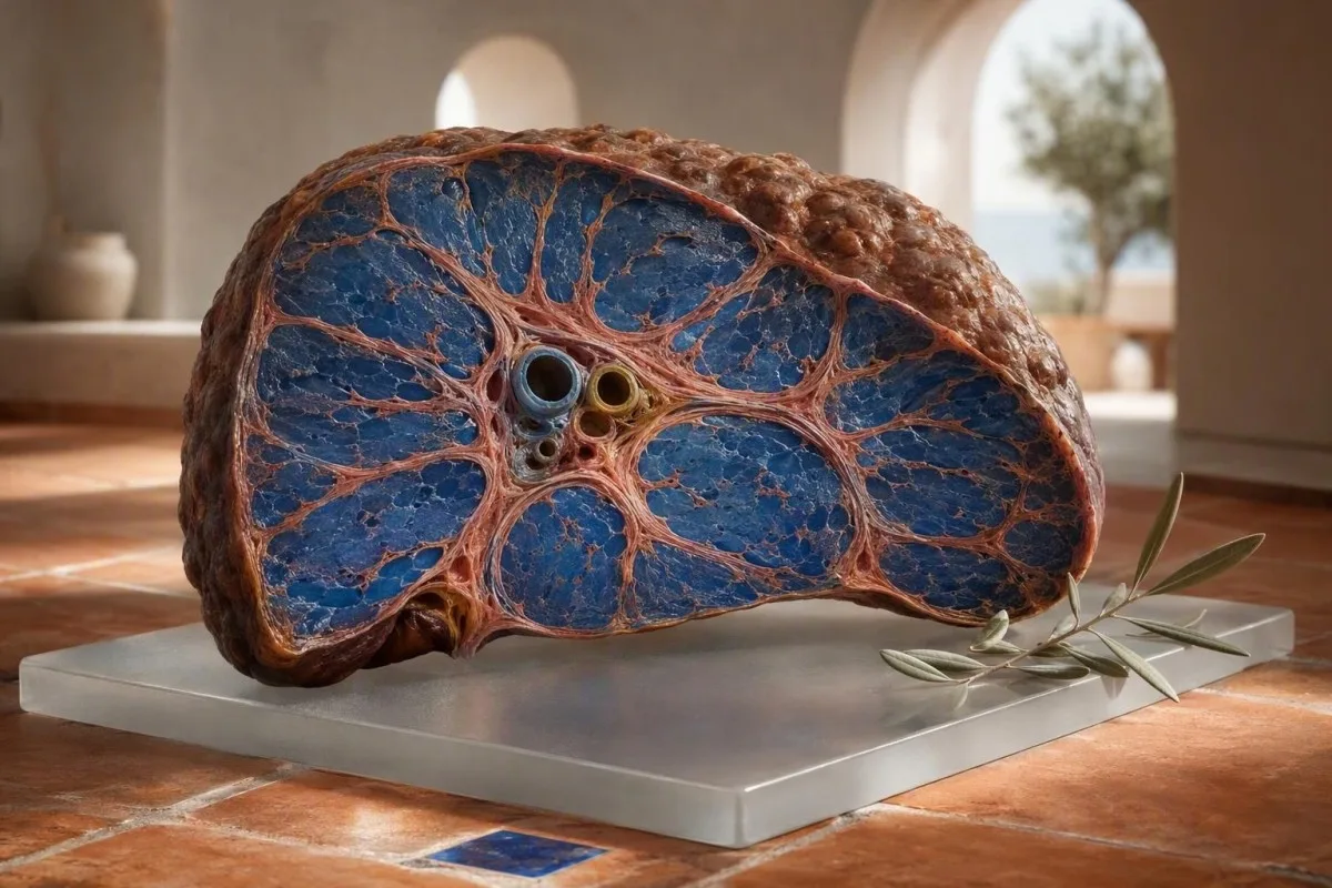
ក្រិនថ្លើមដំណាក់កាលដំបូងច្រើនតែមិនបង្កជាការឈឺចាប់ជាក់ស្តែងទេ៖ ភាពអស់កម្លាំង ការផ្លាស់ប្តូរដំណេក ភាពជាំ រមាស់ និងការថយចុះចំនួនប្លាកែតអាចកើតមានឡើងមុនពេលមានជម្ងឺខាន់លឿង។ ការចង្អុលបង្ហាញដែលមានប្រយោជន៍ជាធម្មតាច្រើនតែជាលំនាំនៃលទ្ធផលតេស្តថ្លើម ការរាប់ចំនួនឈាម និងពិន្ទុនៃជំងឺក្រិនថ្លើម — មិនមែនជាអង់ស៊ីមដែលមិនប្រក្រតីតែមួយទេ។.


- ស្លាកស្នាមស្ងាត់ អាចកើតឡើងជាមួយនឹង ALT និង AST ធម្មតា ជាពិសេសនៅក្នុងជំងឺថ្លើមខ្លាញ់ដែលទាក់ទងនឹងមេតាប៉ូលីស និងជំងឺថ្លើមដែលទាក់ទងនឹងគ្រឿង ។.
- ប្លាកែតទាបជាង 150 × 10⁹/L អាចជាសញ្ញាដំបូងនៃជំងឺលើសឈាមក្នុងប្រពន្ធរន្ធគូថ នៅពេលដែលពួកគេធ្លាក់ចុះជាបន្តបន្ទាប់រួមជាមួយកត្តាហានិភ័យថ្លើម។.
- FIB-4 ក្រោម 1.3 ជាធម្មតាបង្ហាញពីហានិភ័យទាបនៃជំងឺក្រិនថ្លើមដំណាក់កាលខ្ពស់ចំពោះមនុស្សពេញវ័យក្រោម 65 ឆ្នាំ; 2.67 ឬខ្ពស់ជាងនេះ ទាមទារការវាយតម្លៃពីអ្នកឯកទេសជាបន្ទាន់។.
- Albumin ក្រោម 35 g/L ឬ INR កើនឡើងអាចបង្ហាញពីការថយចុះមុខងារសំយោគថ្លើម ទោះបីជា aminotransferases មានកម្រិតមធ្យមក៏ដោយ។.
- AST:ALT លើសពី 1 មិនមែនជាការធ្វើរោគវិនិច្ឆ័យជំងឺក្រិនថ្លើមទេ ប៉ុន្តែវាបង្កឱ្យមានការព្រួយបារម្ភនៅពេលផ្សំជាមួយនឹងជំងឺthrombocytopenia, GGT កើនឡើង, ឬការផ្លាស់ប្តូររូបភាព។.
- Bilirubin លើសពី 34 µmol/L អ្នកដែលមានជំងឺខាន់លឿង ទឹកនោមពណ៌ខ្មៅ ក្តៅខ្លួន ភាន់ច្រឡំ ឬពោះរីកហើម ត្រូវការការវាយតម្លៃពីគ្រូពេទ្យជាបន្ទាន់។.
- អង់ស៊ីមធម្មតា មានន័យថាមិនមានការលេចធ្លាយអង់ស៊ីមសកម្មនៅពេលនោះទេពួកគេមិនបញ្ជាក់ថាស្ថាបត្យកម្មថ្លើមមានលក្ខណៈធម្មតាទេ។.
- ការឈប់ផឹកស្រា ការព្យាបាលហានិភ័យមេតាប៉ូលីស ការថែទាំជំងឺរលាកថ្លើមដោយមេរោគ និងការពិនិត្យថ្នាំ អាចបន្ថយល្បឿន ឬពេលខ្លះបញ្ច្រាស ការឡើងដំបៅនៅដំណាក់កាលដំបូង។.
រោគសញ្ញាដំបូងនៃក្រិនថ្លើមដែលមិនច្បាស់លាស់ដែលសមនឹងទទួលបានការពិនិត្យថ្លើម
រោគសញ្ញាដំបូងនៃជំងឺក្រិនថ្លើមច្រើនតែមិនច្បាស់លាស់ និងកើតឡើងជា intermittent, ជាមួយនឹងអស់កម្លាំង ការថយចុះចំណង់អាហារ ការគេងមិនលក់ រមាស់ ហូរឈាមងាយ និងការថយចុះសមត្ថភាពធ្វើលំហាត់ប្រាណ កើតឡើងយូរមុនពេលស្បែកលឿង ឬការហើម។ បទពិសោធន៍គ្លីនិករបស់ខ្ញុំ លក្ខណៈពិសេសដែលគួរឱ្យព្រួយបារម្ភមិនមែនជាការអស់កម្លាំងមួយរសៀលនោះទេ វាជាការប្រមូលផ្តុំដែលបន្តរយៈពេល 6 ទៅ 12 សប្តាហ៍ចំពោះអ្នកដែលមានជំងឺទឹកនោមផ្អែម ធាត់ ការប៉ះពាល់នឹងជាតិអាល់កុលទៀងទាត់ ហានិភ័យនៃជំងឺរលាកថ្លើម ឬប្រវត្តិគ្រួសារនៃជំងឺថ្លើម។.
មនុស្សម្នាក់នៅតែអាចធ្វើការ ហ្វឹកហាត់ និងមើលទៅមានសុខភាពល្អទាំងស្រុង ខណៈពេលដែលជំងឺក្រិនថ្លើមកើតឡើង។ ខ្ញុំបានឃើញបុគ្គលិកការិយាល័យម្នាក់ អាយុ 48 ឆ្នាំ ដែលមានតែការផ្លាស់ប្តូរគឺភ្ញាក់នៅម៉ោង 3 ទៀបភ្លឺដោយសាររមាស់ និងអស់កម្លាំង។ ALT របស់គាត់មាន 31 IU/L ប៉ុន្តែប្លាកែតបានធ្លាក់ចុះពី 214 ទៅ 136 × 10⁹/L ក្នុងរយៈពេលបួនឆ្នាំ។ និន្នាការនោះបានជំរុញឱ្យមានការធ្វើអេឡាស់ត្រូហ្គាពី (elastography) និងបានរកឃើញជំងឺក្រិនថ្លើមដំណាក់កាលខ្ពស់។.
ជំងឺក្រិនថ្លើមជាធម្មតាមិនបណ្តាលឱ្យមានការឈឺថ្លើមស្រួចស្រាវទេ។. ភ្នាសថ្លើមអាចលាតសន្ធឹងនៅដំណាក់កាលចុងក្រោយ ប៉ុន្តែការឡើងស្នាមជាំដំបូងមានសភាពស្ងប់ស្ងាត់ខាងជីវសាស្ត្រ ដោយសារការឡើងដំបៅកើតឡើងនៅចន្លោះបណ្តាញសរសៃឈាមមីក្រូទស្សន៍។ ការមានអារម្មណ៍ពេញពោះ កាន់តែឆាប់ឆ្អែត ឬហើមកជើង គឺគួរឱ្យព្រួយបារម្ភជាងការឈឺនៅផ្នែកខាងស្តាំដែលមិនច្បាស់លាស់ ជាពិសេសនៅពេលទម្ងន់កើនឡើងលើសពី 2 គីឡូក្រាមក្នុងមួយសប្តាហ៍។.
ផ្ទាំងលទ្ធផល តេស្តឈាមមូលដ្ឋាន កាន់តែមានប្រយោជន៍ច្រើន នៅពេលដែលអាចប្រៀបធៀបជាមួយលទ្ធផលពី 12, 24, និង 48 ខែមុន។.
រោគសញ្ញាដែលមិនគួររង់ចាំការណាត់ជួបធម្មតា
ការភាន់ច្រឡំ ការងងុយដេកនៅពេលថ្ងៃ ការក្អួតឈាម ការបន្ទោររាវខ្មៅ ក្តៅខ្លួនជាមួយនឹងការហើមពោះ ឬជំងឺខាន់លឿងរីកលឿន ត្រូវការការវាយតម្លៃជាបន្ទាន់នៅថ្ងៃតែមួយ។. ទាំងនេះអាចឆ្លុះបញ្ចាំងពីជំងឺក្រិនថ្លើមដែលបាត់បង់សមត្ថភាព ការហូរឈាមក្នុងប្រព័ន្ធរំលាយអាហារ ការឆ្លងមេរោគ ឬការរងរបួសថ្លើមស្រួចស្រាវ ជាជាងការប្រែប្រួលដែលគ្មានគ្រោះថ្នាក់។.
ហេតុអ្វីបានជា ALT និង AST ធម្មតា មិនអាចបដិសេធជំងឺក្រិនថ្លើមបានទេ
ALT និង AST ធម្មតា មិនបានដកចេញនូវជំងឺក្រិនថ្លើម ឬជំងឺក្រិនថ្លើមដែលមានន័យទេ។. ALT និង AST ឆ្លុះបញ្ចាំងពីការលេចធ្លាយអង់ស៊ីមក្នុងកោសិកាដែលមានបច្ចុប្បន្ន ចំណែកជំងឺក្រិនថ្លើមឆ្លុះបញ្ចាំងពីការឡើងស្នាមជាំនៃស្ថាបត្យកម្មដែលប្រមូលផ្តុំ ដែលអាចបង្កឱ្យមានការលេចធ្លាយបន្តិចបន្តួច នៅពេលម៉ាស់កោសិកាថ្លើម និងការរលាកថយចុះ។.
មន្ទីរពិសោធន៍ជាច្រើនប្រើប្រាស់ដែនកំណត់ខាងលើរបស់ ALT ប្រហែល 35 ទៅ 45 IU/L សម្រាប់បុរស និង 25 ទៅ 35 IU/L សម្រាប់ស្ត្រី ទោះបីជាចន្លោះយោងប្រែប្រួលទៅតាមការវិភាគ និងប្រទេសក៏ដោយ។ លទ្ធផល 28 IU/L អាចជារឿងធម្មតានៅលើក្រដាស ប៉ុន្តែនៅតែមិនគួរឱ្យទុកចិត្តចំពោះអ្នកដែលមានជំងឺធាត់ ជំងឺទឹកនោមផ្អែមប្រភេទ 2 ចំនួនប្លាកែត 128 × 10⁹/L និងពិន្ទុ FIB-4 ខ្ពស់។.
ការណែនាំរបស់ AASLD ឆ្នាំ 2023 ចែងថា អាមីណូត្រាស្ហ្វឺរ៉ាស់ (aminotransferases) អាចមានលក្ខណៈធម្មតាចំពោះអ្នកជំងឺដែលមានជំងឺថ្លើមខ្លាញ់មិនមែនអាល់កុលដំណាក់កាលខ្ពស់ ហេតុដូច្នេះហើយ ការកំណត់ហានិភ័យគួរតែរួមបញ្ចូលការធ្វើតេស្តជំងឺក្រិនថ្លើមដែលមិនមានការឈឺចាប់ជាជាងអង់ស៊ីមតែមួយមុខ (Rinella et al., 2023) ។ នេះគឺជាផ្នែកមួយដែលលំនាំមានសារៈសំខាន់ជាងទង់ជាតិដែលមានតម្លៃតែមួយ។.
ការហ្វឹកហាត់យ៉ាងខ្លាំង ការរងរបួសសាច់ដុំ និងថ្នាំមួយចំនួនអាចបង្កើន AST ដោយគ្មានការឡើងស្នាមជាំថ្លើម។ អ្នករត់ម៉ារ៉ាតុនអាយុ 52 ឆ្នាំម្នាក់ដែលមាន AST 89 IU/L បន្ទាប់ពីការប្រណាំងអាចមានប្រភពមកពីសាច់ដុំ ជាពិសេសប្រសិនបើ creatine kinase ខ្ពស់ និង bilirubin, platelets, INR, និង GGT ធម្មតា; របស់យើង ការណែនាំ AST ខ្ពស់ ពន្យល់ពីភាពខុសគ្នានោះ។.
ការធ្វើតេស្តឈាមរកជំងឺក្រិនថ្លើម៖ លំនាំដែលគ្រូពេទ្យពិតជាបារម្ភ
លំនាំតេស្តឈាមជំងឺក្រិនថ្លើមដែលគួរឱ្យព្រួយបារម្ភបំផុតរួមបញ្ចូលប្លាកែតធ្លាក់ចុះ AST លើស ALT, bilirubin ឬ GGT កើនឡើង និង albumin ឬ INR ថយចុះ ជាជាងការកើនឡើងអង់ស៊ីមតែមួយ។. ការធ្វើតេស្តនីមួយៗចាប់យកទិដ្ឋភាពផ្សេងគ្នានៃជីវវិទ្យាថ្លើម៖ ការរងរបួស ការហូរប្រមាត់ សម្ពាធក្នុងសរសៃឈាមក្នុងប្រហោងពោះ ឬការផលិតប្រូតេអ៊ីន។.
ប្លាកែតទាបជាង 150 × 10⁹/L កើតឡើងជាទូទៅក្នុងជំងឺក្រិនថ្លើមដំណាក់កាលខ្ពស់ ដោយសារសម្ពាធក្នុងប្រព័ន្ធច្រាសថ្លើមធ្វើឲ្យកូនកណ្ដុររីកធំ និងបង្កើនការប្រមូលផ្តុំប្លាកែត។ ការរាប់ចំនួន ១៤២ × ១០⁹/លីត្រ មិនមែនជាភស្តុតាងនៃជំងឺក្រិនថ្លើមទេ ប៉ុន្តែនិន្នាការថយចុះពី ២៤០ ទៅ ១៤២ មានសារសំខាន់ខាងគ្លីនិកជាងលទ្ធផលតែមួយប៉ុណ្ណោះ។.
សមាមាត្រ AST:ALT ខ្ពស់ជាង ១.០ អាចកើតឡើងនៅពេលដែលជំងឺក្រិនថ្លើមរីករាលដាល ខណៈដែលសមាមាត្រខ្ពស់ជាង ២.០ ត្រូវបានផ្សារភ្ជាប់ជាប្រពៃណីជាមួយនឹងជំងឺរលាកថ្លើមដែលទាក់ទងនឹងជាតិអាល់កុល ប៉ុន្តែមិនមានភាពរសើបឬភាពជាក់លាក់ទេ។ GGT តែងតែកើនឡើងដោយសារជាតិអាល់កុល ជំងឺថ្លើមខ្លាញ់ដោយសារមេតាបូលីស ជំងឺប្រមាត់ស្ទះ និងផលប៉ះពាល់ពីថ្នាំ។ សូមអានបន្ថែមអំពី អត្ថន័យនៃ GGT មុននឹងសន្មតថាជាតិអាល់កុលជាមូលហេតុ។.
Kantesti AI បកស្រាយលទ្ធផលពិនិត្យថ្លើមដោយថ្លឹងថ្លែងលទ្ធផលដែលតភ្ជាប់គ្នាទាំងនេះទាក់ទងនឹងអាយុ ភេទ លក្ខខណ្ឌដែលបានរាយការណ៍ និងតម្លៃពីមុនជាជាងការផ្តល់ការធ្វើរោគវិនិច្ឆ័យពីសញ្ញាមួយប៉ុណ្ណោះ។. Kantesti គឺជាឧបករណ៍វិភាគតេស្តឈាមដោយ AI ត្រូវបានរចនាឡើងដើម្បីកំណត់អត្តសញ្ញាណគំរូតាមដានដូចជាប្លាកែតទាបរួមជាមួយសមាមាត្រ AST:ALT កើនឡើង ដែលទាមទារការពិនិត្យដោយគ្រូពេទ្យ។.
دامលនៃការធានាសុវត្ថិភាពខុសឆ្គងទូទៅ
ប៊ីលីរុយប៊ីនធម្មតា និងអាល់ប៊ុយមីនធម្មតាអាចកើតឡើងជាមួយនឹងជំងឺក្រិនថ្លើមដែលមានសំណង។. សូចនាករទាំងនោះតែងតែកាន់តែយ៉ាប់យ៉ឺននៅពេលក្រោយ បន្ទាប់ពីថ្លើមបានបាត់បង់ការបម្រុងមុខងារកាន់តែច្រើន ដូច្នេះវាជាការធ្វើតេស្តច្រោះរកស្នាមស្លាកស្នាមដំបូងដែលមិនសូវល្អ។.
FIB-4 និងសញ្ញាដែលមិនជ្រៀតជ្រែកក្នុងការរកឃើញស្លាកស្នាមដែលមានសារៈសំខាន់ខាងគ្លីនិក
FIB-4 គឺជាពិន្ទុច្រោះរកជំងឺក្រិនថ្លើមដែលមានសុពលភាពសម្រាប់ការប្រើប្រាស់ដំបូង ដោយប្រើអាយុ AST ALT និងចំនួនប្លាកែត។. ចំពោះមនុស្សពេញវ័យអាយុក្រោម ៦៥ ឆ្នាំ FIB-4 ទាបជាង ១.៣ ជាទូទៅបង្ហាញពីហានិភ័យទាបនៃជំងឺក្រិនថ្លើមដំណាក់កាលខ្ពស់ ខណៈដែលលទ្ធផល ២.៦៧ ឬខ្ពស់ជាងនេះគួរតែជំរុញឱ្យមានការវាយតម្លៃដោយអ្នកឯកទេស ឬការស្កេនអេឡាសโตហ្គ្រាហ្វី។.
រូបមន្តគឺអាយុ × AST បែងចែកដោយប្លាកែត × ការគណនាការ៉េនៃ ALT ដោយ AST និង ALT ក្នុង IU/L និងប្លាកែតក្នុង × 10⁹/L ។ អាយុផ្លាស់ប្តូរពិន្ទុយ៉ាងច្រើន ៖ EASL ណែនាំកម្រិតហានិភ័យទាបខ្ពស់ជាងមុនគឺ ២.០ សម្រាប់មនុស្សអាយុលើសពី ៦៥ ឆ្នាំ ដោយសារការគណនាតាមអាយុធម្មតាអាចផ្តល់លទ្ធផលវិជ្ជមានក្លែងក្លាយបានបើមិនដូច្នេះទេ (European Association for the Study of the Liver, 2021)។.
ពិន្ទុ FIB-4 គឺជាឧបករណ៍តម្រៀប មិនមែនជាការធ្វើរោគវិនិច្ឆ័យទេ។. ជំងឺរលាកថ្លើមស្រួចស្រាវ ការផឹកស្រាច្រើនថ្មីៗ ការព្យាបាលដោយគីមីบำบัด ឬការធ្លាក់ចុះប្លាកែតបណ្ដោះអាសន្នអាចធ្វើឲ្យវាកើនឡើង។, ដូច្នេះខ្ញុំតែងតែធ្វើតេស្តដែលមិនស្ថិតស្ថេរឡើងវិញក្នុងរយៈពេល ៤ ទៅ ១២ សប្តាហ៍មុននឹងបកស្រាយលទ្ធផលដែលមានព្រំដែនលុះត្រាតែបុគ្គលនោះមានជំងឺខាន់លឿង ឬសញ្ញាគ្រោះថ្នាក់ដទៃទៀត។.
កាន់តេស្ទី គឺជា សេវាកម្មបកស្រាយការធ្វើតេស្តរបស់ AI lab ដែលអាចគណនា និងដាក់បរិបទ FIB-4 នៅពេលដែលលទ្ធផលចាំបាច់មានវត្តមាន ប៉ុន្តែពិន្ទុខ្ពស់ត្រូវការការបញ្ជាក់ផ្នែកវេជ្ជសាស្ត្រដោយការស្កេនអេឡាសโตហ្គ្រាហ្វី ការវាយតម្លៃដោយអ្នកឯកទេស ឬពេលខ្លះការពិនិត្យជាលិកា។ ការពិនិត្យរួមនៃបន្ទះថ្លើមរបស់យើង បង្ហាញពីការធ្វើតេស្តណាដែលមានជាទូទៅ។.
ប្លាកែតទាប៖ សញ្ញាដំបូងនៃជំងឺលើសឈាមក្នុងប្រពន្ធរន្ធគូថ មិនមែនជាការសម្រេចចិត្តទេ
ចំនួនប្លាកែតទាបជាបន្តបន្ទាប់ក្រោម 150 × 10⁹/L អាចជាសញ្ញាដំបូងបំផុតនៃការធ្វើតេស្តឈាមជាប្រចាំនៃជំងឺលើសសម្ពាធឈាមក្នុងប្រហោងឆ្អឹង ប៉ុន្តែវាក៏មានមូលហេតុក្រៅពីថ្លើមជាច្រើនផងដែរ។. ទំនាក់ទំនងនឹងថ្លើមកាន់តែរឹងមាំនៅពេលដែលចំនួនថយចុះជាលំដាប់ហើយកើតមានរួមជាមួយនឹងការរីកធំនៃលំពែង, FIB-4 ខ្ពស់, ឬរូបភាពថ្លើមមិនប្រក្រតី។.
ជំងឺលើសសម្ពាធក្នុងប្រហោងឆ្អឹងផ្លាស់ប្តូរលំហូរឈាមតាមរយៈថ្លើម និងលំពែង។ លំពែងអាចរក្សាប្លាកែតបានកាន់តែច្រើន ដូច្នេះចំនួនពី 110 ទៅ 140 × 10⁹/L អាចកើតមានមុនជំងឺហើមពោះ ឬសរសៃឈាមរីកក្នុងរយៈពេលជាច្រើនឆ្នាំក្នុងជំងឺដែលមានការទូទាត់សងវិញ។ នោះហើយជាមូលហេតុដែលនិន្នាការប្លាកែតត្រូវបានបញ្ចូលទៅក្នុង FIB-4 ជាជាងត្រូវបានចាត់ទុកថាជាសេចក្តីអត្ថាធិប្បាយ CBC ទូទៅ។.
លទ្ធផលទាបមិនមែនសុទ្ធតែឆ្លុះបញ្ចាំងពីការធ្លាក់ចុះពិតប្រាកដទេ។ ការកកនៃប្លាកែតដែលទាក់ទងនឹង EDTA អាចធ្វើឱ្យចំនួនធ្លាក់ចុះខុសឆ្គង ហើយមន្ទីរពិសោធន៍អាចបញ្ជាក់ពីរឿងនេះដោយការពិនិត្យមើលខ្សែឈាម ឬគំរូស៊ីត្រាត។ សូមមើលការពន្យល់របស់យើងអំពី ការកកជាប្លាកែត EDTA. ជំងឺវីរុស, ជំងឺខ្វះប្លាកែតដោយប្រព័ន្ធភាពស៊ាំ, កង្វះហ្វូឡាត, និងជំងឺខួរឆ្អឹងក៏នៅតែអាចកើតមានផងដែរ។.
លោកវេជ្ជបណ្ឌិត Thomas Klein បានរកឃើញថាចំនួនដែលធ្លាក់ចុះ 20% ក្នុងរយៈពេលពីរឆ្នាំច្រើនតែមានប្រយោជន៍ជាងការសួរថាតើវាបានឆ្លងកាត់ដែនកំណត់ទាបបំផុតនៃមន្ទីរពិសោធន៍ហើយឬទេ។ ផ្គូផ្គងវាជាមួយនឹង ប្រភេទនៃការរាប់កោសិកាឈាមពេញលេញ និងរបាយការណ៍អ៊ុលត្រាសោនពីមុនមុននឹងឈានដល់ការសន្និដ្ឋាន។.
Albumin និង INR: ការធ្វើតេស្តឈាមដែលវាស់ស្ទង់ទុនបម្រុងថ្លើម
ការថយចុះនៃអាល់ប៊ុយមីន និងការកើនឡើងនៃ INR អាចជាសញ្ញានៃការថយចុះមុខងារសំយោគរបស់ថ្លើម ជាពិសេសនៅពេលដែលវាកើតឡើងជាមួយគ្នា។. អាល់ប៊ុយមីនក្រោម 35 g/L ឬ INR លើសពី 1.3 ដោយគ្មានការព្យាបាលដោយថ្នាំប្រឆាំងការកកឈាមទាមទារឱ្យមានការពិនិត្យព្យាបាលទាន់ពេលវេលា ទោះបីជាលទ្ធផលទាំងពីរមិនបានចង្អុលបង្ហាញពីជំងឺក្រិនថ្លើមក៏ដោយ។.
អាល់ប៊ុយមីនមានអាយុពាក់កណ្ដាលប្រហែល 20 ថ្ងៃ ដូច្នេះវាផ្លាស់ប្តូរយឺតៗ ហើយច្រើនតែនៅតែធម្មតាក្នុងជំងឺក្រិនថ្លើមដែលមានការទូទាត់សងវិញ។ ការខ្វះអាហារូបត្ថម្ភ, ការបាត់បង់ប្រូតេអ៊ីនក្នុងតម្រងនោម, ជំងឺប្រព័ន្ធធ្ងន់ធ្ងរ, ការមានផ្ទៃពោះ, និងការហើមដោយសារសារធាតុរាវច្រើនពេកក៏អាចធ្វើឱ្យអាល់ប៊ុយមីនទាបផងដែរ។ មគ្គុទេសក៍អាល់ប៊ុមីន និងហ្គ្លូប៊ុលីន ជួយបែងចែកលទ្ធភាពទាំងនេះ។.
INR ឆ្លុះបញ្ចាំងពីសកម្មភាពកត្តាកកឈាមដែលពឹងផ្អែកលើវីតាមីន K ដែលភាគច្រើនត្រូវបានផលិតដោយថ្លើម។. INR 1.5 ចំពោះមនុស្សដែលមិនបានប្រើថ្នាំវ៉ាហ្វារីនគឺគួរឱ្យបារម្ភជាងការកើនឡើងបន្តិចបន្តួចនៃ ALT, ប៉ុន្តែកង្វះវីតាមីន K ដោយសារការស្រូបយកមិនបានល្អ, ការប្រើថ្នាំអង់ទីប៊ីយូទិក, ឬជំងឺប្រមាត់ក៏អាចធ្វើឱ្យវាកាន់តែយូរហើយអាចព្យាបាលបានផងដែរ។.
Kantesti AI គឺជាអ្វីមួយដែល ឧបករណ៍វិភាគតេស្តឈាមដោយ AI ដែលបង្ហាញពីគំរូអាល់ប៊ុយមីន, ប៊ីលីរុយ, INR, និងប្លាកែតរួមគ្នាដោយសារមុខងារសំយោគមិនត្រូវបានចាប់យកដោយ ALT តែមួយទេ។ សម្រាប់បរិបទបច្ចេកទេសស្តីពីការវិភាគការកកឈាម សូមពិនិត្យមើល ការធ្វើតេស្តការកកឈាម.
ជម្ងឺខាន់លឿង រមាស់ ទឹកនោមពណ៌ងងឹត និងលាមកស្លេក៖ អ្វីដែលពួកគេអាចមានន័យ
ជំងឺ、尿呈深茶色、粪便颜色浅、全身瘙痒可能表明胆汁流动或胆红素处理受损,应及时评估。. 总胆红素高于 34 µmol/L(约 2 mg/dL)通常在日光下会显示为黄色,尽管肤色和光线会影响检测。.
尿液呈深色通常是由结合胆红素引起的,而不是非结合胆红素。. 因此,尿液试纸检测胆红素呈阳性会提示临床医生注意肝胆疾病或梗阻,而不是单纯的红细胞分解;我们的 មគ្គុទេសក៍ bilirubin ក្នុងទឹកនោម 解释了接下来的检查。.
粪便颜色浅或呈陶土色意味着进入肠道的色素较少。这可能与胆结石、药物性胆汁淤积、胰腺或胆管疾病或晚期肝功能障碍有关,因此不能安全地将其标记为排毒反应;请参阅 មូលហេតុលាមកស្លេក.
胆汁淤积引起的瘙痒在胆红素明显升高之前可能很严重。它通常影响手掌和脚底,并在夜间加重;临床医生通常会检查 ALP、GGT、胆红素各组分、药物暴露情况、相关的妊娠状况以及胆管的超声检查。.
ALP, GGT, និង bilirubin: ការស្គាល់លំនាំនៃជំងឺខាន់លឿង
胆汁淤积性肝功能试验模式是指碱性磷酸酶和 GGT 的升高幅度大于 ALT 和 AST,通常伴有直接胆红素升高。. 这种模式应促使寻找胆管梗阻、药物损伤、自身免疫性胆汁淤积性疾病或浸润性肝病,而不是自动假定为肝硬化。.
ALP 由胆管组织产生,但也由骨骼、胎盘和肠道产生。. ALP 升高而 GGT 正常可能来源于骨骼, ,尤其是在骨折后、生长期或在កង្វះវីតាមីន D 时,而 ALP 和 GGT 均升高则支持来源于肝胆系统;我们的 ALP 和 GGT 解释 ផ្តល់ឧទាហរណ៍ជាក់ស្តែង។.
R 比率有助于对急性酶模式进行分类:ALT 除以其正常值上限,再除以 ALP 除以其正常值上限。R 比率大于 5 表明肝细胞损伤,小于 2 表明胆汁淤积,2 到 5 为混合性—这对于评估药物作用有用,但并非独立诊断。.
Kantesti AI 使用上传报告中的实验室参考限值,而不是强制规定通用的 ALP 截止值,因为范围各不相同。 胆汁淤积血液检测指南 有助于在与医生讨论前做好准备。.
ការរកឃើញមូលហេតុមានសារៈសំខាន់ដូចទៅនឹងការរកឃើញស្លាកស្នាមដែរ
肝硬化是一个终点,而不是一种单一疾病:酒精、代谢性脂肪肝、乙型或丙型肝炎、自身免疫性疾病、铁过载和一些药物都可能导致肝硬化。. 确定病因可以改变治疗、家庭筛查、癌症监测以及减缓进一步纤维化的机会。.
代谢风险值得直接讨论。中心性肥胖、甘油三酯高于 1.7 mmol/L 或 150 mg/dL、高血压和 2 型糖尿病会增加脂肪性肝病的可能性,即使饮酒量很少。 代谢综合征检查表 可以阐明相关风险。.
កម្រិត Ferritin ខ្ពស់ជាង 300 ng/mL ចំពោះបុរស ឬស្ត្រីអស់រដូវ មិនគ្រប់គ្រាន់ដើម្បីធ្វើរោគវិនិច្ឆ័យការលើសជាតិដែកទេ។. ការតិតៀត Transferrin ខ្ពស់ជាង 45% ជាប្រចាំ គឺមានភាពជាក់លាក់ជាងសម្រាប់ជំងឺ hemochromatosis តំណពូជ ហើយជាធម្មតាតម្រូវឱ្យមានការធ្វើតេស្តហ្សែន HFE ក្នុងបរិបទគ្លីនិកសមស្រប។ សូមមើល ការណែនាំអំពីរោគសញ្ញា hemochromatosis.
ប្រវត្តិនៃការប្រើប្រាស់ថ្នាំ និងអាហារបំប៉នត្រូវបានគេមើលរំលងជាប្រចាំ។ ការប្រើសារាយតែបៃតងក្នុងកម្រិតខ្ពស់ ភ្នាក់ងារ anabolic ផលិតផលសម្រកទម្ងន់ដែលមានច្រើនមុខ ក៏ដូចជា amiodarone, methotrexate និងថ្នាំអង់ទីប៊ីយូទិចមួយចំនួនអាចធ្វើឱ្យលទ្ធផលតេស្តថ្លើមប្រែប្រួលបាន ខណៈពេលដែលការសម្អាតថ្លើមតាមការអះអាងអាចពន្យារពេលការវាយតម្លៃត្រឹមត្រូវ។ ការពិនិត្យឡើងវិញអំពីសុវត្ថិភាពនៃអាហារបំប៉នរបស់យើង គឺប្រុងប្រយ័ត្នជាពិសេសចំពោះរឿងនេះ។ គឺមានការប្រុងប្រយ័ត្នជាពិសេសចំពោះរឿងនេះ។.
ជំងឺរលាកថ្លើមដោយមេរោគគួរត្រូវបានធ្វើតេស្ដ មិនមែនការស្មានពីរោគសញ្ញាទេ។
ជំងឺរលាកថ្លើមប្រភេទ C រ៉ាំរ៉ៃអាចគ្មានរោគសញ្ញាអស់ជាច្រើនទសវត្សរ៍ ខណៈពេលដែលការវិវត្តនៃការប្រេះស្រាំនៅតែបន្ត។. ការធ្វើតេស្ដអង្គបដិប្រាណកំណត់ការប៉ះពាល់ ចំណែកការធ្វើតេស្ដរកមេរោគ HCV RNA បញ្ជាក់ពីការឆ្លងមេរោគសកម្ម។ អត្ថបទរបស់យើង មគ្គុទេសក៍តេស្ត hepatitis C ពន្យល់ថាហេតុអ្វីបានជាតេស្ដទាំងពីរមានសារសំខាន់។.
អ្វីដែលកើតឡើងបន្ទាប់ពីលទ្ធផលតេស្តឈាមរកជំងឺក្រិនថ្លើមមិនប្រក្រតី
លំនាំប្រេះស្រាំមិនប្រក្រតីជាទូទៅនឹងនាំឱ្យមានការធ្វើតេស្ដឈាមឡើងវិញ ការពិនិត្យអ៊ុលត្រាសោន និងការពិនិត្យភាពយឺតនៃជាលិកា (elastography) ជាជាងការធ្វើរោគវិនិច្ឆ័យជំងឺក្រិនថ្លើមភ្លាមៗ។. Elastography គឺការប៉ាន់ប្រមាណភាពរឹងនៃថ្លើមជាឯកតា kilopascals ហើយមានភាពជឿជាក់បំផុតនៅពេលដែលការរលាកស្រួច ការស្ទះ និងការប៉ះពាល់ជាតិអាល់កុលខ្លាំងនាពេលថ្មីៗត្រូវបានពិចារណា។.
អ្នកព្យាបាលជាទូទៅនឹងធ្វើតេស្ដ AST, ALT, ALP, GGT, bilirubin, albumin, INR និងការពិនិត្យឈាមពេញលេញឡើងវិញក្នុងរយៈពេលប៉ុន្មានសប្តាហ៍ ប្រសិនបើលទ្ធផលមិនរំពឹងទុកទេ។ ពួកគេអាចបន្ថែមការធ្វើតេស្ដជំងឺរលាកថ្លើម B និង C, ferritin ជាមួយនឹងការតិតៀត transferrin, immunoglobulins, autoantibodies, ការធ្វើតេស្ដ coeliac និង ceruloplasmin ចំពោះមនុស្សវ័យក្មេង ចំណែកបញ្ជីជាក់លាក់គឺអាស្រ័យលើអាយុ និងប្រវត្តិហានិភ័យ។.
ការណែនាំស្តីពីការធ្វើតេស្ដដោយមិនចាំបាច់វះកាត់ឆ្នាំ 2021 របស់ EASL គាំទ្រវិធីសាស្ត្រពីរជំហាន៖ ដំបូង FIB-4 បន្ទាប់មក transient elastography ឬការធ្វើតេស្ដឈាមទីពីរសម្រាប់អ្នកដែលមានហានិភ័យខ្ពស់ជាងកម្រិតទាប (European Association for the Study of the Liver, 2021) ។ ភាពរឹងនៃថ្លើមមិនដូចទៅនឹងការប្រេះស្រាំទេ។ ការស្ទះដោយជំងឺខ្សោយបេះដូង ការស្ទះទឹកប្រមាត់ និងជំងឺរលាកថ្លើមស្រួចស្រាវអាចបង្កើនវាជាបណ្ដោះអាសន្ន។.
កាន់តេស្ទី គឺជា វេទិកាបកស្រាយសូចនាករជីវសាស្ត្រដោយ AI ដែលអាចរៀបចំបញ្ជីដែលបានផ្ទុកឡើងទៅជាសំណួរសម្រាប់ការណាត់ជួបបន្ទាប់ រួមទាំងតម្លៃណាដែលបានផ្លាស់ប្តូរ និងមូលហេតុដែលអាចត្រូវតែដកចេញ។ វិធីសាស្ត្រព្យាបាលរបស់យើងត្រូវបានពិពណ៌នានៅក្នុង ទិដ្ឋភាពទូទៅនៃការធ្វើឱ្យមានសុពលភាពផ្នែកវេជ្ជសាស្ត្រ, ប៉ុន្តែវាមិនជំនួសការពិនិត្យ ឬការបញ្ជាទិញរូបភាពទេ។.
គ្រឿង, ហានិភ័យមេតាប៉ូលីស, និងពេលណាត្រូវធ្វើតេស្តថ្លើមឡើងវិញ
ការកាត់បន្ថយជាតិអាល់កុល និងការព្យាបាលហានិភ័យមេតាបូលីសអាចធ្វើឱ្យអង់ស៊ីមថ្លើមប្រសើរឡើងក្នុងរយៈពេលប៉ុន្មានសប្តាហ៍ ប៉ុន្តែហានិភ័យនៃការប្រេះស្រាំមិនគួរត្រូវបានវិនិច្ឆ័យពីលទ្ធផល ALT ល្អតែមួយទេ។. GGT តែងតែថយចុះក្នុងរយៈពេល 2 ទៅ 6 សប្តាហ៍ បន្ទាប់ពីការបញ្ឈប់ជាតិអាល់កុល ចំណែកការផ្លាស់ប្តូរចំនួនប្លាកែត និងពិន្ទុប្រេះស្រាំអាចចំណាយពេលយូរជាងនេះ ហើយអាស្រ័យលើដំណាក់កាលជំងឺដែលមានមូលដ្ឋាន។.
មិនមានកម្រិតអាល់កុលដែលមានសុវត្ថិភាពជាសកលសម្រាប់អ្នកដែលមានជំងឺក្រិនថ្លើមដែលបានបង្កើតឡើងទេ។ ការតមគឺជាការណែនាំស្តង់ដារដោយសារតែសូម្បីតែក្នុងបរិមាណតិចតួចក៏អាចធ្វើឱ្យមានភាពលើសឈាមក្នុងប្រព័ន្ធពោះវៀនកាន់តែអាក្រក់ទៅៗ ឬរំខានដល់ការជាសះស្បើយផងដែរ។ សម្រាប់អ្នកដែលមិនមានជំងឺក្រិនថ្លើម ការទទួលទានដែលបានរាយការណ៍នៅតែត្រូវការបរិបទ៖ លំនាំការផឹកច្រើនទម្ងន់ខ្លួន ជំងឺទឹកនោមផ្អែម និងការប្រើប្រាស់ថ្នាំកែប្រែហានិភ័យយ៉ាងច្រើន។.
ក្នុងជំងឺថ្លើមមេតាបូលីស ការកាត់បន្ថយទម្ងន់ខ្លួនដោយនិរន្តរភាព ចំនួន 5% ទៅ 10% គឺទាក់ទងនឹងការប្រសើរឡើងនៃជំងឺ steatohepatitis ហើយអាចធ្វើឱ្យប្រសើរឡើងនូវការប្រេះស្រាំក្នុងអ្នកជំងឺខ្លះ ទោះបីជាលទ្ធផលរបស់បុគ្គលម្នាក់ៗប្រែប្រួលក៏ដោយ។ ការតមអាហារខ្លាំងអាចធ្វើឱ្យ ALT កើនឡើងជាបណ្ដោះអាសន្ន ដូច្នេះត្រូវបកស្រាយបញ្ជីរួមជាមួយនឹងការផ្លាស់ប្តូរទម្ងន់នាពេលថ្មីៗនេះ។ អត្ថបទរបស់យើងអំពី ክብደት ከቀነሰ በኋላ የጉበት ኢንዛይሞች (liver enzymes after weight loss) ពន្យល់ថាហេតុអ្វី។.
Kantesti AI trend analysis គឺមានព័ត៌មានច្រើនបំផុតនៅពេលដែលការធ្វើតេស្ដត្រូវបានធ្វើឡើងនៅចំណុចប្រៀបធៀបបាន – ជាពិសេសការជៀសវាងការហាត់ប្រាណខ្លាំងរយៈពេល 48 ម៉ោង និងការកត់សម្គាល់អំពីជាតិអាល់កុល ជំងឺ និងអាហារបំប៉នថ្មី។ .
រឿងប្រឌិតអំពីអាហារ និងអាហារបំប៉នដែលអាចពន្យារពេលការថែទាំថ្លើម
. .
, .
ការណែនាំអំពីអាហារសម្រាប់ថ្លើម .
, .
នៅពេលរោគសញ្ញាក្រិនថ្លើមប្រែជាបន្ទាន់
. .
. .
.
បញ្ជីត្រួតពិនិត្យការណាត់ជួបថ្លើមជាក់ស្តែងដែលផ្លាស់ប្តូរការសម្រេចចិត្ត
. .
មគ្គុទេសក៍ប្រៀបធៀបលទ្ធផលជាប់ជាប់ .
សួរសំណួរផ្ទាល់ចំនួនបួន: តើអ្វីជាមូលហេតុដែលទំនងជាមាន? តើដំណាក់កាលនៃការកកឈាមត្រូវបានសង្ស័យ? តើខ្ញុំត្រូវការការបញ្ជូនទៅផ្នែកពិនិត្យអេឡាស់ស្ទិកក្រាហ្វ ឬផ្នែកជំងឺថ្លើមទេ? រោគសញ្ញាណាខ្លះដែលគួរនាំឱ្យខ្ញុំទៅបន្ទាន់ព្យាបាល? ប្រសិនបើជំងឺក្រិនថ្លើមត្រូវបានបញ្ជាក់ សូមសួរអំពីការតាមដានអេកូអ៊ុលត្រាសោនរៀងរាល់ប្រាំមួយខែសម្រាប់ជំងឺមហារីកថ្លើម និងតើត្រូវការការពិនិត្យដោយកាមេរ៉ាបញ្ចូលក្នុងបំពង់អាហារទេ ដើម្បីវាយតម្លៃសរសៃឈាមរីក។.
គិតត្រឹមថ្ងៃទី ៤ ខែកញ្ញា ឆ្នាំ ២០២៦ វេជ្ជបណ្ឌិតរបស់យើងនៅតែណែនាំឲ្យចាត់ទុកការបកស្រាយដែលបង្កើតដោយ AI ជាការរៀបចំ មិនមែនជាការធ្វើរោគវិនិច្ឆ័យ។ ការងារព្យាបាលរបស់ Kantesti ត្រូវបានពិនិត្យដោយមានការត្រួតពិនិត្យពី ក្រុមប្រឹក្សាប្រឹក្សាវេជ្ជសាស្ត្រ, ហើយ ការសម្រេចចិត្តចុងក្រោយអំពីជំងឺក្រិនថ្លើម គឺស្ថិតនៅជាមួយគ្រូពេទ្យព្យាបាលដែលអាចពិនិត្យអ្នក និងបញ្ជាការធ្វើតេស្តបញ្ជាក់។.
សំណួរដែលសួរញឹកញាប់
តើអាចកើតជំងឺក្រិនថ្លើមដោយមានអង់ស៊ីមថ្លើមធម្មតាបានទេ?
បាទ/ចាស។ ALT និង AST ធម្មតាមិនអាចច្រានចោលជំងឺក្រិនថ្លើមបានទេ ព្រោះអង់ស៊ីមទាំងនេះឆ្លុះបញ្ចាំងពីការខូចខាតកោសិកាថ្លើមបច្ចុប្បន្នជាជាងបរិមាណជាលិកាស្លាកស្នាមដែលបានបង្កើតឡើង។ ជំងឺក្រិនថ្លើមដែលមានសំណងអាចបង្ហាញដោយមាន ALT និង AST ក្នុងចន្លោះឯកសារយោងនៃមន្ទីរពិសោធន៍ ជាពិសេសក្នុងជំងឺថ្លើមមេតាបូលីត។ ចំនួនប្លាកែតក្រោម 150 × 10⁹/L, FIB-4 2.67 ឬច្រើនជាងនេះ, អេឡាស្ត្រូក្រាហ្វីមិនប្រក្រតី, អាល់ប៊ុយមីនទាប, ឬ INR កើនឡើងអាចផ្តល់នូវសញ្ញាមានប្រយោជន៍ជាងអង់ស៊ីមតែប៉ុណ្ណោះ។.
តេសញ្ញាព្រមានដំបូងនៃជំងឺក្រិនថ្លើមមានអ្វីខ្លះ?
រោគសញ្ញាដំបូងនៃជំងឺក្រិនថ្លើមអាចរួមមាន អស់កម្លាំង