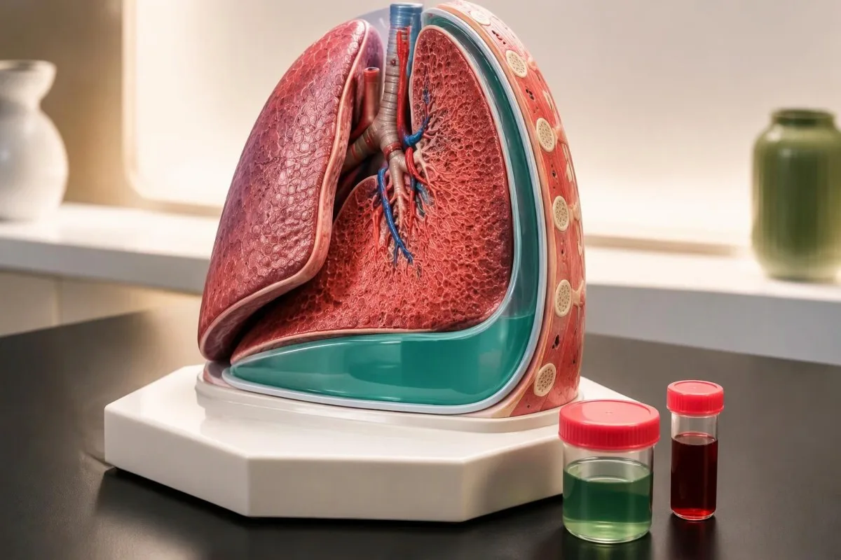
Pleural fluid analysis uses Light’s criteria: fluid is exudative if its protein/serum ratio is >0.5, LDH/serum ratio is >0.6, or LDH exceeds two-thirds of the serum laboratory’s upper limit. Diuretics can create pseudoexudates; classification guides investigation but does not diagnose cancer or infection.

Key takeaways
- Light’s criteria classify an effusion as exudative when any 1 of 3 thresholds is exceeded; all 3 must be negative for a transudative classification.
- Protein ratio means pleural-fluid total protein divided by serum total protein; a result >0.5 meets an exudate criterion.
- LDH ratio means pleural-fluid LDH divided by serum LDH; a result >0.6 meets an exudate criterion.
- LDH upper limit refers to the laboratory’s serum reference limit, not the patient’s serum result; with an upper limit of 240 IU/L, the fluid threshold is >160 IU/L.
- Diuretics can concentrate cardiac or hepatic pleural fluid enough to cross an exudate threshold without changing the underlying cause.
- Albumin gradient >1.2 g/dL, or >12 g/L, supports a transudative mechanism when heart failure remains likely despite positive Light’s criteria.
- Pleural pH ≤7.20 in a suspected parapneumonic effusion generally supports drainage when ultrasound shows safely accessible fluid.
- Additional findings such as lymphocytes, high LDH, low glucose, or an elevated tumor marker direct testing; none independently establishes cancer or a specific infection.
What does pleural fluid analysis actually classify?
Pleural fluid analysis classifies fluid around the lung as an exudate or transudate, using 3 biochemical criteria to help identify its likely mechanism. An exudate suggests local tissue or lymphatic involvement; a transudate more often reflects pressure or protein-balance changes elsewhere in the body.
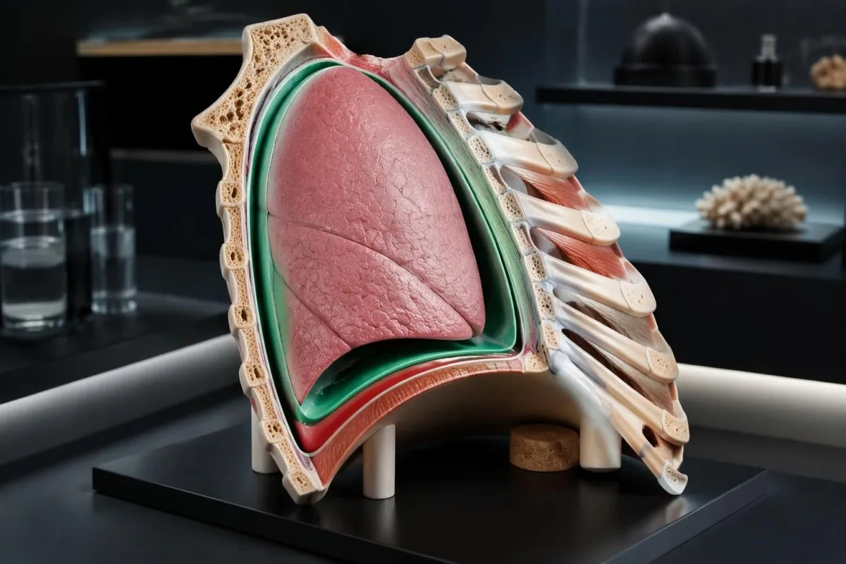
An effusion occupies the pleural space rather than the lung’s air sacs, so it can restrict expansion even when the airways remain open. In a hypothetical person with 800 mL of fluid, breathlessness depends on accumulation speed, underlying lung function, and heart disease—not simply the measured volume.
The exudate vs transudate distinction describes how fluid accumulated, not whether it is harmless or dangerous. A transudate from severe heart failure may require urgent treatment, while an exudate can follow a treatable pneumonia; meeting 1 criterion does not establish either diagnosis or severity.
Kantesti is an AI blood test analyzer that helps explain accompanying serum protein and LDH results, while pleural-fluid classification requires correctly identified specimens and clinician review of all 3 criteria. Our organizational background explains the service; an AI explanation cannot replace assessment of an unexplained effusion.
A new unilateral effusion deserves attention even when its chemistry suggests a transudate, because 2 causes can coexist—for example, heart failure and pneumonia. Our breathlessness testing guide explains why routine blood results cannot determine whether fluid, airway disease, anemia, or a clot is responsible.
What are the three Light’s criteria thresholds?
Light's criteria identify an exudate if the pleural-fluid/serum protein ratio is >0.5, the pleural-fluid/serum LDH ratio is >0.6, or pleural-fluid LDH exceeds two-thirds of the serum LDH upper reference limit. These are alternative tests: any 1 positive criterion is sufficient.
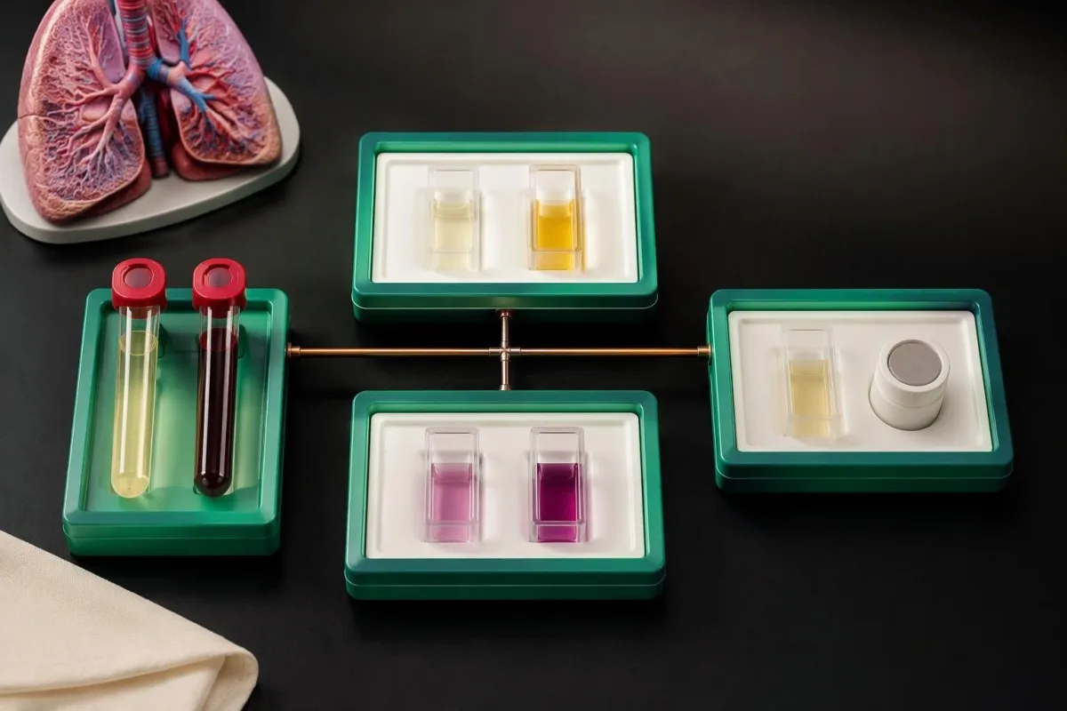
The protein ratio compares total protein in 2 compartments rather than interpreting fluid protein alone. For example, fluid protein of 3.2 g/dL divided by serum protein of 6.0 g/dL equals 0.53, meeting the protein criterion; the same fluid result divided by 7.0 g/dL equals 0.46.
Lactate dehydrogenase, or LDH, contributes 2 separate criteria because enzyme activity in fluid and serum carries different information. The meaning of LDH extends beyond cancer: LDH can increase with cellular injury, inflammatory activity, and specimen damage, so its diagnostic specificity is limited.
The original Light et al. study, published in Annals of Internal Medicine in 1972, established this combined approach to distinguish transudates from exudates. Its 3-part design prioritizes identifying exudates; subsequent experience shows that this sensitivity comes with false-exudate classifications, especially after treatment of heart failure.
The formal inequalities are strictly greater than: a protein ratio exactly 0.50 or an LDH ratio exactly 0.60 does not exceed that particular threshold. However, a displayed ratio rounded to 2 decimal places can conceal a slightly higher underlying value, so near-boundary results should be checked against the laboratory’s unrounded calculation.
| Level | Range | What it means |
|---|---|---|
| Protein ratio criterion | Pleural protein ÷ serum protein >0.5 | Meets one exudate criterion; both measurements must use compatible units. |
| LDH ratio criterion | Pleural LDH ÷ serum LDH >0.6 | Meets one exudate criterion; uses the patient’s actual serum LDH. |
| Absolute pleural LDH criterion | Pleural LDH > two-thirds of serum LDH upper reference limit | Meets one exudate criterion; uses the laboratory reference limit, not the patient’s serum LDH. |
| All criteria negative | None of the 3 thresholds exceeded | Biochemical classification is transudative; clinical findings may still require investigation. |
Why do paired serum results, units, and timing matter?
Paired pleural-fluid and serum measurements reduce classification errors by comparing samples collected close together, preferably during the same assessment. The 2 ratios require the patient’s serum results, while the third criterion requires the laboratory’s serum LDH upper reference limit.
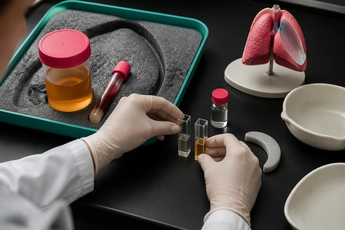
A unit mismatch can create a tenfold error because 1 g/dL equals 10 g/L. Pleural protein of 30 g/L and serum protein of 6 g/dL produce a ratio of 0.50 after conversion—not 5.0; our serum proteins guide distinguishes total protein from albumin, which is a different measurement.
The LDH reference limit varies by laboratory method, so there is no single universal pleural LDH cutoff. An upper serum limit of 225 IU/L gives a fluid threshold of >150 IU/L, whereas an upper limit of 300 IU/L gives >200 IU/L; borrowing another laboratory’s range can change the classification.
Hemolysis can raise LDH and distort a result near 0.6, particularly when specimen damage affects one sample more than the other. A high hemolysis index warrants checking laboratory comments; visibly blood-stained pleural fluid and damaged serum are not interchangeable explanations.
An incomplete report may establish an exudate through the third criterion if pleural LDH and the correct serum upper limit are available, but it cannot establish that all 3 criteria are negative. At Kantesti, we encourage checking specimen names and collection times before interpreting ratios; a serum result from 2 weeks earlier may reflect a different treatment state.
How do you calculate Light’s criteria on your report?
Pleural fluid results interpretation starts by calculating both ratios and the laboratory-specific LDH threshold, then checking whether any result exceeds its cutoff. The following 3 examples are hypothetical and use a serum LDH upper reference limit of 240 IU/L, giving a fluid threshold of 160 IU/L.
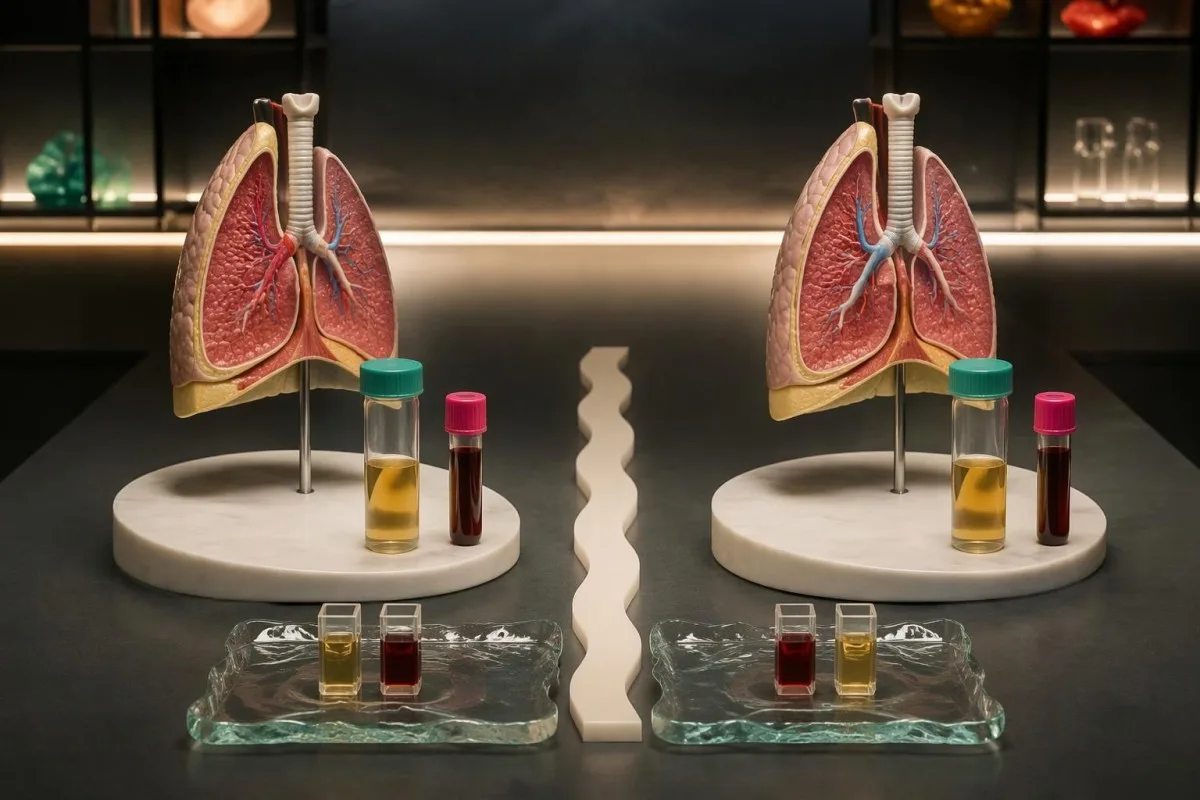
Example 1: a transudative pattern. Pleural protein 2.0 g/dL divided by serum protein 6.5 g/dL gives 0.31; pleural LDH 90 IU/L divided by serum LDH 200 IU/L gives 0.45. Fluid LDH of 90 IU/L is also below 160 IU/L, so none of the criteria is positive.
Example 2: one positive criterion. Pleural protein 3.4 g/dL and serum protein 6.0 g/dL give 0.57, while pleural LDH 120 IU/L and serum LDH 250 IU/L give 0.48. Fluid LDH remains below 160 IU/L, but the positive protein ratio alone classifies the effusion as exudative.
Example 3: absolute LDH changes the answer. Pleural protein 2.5 g/dL and serum protein 6.5 g/dL give 0.38; pleural LDH 180 IU/L and serum LDH 400 IU/L give 0.45. Although both ratios are negative, 180 IU/L exceeds the laboratory-derived 160 IU/L threshold, making the classification exudative.
The number of positive criteria is not a validated severity score: 3 positives do not mean an effusion is three times more dangerous than 1 positive. Our LDH fraction interpretation guide explains another limitation—additional enzyme characterization does not, by itself, identify the cause of pleural fluid.
Why can diuretics make a transudate look exudative?
Diuretics can produce a pseudoexudate by removing water faster than protein and LDH leave pleural fluid, causing concentrations or ratios to cross Light’s thresholds. A mildly positive result—such as a protein ratio of 0.53—may therefore coexist with a cardiac transudative mechanism.
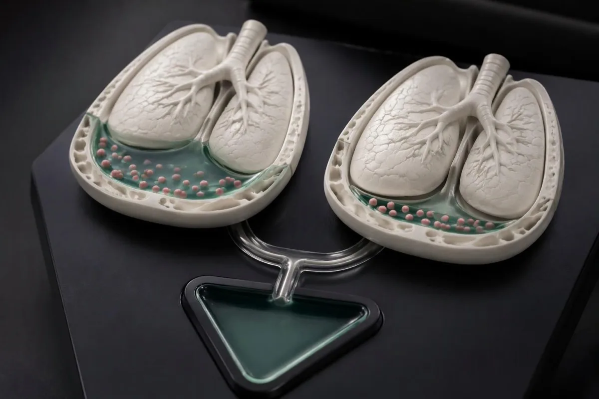
Pseudoexudate means a misleading biochemical classification, not an imaginary effusion or an incorrect sample collection. The mechanism is especially relevant after several days of diuretic treatment, but there is no reliable 24-hour window or universal medication dose that guarantees when an effusion will cross a threshold.
In Bielsa et al.’s 2012 Respirology study, Light’s criteria misclassified 29% of cardiac effusions and 18% of hepatic hydrothoraces as exudates. Those figures describe the study’s populations, not every hospital or every treated patient; nevertheless, they explain why a positive criterion cannot automatically overturn a well-supported heart-failure diagnosis.
A treatment timeline matters when the report is borderline: document when diuretics began, weight change, kidney function, and whether breathlessness improved. Our urine sodium interpretation guide describes another test affected by diuretics; a value obtained 2 hours after a dose is not equivalent to an untreated baseline.
A suspected pseudoexudate still needs clinical safeguards because 2 conditions can coexist. Fever, pleuritic pain, loculations, persistent unilateral fluid, or concerning imaging should not be dismissed as medication effects; do not stop prescribed diuretics solely to obtain a chemically cleaner classification.
When do albumin gradients and NT-proBNP help?
A serum-minus-pleural albumin gradient >1.2 g/dL supports a transudative mechanism when Light’s criteria suggest an exudate but heart failure remains clinically likely. A protein gradient >3.1 g/dL offers additional support, although neither result excludes a second cause.
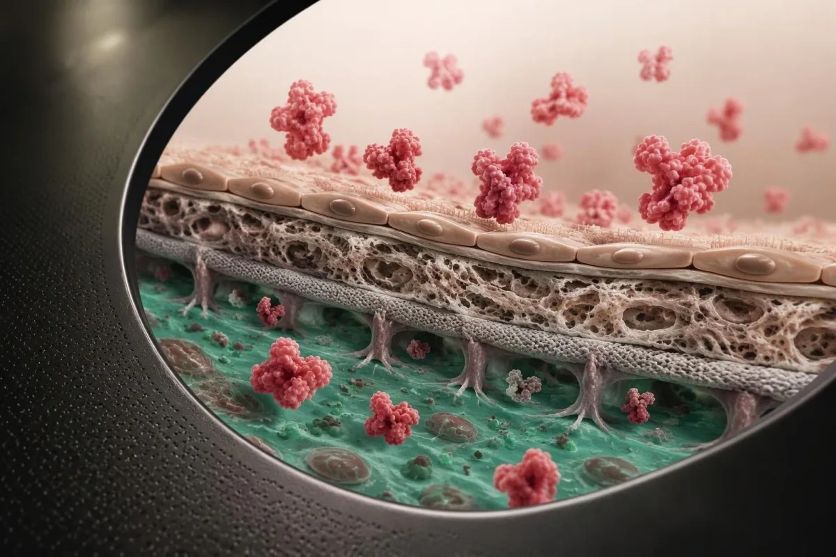
Albumin gradients use subtraction, not division: serum albumin minus pleural albumin. In a hypothetical patient, serum albumin of 3.6 g/dL and fluid albumin of 1.9 g/dL give 1.7 g/dL, exceeding 1.2 g/dL; calculate from matched samples rather than substituting total protein for albumin.
Albumin performed better than the protein gradient for misclassified cardiac effusions in Bielsa et al. (2012): the >1.2 g/dL albumin threshold correctly identified 83% of false cardiac exudates in that study. It did not recover every case, and the same approach was less successful for hepatic hydrothorax, so clinical context remains necessary.
Serum NT-proBNP can support heart failure as an effusion’s cause, but age, kidney dysfunction, atrial fibrillation, and concurrent illness influence its interpretation. The 2023 British Thoracic Society guideline supports its selective use for suspected cardiac effusions; our heart-failure biomarker guide explains why a risk marker and a diagnostic assessment are different tools.
Kantesti is an AI blood test interpretation platform that helps contextualize serum albumin, kidney function, and cardiac biomarkers; explaining a >1.2 g/dL gradient requires correctly supplied pleural measurements as well. Our platform’s blood-test role should not be mistaken for validated standalone analysis of pleural specimens or a decision about drainage.
| Level | Range | What it means |
|---|---|---|
| Albumin gradient supporting transudation | Serum albumin − pleural albumin >1.2 g/dL | Useful when a cardiac pseudoexudate is suspected; equivalent to >12 g/L. |
| Protein gradient supporting transudation | Serum protein − pleural protein >3.1 g/dL | Supplementary evidence in selected cases; equivalent to >31 g/L. |
| Gradient below supportive threshold | Albumin ≤1.2 g/dL or protein ≤3.1 g/dL | Does not provide this biochemical support for transudation; does not independently establish a cause. |
Which causes become more likely after classification?
Transudates commonly accompany heart failure, hepatic hydrothorax, and some protein-losing states; exudates commonly accompany pneumonia, malignancy, pulmonary embolism, and inflammatory pleural disease. These 2 categories organize the differential diagnosis rather than determining which disease a person has.
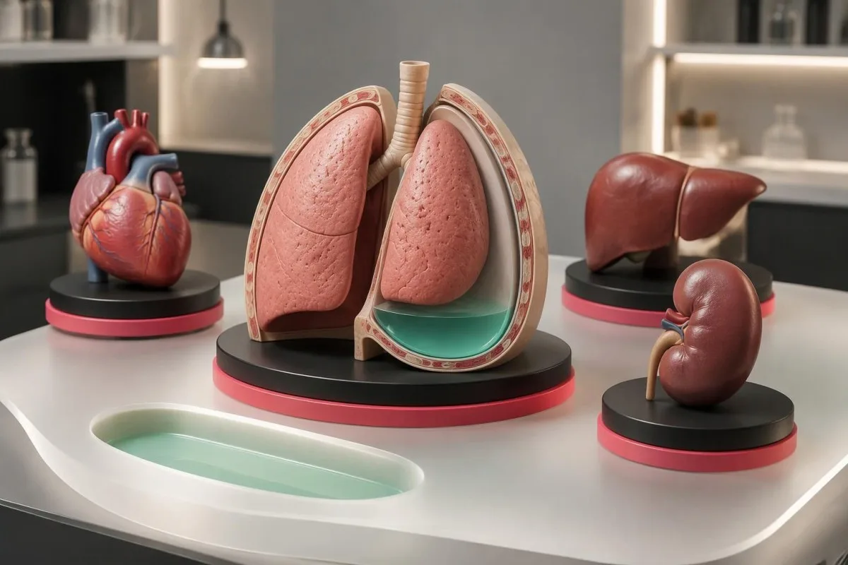
Hepatic hydrothorax can occur when abdominal fluid passes through small diaphragmatic defects, often producing a right-sided effusion. A person may have substantial pleural fluid without obvious ascites on examination; the signs of cirrhosis provide context, but neither fluid protein nor 1 liver enzyme result establishes this mechanism.
Nephrotic protein loss can reduce circulating albumin and contribute to edema and effusions, but low albumin alone is an incomplete explanation. Investigating persistent foamy urine and protein loss may include a urine protein ratio; a serum albumin of 2.0 g/dL does not identify whether the problem is urinary loss, liver synthesis, inflammation, or several factors.
Pulmonary embolism can cause an exudative effusion, sometimes with blood-stained fluid, and deserves consideration when symptoms fit even if another explanation seems plausible. A new effusion accompanied by sudden breathlessness and 1 swollen leg calls for assessment of clot risk rather than waiting for a definitive fluid pattern.
Mixed-cause effusions explain why a convincing transudative pattern cannot close every investigation. For example, a hypothetical person with bilateral fluid from heart failure may develop pneumonia on 1 side; an asymmetric clinical course, persistent fever, or failure to improve should prompt reconsideration rather than forcing all findings into one biochemical category.
What do neutrophils, lymphocytes, eosinophils, and ADA suggest?
Pleural differential counts guide investigation: neutrophil predominance often accompanies acute processes, while lymphocyte predominance can accompany tuberculosis, malignancy, or chronic effusions. Eosinophils ≥10% define an eosinophilic effusion, but no cell pattern independently confirms or excludes cancer or infection.
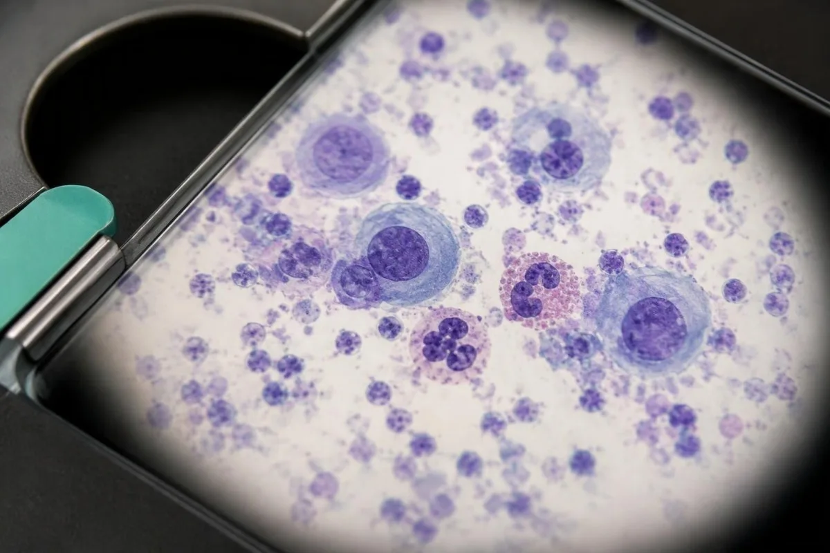
Neutrophil predominance raises concern for a recent inflammatory process, particularly alongside pneumonia, but pulmonary embolism and other conditions can produce the same pattern. A result of 80% neutrophils has a different meaning when paired with fever, low pleural pH, and a septated collection than when these findings are absent.
Lymphocyte predominance does not equal tuberculosis: a hypothetical result of 85% lymphocytes can also occur with lymphoma, other malignancy, chylothorax, or a longstanding cardiac effusion. An IGRA result assesses immune recognition of tuberculosis antigens; even a positive result cannot establish that pleural fluid contains active tuberculosis.
Pleural adenosine deaminase, or ADA, around or above 40 U/L is a commonly used supportive tuberculosis threshold, but assays, populations, and local prevalence differ. Empyema, lymphoma, and some inflammatory conditions can also elevate ADA; the British Thoracic Society guideline (Roberts et al., 2023) emphasizes prevalence-sensitive interpretation and tissue sampling for culture when pleural tuberculosis is suspected.
Eosinophilic pleural fluid can follow air or blood entering the space, medication effects, parasites, or malignancy. The ≥10% definition should not be confused with a peripheral high eosinophil count, and eosinophilia is not a reliable cancer exclusion test.
When do pleural pH, glucose, and LDH affect drainage?
In a suspected parapneumonic effusion, pleural pH ≤7.20 indicates a high risk of complicated infection and generally supports drainage if fluid is safely accessible. Values >7.20 but <7.40 require additional assessment; these management thresholds are different from Light’s classification thresholds.
The 2023 British Thoracic Society guideline recommends considering drainage for intermediate-pH effusions when LDH is >900 IU/L, particularly with persistent fever, a large collection, glucose ≤4.0 mmol/L, pleural enhancement, or ultrasound septations (Roberts et al., 2023). LDH >900 IU/L in this pathway is a risk-management clue, not a universal infection diagnostic threshold.
Pleural pH ≥7.40 suggests a low risk of complicated parapneumonic infection and does not, by itself, indicate immediate drainage. Conversely, low pH can occur in rheumatoid pleuritis or malignancy, so the ≤7.20 recommendation must be applied to a compatible clinical setting rather than every acidic effusion.
Collection errors alter pH: air exposure can raise it, local-anesthetic contamination can lower it, and delayed testing can make the result unreliable. Pleural pH should be measured promptly—ideally within 1 hour—using appropriate anaerobic collection and a blood-gas analyzer, not dipstick paper; our blood-gas interpretation guide concerns blood rather than pleural-fluid reference values.
Low glucose supports further investigation but is not specific for bacteria: rheumatoid disease, malignancy, and tuberculosis can also reduce it. If prompt pH measurement is unavailable, pleural glucose <3.3 mmol/L, about 60 mg/dL, can support concern for complicated infection in the appropriate setting; the patient’s serum glucose and imaging still matter.
| Level | Range | What it means |
|---|---|---|
| High-risk parapneumonic pH | pH ≤7.20 | Generally supports intercostal drainage if a safely accessible collection is present. |
| Intermediate-risk parapneumonic pH | pH >7.20 and <7.40 | Assess LDH, glucose, fever, fluid volume, CT findings, and ultrasound septations. |
| Lower-risk parapneumonic pH | pH ≥7.40 | No immediate drainage indication from pH alone; reassess if the clinical course changes. |
| Supporting intermediate-risk LDH | LDH >900 IU/L | May support drainage in the intermediate-pH pathway alongside other concerning findings. |
Can negative cultures rule out pleural infection?
Negative pleural cultures do not rule out infection, especially after antibiotics or with low organism numbers. Frank pus or a credible positive pleural Gram stain or culture provides stronger direct evidence than the 3 Light’s criteria, while possible contaminants require clinical interpretation.
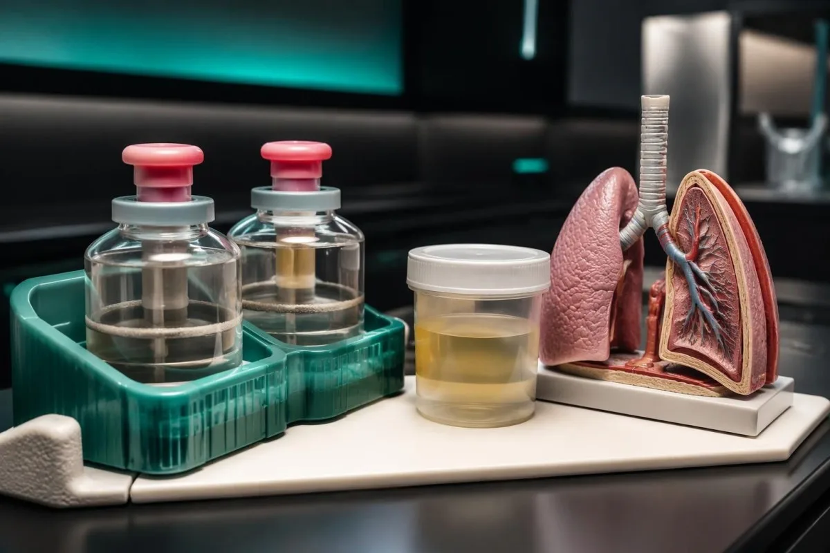
Culture yield depends on handling, so suspected pleural infection should prompt appropriate laboratory submission rather than chemistry testing alone. The British Thoracic Society guideline (Roberts et al., 2023) recommends sending fluid in both a sterile container and aerobic and anaerobic culture bottles, commonly using 5–10 mL for bottle inoculation when available.
Prior antibiotics can suppress culture growth without sterilizing the pleural space or resolving a loculated collection. A hypothetical sample obtained after 3 treatment days may be culture-negative despite fever, low pH, and ultrasound septations; clinicians should not delay necessary antibiotics solely to improve laboratory yield.
Blood inflammatory markers complement, but cannot replace, fluid assessment and imaging. Our WBC and CRP comparison explains discordant responses; a person with 1 normal white-cell count may still have a clinically significant pleural infection, especially with immune suppression or an early sample.
Procalcitonin is not a validated standalone exclusion test for localized pleural infection. At Kantesti, we distinguish a laboratory signal from a diagnosis; our procalcitonin interpretation guide explains why a serum value below 0.25 ng/mL cannot override convincing pleural-fluid and imaging findings.
What can pleural cytology tell you about cancer?
Positive malignant cytology can establish malignant involvement of the pleural space, but negative cytology does not exclude it. High protein, high LDH, lymphocyte predominance, or low glucose are investigation clues—not cancer diagnoses—even when 2 or 3 findings occur together.
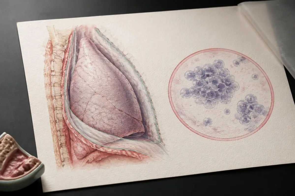
Sample volume and processing affect cytology usefulness, although simply sending more fluid cannot overcome every limitation. The British Thoracic Society guideline recommends submitting 25–50 mL when evaluating suspected malignant effusion and using appropriate cytological preparation, including a cell block where available (Roberts et al., 2023).
Cytology sensitivity varies by cancer type, with some cancers releasing recognizable cells more readily than others. Mesothelioma is a notable setting where fluid cytology can be insufficient; after 1 negative result, suspicious imaging or a persistent unexplained effusion may justify image-guided pleural biopsy or thoracoscopy rather than assuming reassurance.
Chest imaging and symptoms determine the next step after negative cytology, not the number of positive Light’s criteria. Persistent cough, unexplained weight loss, pleural thickening, or recurrent unilateral fluid may warrant a specialist pathway; our persistent cough warning signs explain why symptoms require assessment even without a diagnostic blood result.
Serum or pleural tumor markers should not substitute for cytology or tissue diagnosis. Our CEA result limitations describe noncancer causes of elevation; a hypothetical CEA of 12 ng/mL neither proves a malignant effusion nor locates its origin, and routine tumor-marker panels are not recommended to establish pleural malignancy.
When are triglycerides, amylase, or hematocrit useful?
Targeted pleural tests investigate particular mechanisms: triglycerides and chylomicrons assess chylothorax, amylase suggests pancreatic or esophageal causes in context, and a fluid-to-blood hematocrit comparison assesses hemothorax. Pleural triglycerides >110 mg/dL strongly support chylothorax, but appearance alone is unreliable.
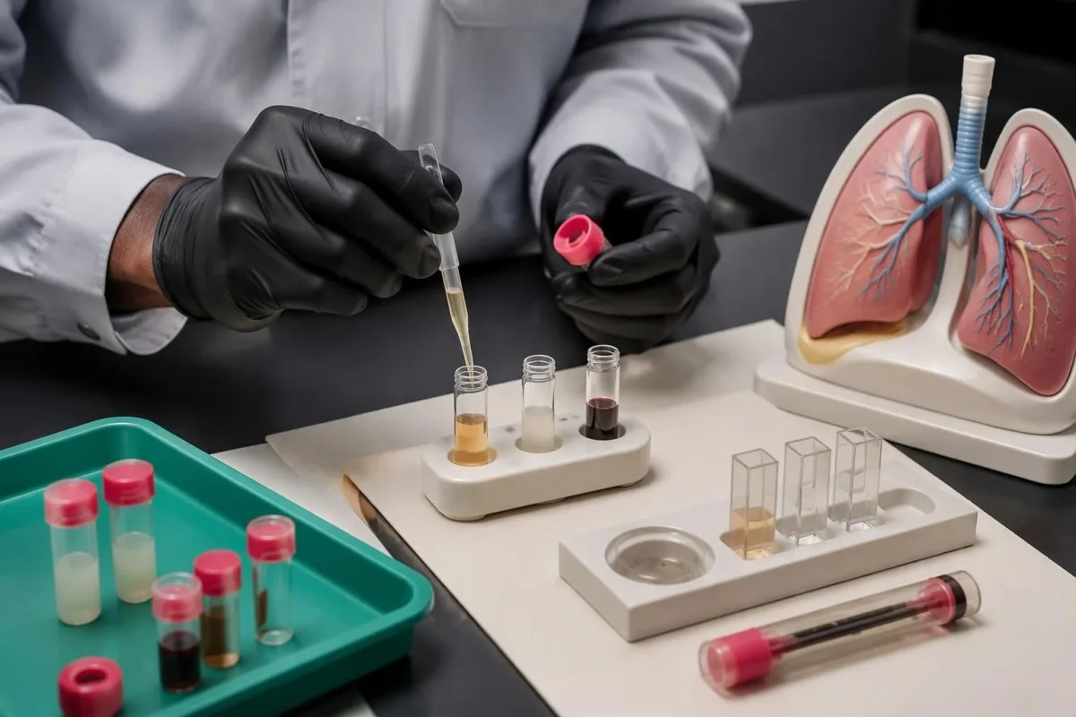
Chylothorax results from lymphatic fluid entering the pleural space, and not every sample looks milky. Triglycerides between 50 and 110 mg/dL often prompt chylomicron testing; a value <50 mg/dL makes chylothorax less likely but may be misleading during fasting or poor intake, especially when clinical suspicion remains high.
Pseudochylothorax usually reflects a longstanding cholesterol-rich effusion rather than fresh lymphatic leakage. Pleural cholesterol >200 mg/dL and cholesterol crystals can support that distinction; our prealbumin and nutrition guide explains why poor nutritional status cannot be diagnosed from 1 protein marker or assumed to explain cloudy fluid.
Amylase-rich pleural fluid can accompany pancreatic disease, esophageal rupture, or some malignancies; an elevation is a direction for investigation, not a pancreatic diagnosis. A pleural/serum amylase ratio >1 or fluid amylase above the serum reference limit is commonly used to identify this pattern, with isoenzyme testing sometimes helping distinguish pancreatic from salivary sources.
Hemothorax conventionally means pleural hematocrit at least 50% of peripheral blood hematocrit, so paired hematocrit measurements matter. A ratio of 25–50% can still be concerning after dilution over time; visibly reddish fluid alone cannot distinguish hemothorax from lesser blood admixture, malignancy, pulmonary embolism, or collection-related contamination.
| Level | Range | What it means |
|---|---|---|
| Triglycerides strongly supporting chylothorax | >110 mg/dL, approximately >1.24 mmol/L | Supports chylothorax; chylomicron detection provides direct confirmation. |
| Intermediate triglycerides | 50–110 mg/dL, approximately 0.56–1.24 mmol/L | Consider chylomicron testing when the clinical setting is compatible. |
| Lower triglycerides | <50 mg/dL, approximately <0.56 mmol/L | Makes chylothorax less likely, but fasting and poor intake can complicate interpretation. |
| Conventional hemothorax definition | Pleural hematocrit ≥50% of peripheral blood hematocrit | Requires prompt assessment of the cause and appropriate management. |
What should you ask next, and when is care urgent?
The next step is to connect all 3 Light’s criteria with symptoms, treatment timing, ultrasound or CT, microbiology, and cytology—not to interpret a flagged number alone. Severe or rapidly worsening breathlessness, fainting, confusion, or sudden chest pain requires urgent assessment regardless of the fluid’s biochemical category.

Bring the complete report, including pleural and serum protein, pleural and serum LDH, the serum LDH upper reference limit, and collection times. These 5 numerical inputs allow checking all 3 criteria; also ask whether pH collection was reliable and whether any laboratory result remains pending.
Ask what remains unexplained if the effusion is persistent, recurrent, or unilateral despite treatment. A second clinician review or specialist procedure may be appropriate after 1 nondiagnostic aspiration, but the choice depends on imaging and likely cause; our clinical validation information distinguishes technical interpretation from decisions requiring direct medical assessment.
Kantesti is an AI lab test interpretation service that helps organize accompanying blood results and questions, not a replacement for deciding whether a pleural collection needs drainage. Our responsible AI principles explain why 1 AI-generated interpretation must not be used to exclude cancer, infection, or pulmonary embolism.
Urgent symptoms override reassuring chemistry, especially after aspiration if breathlessness or chest pain suddenly worsens, because complications such as pneumothorax require examination and imaging. In the UK, call 999 for severe symptoms; elsewhere use the local emergency number, and do not wait for the next laboratory update—our chest pain testing guide explains why blood tests alone are insufficient.
Research publications and the limits of the evidence
Clinical guidance for pleural fluid analysis rests on the original 1972 Light’s criteria study, subsequent misclassification research, and disease-specific recommendations such as the 2023 British Thoracic Society guideline. The 2 additional DOI-linked laboratory publications below are contextual reading, not validation studies of pleural-fluid diagnosis.
Light et al. (1972) provides the foundation for the 3 biochemical criteria, while Bielsa et al. (2012) explains why cardiac and hepatic transudates can be misclassified. These publications answer different questions: a classification study does not validate a cancer test, and a gradient study does not determine whether an infected collection needs drainage.
The 2023 British Thoracic Society guideline, by Roberts and colleagues, connects fluid analysis with ultrasound-guided procedures, cytology, microbiology, and infection-management pathways. Its pH ≤7.20 recommendation applies to suspected parapneumonic disease; moving that number into another clinical setting without its accompanying conditions can produce an inappropriate treatment conclusion.
Uncertainty remains greatest near thresholds, with mixed causes, altered specimens, and partially treated effusions. A protein ratio of 0.51 cannot carry the same diagnostic weight as direct malignant cytology or credible positive microbiology; practical interpretation should state what the result supports, what it cannot exclude, and which next test could change management.
DOI registration identifies a publication, but does not establish peer review, clinical accuracy, or endorsement by a guideline body. Kantesti’s 2 listed publications address broader laboratory education—including LDH and viral testing—and should not be cited as evidence for pleural-fluid thresholds; our medical content standards explain the distinction between educational material and clinical evidence.
Frequently asked questions
What are Light’s criteria for pleural fluid?
Light’s criteria classify pleural fluid as exudative if its protein/serum protein ratio is >0.5, its LDH/serum LDH ratio is >0.6, or its LDH exceeds two-thirds of the laboratory’s serum LDH upper reference limit. Any 1 positive criterion is sufficient. All 3 must be negative for a transudative biochemical classification. The criteria identify a fluid pattern, not its specific cause.
Does an exudative pleural effusion mean cancer?
An exudative pleural effusion does not mean cancer: pneumonia, pulmonary embolism, tuberculosis, and inflammatory disease can also produce this pattern. Meeting 1, 2, or 3 Light’s criteria does not measure cancer probability or severity. Malignant cytology can establish malignant involvement, but negative cytology does not exclude it. Persistent unexplained fluid or suspicious imaging may require pleural tissue sampling.
Can diuretics affect Light’s criteria?
Diuretics can concentrate pleural protein and LDH, making a cardiac or hepatic transudate meet Light’s criteria for an exudate. This pattern is called a pseudoexudate. A serum-minus-pleural albumin gradient >1.2 g/dL can support a transudative mechanism when heart failure remains likely. The gradient does not exclude pneumonia, malignancy, or another condition occurring at the same time.
Why do I need a serum sample with pleural fluid analysis?
A serum sample supplies the denominator for the pleural-fluid protein and LDH ratios, whose exudate thresholds are >0.5 and >0.6 respectively. Samples should be collected close together so treatment or illness changes do not distort the comparison. The third criterion uses the laboratory’s serum LDH upper reference limit, not the patient’s actual LDH result. Without complete inputs, clinicians may identify a positive criterion but cannot reliably establish that all 3 are negative.
What does a pleural fluid pH of 7.2 mean?
A pleural-fluid pH of 7.20 or lower supports a high risk of complicated parapneumonic infection when pneumonia or pleural infection is suspected. The 2023 British Thoracic Society guideline generally supports drainage if the collection is safely accessible. Low pH can also occur with rheumatoid pleuritis or malignancy, so the clinical setting matters. Air exposure, local-anesthetic contamination, and delayed analysis can make the measurement misleading.
Can a negative pleural fluid culture rule out infection?
A negative pleural-fluid culture cannot rule out infection, particularly after antibiotics or when organism numbers are low. Clinicians combine microbiology with symptoms, ultrasound or CT, pleural pH, glucose, and LDH. Pleural pH ≤7.20 in a compatible parapneumonic effusion may support drainage even when culture is negative. Frank pus or credible positive microbiology provides stronger direct evidence than a nonspecific protein or LDH elevation.
Get your blood test results explained
Upload your lab report and Kantesti's AI explains each biomarker in plain language within seconds. Used by more than 2 million people worldwide.
References
- Klein, T., Mitchell, S., & Weber, H. (2026). Nipah Virus Blood Test: Early Detection & Diagnosis Guide 2026. Kantesti AI Medical Research. DOI
- Klein, T., Mitchell, S., & Weber, H. (2026). B Negative Blood Type, LDH Blood Test & Reticulocyte Count Guide. Kantesti AI Medical Research. DOI
- Light RW et al. (1972). Pleural effusions: the diagnostic separation of transudates and exudates. Annals of Internal Medicine. DOI
- Bielsa S et al. (2012). Solving the Light's criteria misclassification rate of cardiac and hepatic transudates. Respirology. DOI
- Roberts ME et al. (2023). British Thoracic Society Guideline for pleural disease. Thorax. DOI
- 2M+Tests analysed
- 127+Countries
- 100+Languages
About the medical team


E-E-A-T Trust Signals
Experience
Physician-led clinical review of lab interpretation workflows.
Expertise
Laboratory medicine focus on how biomarkers behave in clinical context.
Authoritativeness
Medically reviewed by Dr. Thomas Klein, with contributions from Dr. Sarah Mitchell and Prof. Dr. Hans Weber.
Trustworthiness
Evidence-based interpretation with clear follow-up pathways to reduce alarm.


Medical disclaimer
This article is for educational purposes only and does not constitute medical advice. Always consult a qualified healthcare provider for diagnosis and treatment decisions.
Published · Contact the editorial team
Published by Kantesti Ltd, registered in England & Wales, company no. 17090423, London, United Kingdom.
