LDH fractions can occasionally narrow the clinical question, but they rarely identify one organ or diagnosis on their own. The most reliable interpretation combines the pattern with sample quality, symptoms, and more specific tests.


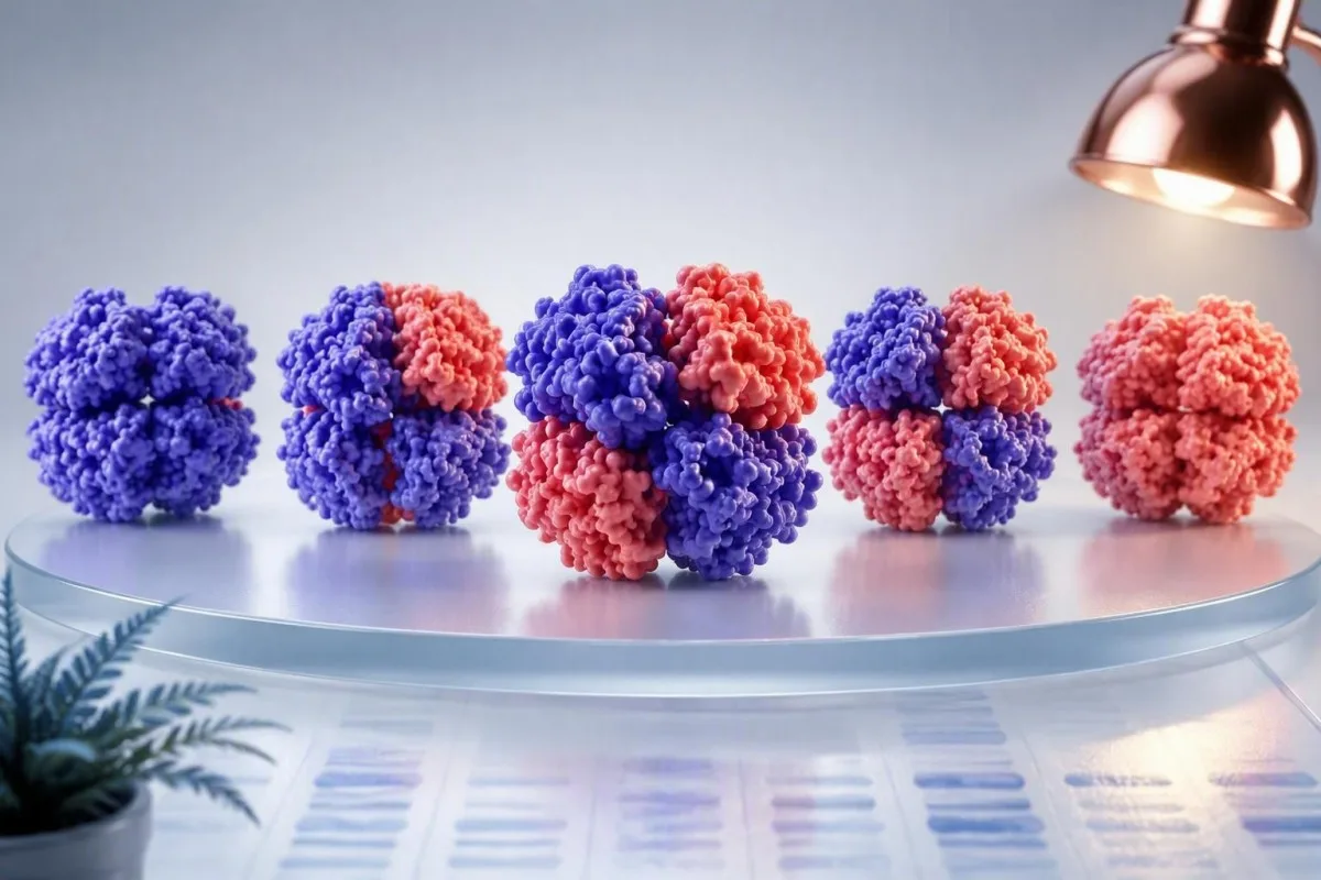
- LDH fractions are five enzyme forms created from different combinations of H and M subunits; their distribution varies by laboratory method.
- LDH-1 vs LDH-2 reversal, historically called an LDH flip, can occur with red-cell breakdown, but it does not prove a heart attack.
- LDH-5 predominance may support liver or skeletal-muscle release when ALT, AST, CK, and the clinical story agree.
- Total LDH above 2 times the laboratory upper limit is nonspecific and should trigger context-based follow-up rather than a diagnosis.
- Sample hemolysis can falsely elevate total LDH and disproportionately increase LDH-1 and LDH-2.
- High-sensitivity troponin has replaced LDH isoenzymes for suspected acute myocardial infarction in modern practice.
- Macro-LDH can cause a persistent unexplained elevation and is usually benign after serious causes are excluded.
- Kantesti AI reads LDH alongside CBC, bilirubin, haptoglobin, CK, aminotransferases, and time trends rather than treating one fraction as a verdict.
What LDH isoenzyme patterns can actually show
LDH isoenzyme patterns can suggest the broad tissue source of enzyme release, but they cannot reliably diagnose a heart attack, liver disease, cancer, or hemolysis by themselves. A rise in LDH-1 and LDH-2 may fit red-cell turnover; LDH-5 may fit liver or muscle injury; yet overlap is substantial and modern clinicians usually confirm the hypothesis with more specific markers.
As of September 17, 2026, many hospital laboratories no longer offer an LDH isoenzyme test routinely because total LDH, clinical examination, imaging, and targeted biomarkers usually answer the question faster. A typical adult total-LDH upper limit is roughly 220 to 250 U/L, but the laboratory’s own reference interval—not an internet cutoff—governs interpretation.
In my 15 years of clinical practice, I have found that a pattern is most useful when it resolves a real fork in the road. For example, a patient with LDH 680 U/L, bilirubin 48 µmol/L, reticulocytosis, and low haptoglobin needs a hemolysis work-up; an LDH fraction result adds texture, not the diagnosis. Our hematology marker guide explains why red-cell turnover is always a multi-test question.
Why the pattern is only a clue
LDH is present in nearly every metabolically active tissue, so an abnormal result records cellular stress or release rather than a named disease. The clinical value rises when the fraction pattern, the magnitude above the upper limit, and a same-day companion marker point in the same direction.
The five LDH fractions and their biochemical origins
The five LDH fractions are tetramers made from H and M subunits: LDH-1 is H4, LDH-2 is H3M1, LDH-3 is H2M2, LDH-4 is H1M3, and LDH-5 is M4. This chemistry explains their tissue tendencies but does not make them organ-specific.
H-rich fractions predominate in tissues that depend heavily on aerobic metabolism, including myocardium, kidney cortex, and erythrocytes. M-rich fractions are more abundant in liver and skeletal muscle, where anaerobic glycolysis is comparatively prominent during high demand.
The fraction percentage is a relative measurement, which creates a quiet trap: one fraction can look low simply because another fraction rose. I ask for both the total LDH value and the percentage or absolute activity of each fraction before drawing conclusions. What out-of-range flags mean is often less alarming than the clinical context suggests.
Kantesti is an AI blood test analyzer that interprets LDH results as a relationship among enzymes, cell counts, bilirubin, and prior values rather than as an isolated tissue map. That approach matters because the same LDH-5 rise can follow a hard workout, medication-related muscle injury, or hepatocellular injury.
Typical LDH fraction distribution in a clean sample
In many adult serum methods, LDH-2 is normally the largest fraction and LDH-1 is second, although published percentage ranges differ by electrophoresis or electrophoretic separation method. A report’s printed reference interval is therefore more trustworthy than a generic online chart.
One commonly used approximate serum distribution is LDH-1 17% to 27%, LDH-2 27% to 37%, LDH-3 18% to 25%, LDH-4 8% to 16%, and LDH-5 6% to 16%. These percentages are not universal diagnostic cutoffs, and a laboratory may report a different interval after validating a different analytic system.
A result near a boundary is weak evidence. A shift from LDH-1 of 26% to 28%, for example, is far less meaningful than a clear LDH-1 predominance combined with an elevated hemolysis index, falling hemoglobin, and rising indirect bilirubin. CBC indices and anemia patterns often provide the actionable clue first.
LDH-1 vs LDH-2: what an LDH flip means
An LDH flip means LDH-1 exceeds LDH-2, and the commonest explanation is red-cell release in the sample or in the patient—not myocardial infarction. This reversal is a pattern worth checking, not a standalone diagnosis.
Erythrocytes contain large amounts of LDH-1 and LDH-2. Even modest in-vitro hemolysis during collection, transport, or delayed processing can create a misleading LDH-1-predominant result; the laboratory’s hemolysis index may be more revealing than a repeated fraction assay.
Historically, an LDH flip appearing 24 to 48 hours after chest pain supported myocardial necrosis and could remain abnormal for 7 to 10 days. That timing is now largely of historical interest: the Fourth Universal Definition of Myocardial Infarction identifies cardiac troponin as the preferred biomarker for myocardial injury (Thygesen et al., 2018). Troponin I and T differences are much more relevant to acute care today.
Dr. Thomas Klein’s practical rule is simple: if the tube was visibly hemolyzed or the lab flags interference, repeat total LDH before interpreting an LDH-1 vs LDH-2 ratio. A clean redraw can prevent a cascade of unnecessary cardiac testing.
LDH-3 predominance and the lung misconception
LDH-3 predominance is sometimes associated with lung tissue, spleen, platelets, and lymphoid cells, but it is too nonspecific to diagnose pulmonary disease. An isolated LDH-3 shift does not diagnose pulmonary embolism, pneumonia, or lymphoma.
Older teaching linked LDH-3 with lung injury because this fraction is relatively abundant in pulmonary tissue. In a breathless patient, however, oxygen saturation, ECG, chest imaging, D-dimer used appropriately, and troponin are clinically superior tools; LDH has no validated threshold for ruling in pulmonary embolism.
I occasionally see a modest LDH-3 proportion after a difficult collection with platelet activation or after a generalized viral illness. The absolute total LDH and trajectory over 48 to 72 hours matter more than a single percentage. For symptom-led testing, see our shortness-of-breath lab guide.
A sustained rise should not be dismissed, particularly when it accompanies weight loss, night sweats, enlarged nodes, anemia, or abnormal smear findings. Still, LDH is a severity marker in those settings—not a tissue biopsy in a tube.
LDH-4 and LDH-5: liver and muscle clues
LDH-4 and LDH-5 can rise when liver cells or skeletal muscle release enzyme, but neither fraction can distinguish those sources reliably without ALT, AST, CK, and history. LDH-5 is most persuasive when it moves in parallel with one of those targeted markers.
A patient with LDH-5 predominance, ALT 180 U/L, AST 145 U/L, and normal CK is more likely to have a hepatocellular pattern than a primary muscle pattern. Conversely, CK above 1,000 U/L after unaccustomed exercise, statin exposure, or trauma makes muscle release more plausible, especially if ALT is only mildly increased.
Here is the nuance many results pages miss: AST is found in muscle and liver, while ALT is more liver-weighted but still not perfectly specific. A 52-year-old marathon runner with AST 89 U/L, CK 1,600 U/L, and mild LDH elevation may simply need hydration and a 5- to 7-day exercise pause before repeat testing. High CK warning signs help separate this from urgent rhabdomyolysis.
Kantesti is an AI-powered blood test analysis tool used by more than 2 million people across 127+ countries, and it flags LDH-5 beside CK, aminotransferases, bilirubin, medication exposure, and recent exertion. It does not label a liver diagnosis from an isoenzyme fraction alone.
How an LDH isoenzyme test is performed and reported
An LDH isoenzyme test separates enzyme forms by electrophoretic mobility or related methods and reports a percentage, activity, or both. Pre-analytical handling is unusually influential because LDH leaks readily from cellular elements after collection.
Many laboratories use serum or heparinized plasma, but their accepted specimen type and processing window differ. A prolonged tourniquet, vigorous tube mixing, pneumatic transport, heat exposure, or delayed separation can raise LDH before the assay begins; the number may be analytically correct and clinically false.
If an unexpected LDH value is less than 1.5 times the upper limit and the person feels well, I commonly repeat a properly handled sample before ordering broad investigations. That is particularly sensible after strenuous exercise, a recent injection, or a difficult sample collection. Repeat testing after hemolysis covers practical redraw timing.
Kantesti AI uses extraction checks and reference-range matching when reading uploaded reports; clinical methodology and thresholds are reviewed through our medical validation standards. A photo or PDF should never override the original laboratory report if a value appears inconsistent.
Hemolysis, macro-LDH, and misleading persistent results
Hemolysis is the most common technical reason for a surprising LDH elevation, while macro-LDH is an uncommon biological explanation for persistently high LDH in a well person. Both can mimic significant disease if the result is read without laboratory context.
In-vivo hemolysis usually produces a coherent set of findings: rising unconjugated bilirubin, low haptoglobin, increased reticulocytes, anemia, and sometimes red-cell fragments on a film. A raised LDH by itself cannot establish hemolysis, because liver injury, infection, exercise, and malignancy can produce the same total number.
Macro-LDH occurs when LDH binds an immunoglobulin or forms a high-molecular-weight complex that clears slowly. It may keep total LDH elevated for months or years with a stable pattern and no symptoms; polyethylene-glycol precipitation or specialized electrophoresis can support the diagnosis after a clinician excludes more consequential causes.
The practical payoff is large. A stable LDH around 400 U/L for 18 months with normal CBC, bilirubin, ALT, CK, renal function, and imaging is a very different problem from a new increase from 190 to 900 U/L in 2 weeks. Low haptoglobin beyond hemolysis adds the other side of that assessment.
Why modern heart care no longer relies on LDH fractions
High-sensitivity cardiac troponin, measured serially with symptoms and ECG findings, is far more specific for myocardial injury than LDH isoenzymes. A normal or abnormal LDH fraction pattern should never delay emergency evaluation for chest pain.
The American and international myocardial infarction definition requires a rise and/or fall in cardiac troponin with at least one value above the assay’s 99th-percentile upper reference limit, plus evidence of ischemia (Thygesen et al., 2018). LDH can rise after muscle injury, hemolysis, liver disease, or malignancy, so it cannot meet that diagnostic role.
Troponin itself is not synonymous with a blocked coronary artery; myocarditis, tachyarrhythmia, severe hypertension, pulmonary embolism, and kidney disease may also elevate it. But it is still vastly more cardiac-specific than an LDH flip, and serial change over 1 to 3 hours adds diagnostic power.
Seek urgent care for chest pressure, breathlessness, fainting, sweating, or pain radiating to the arm, jaw, back, or upper abdomen—especially if symptoms are new or persistent for more than 10 minutes. Chest-pain blood testing explains why home interpretation is not safe in that situation.
Using LDH in a real hemolysis work-up
For suspected hemolysis, LDH is best interpreted with hemoglobin, reticulocytes, haptoglobin, bilirubin, direct antiglobulin testing when indicated, and a peripheral film. The combination is more informative than any one result, including the isoenzymes.
A convincing hemolytic pattern often includes LDH above the laboratory upper limit, haptoglobin below the assay range, indirect hyperbilirubinemia, and reticulocytosis. Reticulocytes may remain low, however, when marrow reserve is limited by iron deficiency, B12 deficiency, infection, kidney disease, or recent chemotherapy; physiology is rarely as tidy as teaching diagrams.
Mechanical hemolysis from a prosthetic valve, microangiopathic hemolysis, autoimmune hemolysis, G6PD-related oxidant stress, and inherited membrane disorders need different follow-up pathways. Schistocytes on a blood film with thrombocytopenia and neurological, renal, or gastrointestinal symptoms is an urgent combination. Read our guide to urgent schistocyte findings rather than waiting on isoenzyme results.
Dr. Thomas Klein advises against starting iron merely because LDH is high and hemoglobin is low. Iron deficiency and hemolysis can coexist, but ferritin, transferrin saturation, MCV, and reticulocyte hemoglobin clarify whether replacement is appropriate.
LDH in cancer: burden and prognosis, not a screening test
LDH can reflect cell turnover and tumor burden in some cancers, especially aggressive lymphomas and germ-cell tumors, but it cannot detect cancer reliably in an otherwise well person. A normal LDH does not exclude cancer, and an elevated LDH does not diagnose it.
In diffuse large B-cell lymphoma, LDH above the local upper limit is one component of the International Prognostic Index; it helps estimate risk alongside age, stage, performance status, and extranodal involvement. It is a population-level prognostic variable, not proof that a person has lymphoma or a measure of a single tumor’s size.
LDH is also followed in selected germ-cell tumor pathways, but clinicians interpret it with AFP, beta-hCG, imaging, and pathology. A mildly elevated total LDH of 280 U/L with a normal examination and normal other tests is far more often explained by common non-malignant factors than occult cancer.
Persistent unexplained elevation deserves a measured review: compare prior values, repeat a clean sample, inspect CBC and liver tests, review medicines and exercise, then investigate symptoms and examination findings. Our blood-cancer test pathway describes the tests that carry more diagnostic weight.
LDH during infection, inflammation, and severe illness
LDH often rises during severe infection, hypoxia, widespread tissue stress, and critical illness, but it measures injury burden rather than identifying the cause. A very high result should accelerate clinical assessment when symptoms are significant, not prompt self-diagnosis from the number.
During the COVID-19 era, LDH was associated with illness severity in hospitalized cohorts, but associations did not make it a useful stand-alone triage test. The same principle applies to many acute illnesses: oxygenation, blood pressure, lactate, kidney function, CRP, cultures, and focused imaging usually determine management.
LDH above 1,000 U/L is not automatically an emergency, but the causes can include major hemolysis, severe liver injury, rhabdomyolysis, ischemia, hematologic malignancy, or laboratory artifact. The urgency comes from the accompanying syndrome: confusion, jaundice, dark urine, marked weakness, reduced urine, severe abdominal pain, or breathlessness need prompt assessment.
Kantesti AI is an AI lab test interpretation service that can organize a complex report and identify discordant patterns, but it cannot examine a patient or replace emergency assessment. For a related physiology question, see high lactate beyond sepsis.
How Kantesti reads LDH results in clinical context
Kantesti interprets LDH by comparing the current result with your laboratory range, prior measurements, specimen comments, and related biomarkers. The useful question is usually “what changed with LDH?” rather than “which organ owns this fraction?”
Kantesti AI matches LDH with CBC indices, reticulocytes, bilirubin, haptoglobin, ALT, AST, ALP, GGT, CK, creatinine, and inflammatory markers when they are available on the uploaded report. A 30% rise from a personal baseline may deserve attention even when both values sit near a broad reference limit.
A trend also distinguishes transient exertional release from a progressive process. If an LDH of 330 U/L returns to 205 U/L after 7 days without intense exercise, dehydration, or acute illness, that trajectory is reassuring; if it rises on repeat, the next test should be hypothesis-driven rather than an indiscriminate panel.
Our AI trend analysis workflow is designed to make those comparisons legible before a clinician visit. We apply privacy-focused, GDPR-aligned handling, but a result explanation remains educational and should be discussed with the clinician who knows your symptoms and medicines.
Next steps after an abnormal LDH isoenzyme result
An unexpected LDH isoenzyme pattern usually warrants confirmation of sample quality, review of total LDH and companion tests, and repeat testing within days to weeks if symptoms are absent. Urgent symptoms or rapidly changing results need same-day medical assessment.
Bring the complete report, including the laboratory reference interval and comments such as “hemolyzed specimen,” rather than only the flagged LDH value. Ask whether repeat total LDH, CBC with reticulocytes, bilirubin, haptoglobin, ALT, AST, CK, creatinine, or troponin is appropriate for your situation; the right next test depends on the suspected source.
Do not use LDH fractions to monitor a workout, a supplement, or “detox” program in isolation. Exercise can raise LDH for 24 to 72 hours, and supplements may cause liver or muscle effects that require conventional safety testing, not interpretation of one fraction. Liver test abbreviations can help you prepare better questions.
For unexplained patterns, Kantesti’s clinical content is produced with physician oversight from our Medical Advisory Board. The bottom line is reassuring but firm: LDH isoenzymes can point a clinician toward a system; they cannot replace specific tests, a physical examination, or urgent care when red flags are present.
Frequently Asked Questions
What does an LDH isoenzyme pattern tell you?
An LDH isoenzyme pattern can suggest whether the released enzyme is relatively H-rich, as in erythrocytes and myocardium, or M-rich, as in liver and skeletal muscle. LDH-1 is commonly about 17% to 27% and LDH-2 about 27% to 37% of total activity in many adult serum methods, but each laboratory sets its own interval. The pattern cannot identify one organ with certainty because tissues overlap and a hemolyzed specimen can distort results. Clinicians confirm a suspected source with targeted tests such as troponin, CK, ALT, bilirubin, haptoglobin, or imaging.
Is LDH-1 higher than LDH-2 always a heart attack?
No. LDH-1 higher than LDH-2, sometimes called an LDH flip, is not a reliable heart-attack diagnosis and is commonly caused by red-cell breakdown in the tube or in the body. Acute myocardial infarction is assessed with serial high-sensitivity troponin, ECG findings, symptoms, and evidence of ischemia rather than LDH fractions. Troponin values above an assay’s 99th-percentile upper reference limit require clinical interpretation, particularly when they rise or fall over serial tests. New chest pressure, breathlessness, sweating, fainting, or radiating pain needs urgent assessment regardless of LDH.
What causes an elevated LDH-5 fraction?
An elevated LDH-5 fraction may occur with hepatocellular injury or skeletal-muscle release because LDH-5 is the M4 form of the enzyme. ALT and AST elevation with normal CK supports a liver-predominant pattern, whereas CK above roughly 1,000 U/L after exertion, medication exposure, or muscle injury makes skeletal muscle more likely. LDH-5 cannot distinguish these sources by itself. A repeat sample after 5 to 7 days without intense exercise is often useful when the clinical situation is stable.
Can exercise raise LDH and change LDH fractions?
Yes. Strenuous or unaccustomed exercise can raise total LDH, AST, and CK for 24 to 72 hours, and muscle contribution may increase the relative M-rich fractions. In a well person, clinicians often repeat testing after at least 5 to 7 days without high-intensity exercise if the LDH elevation is mild, such as less than 1.5 times the laboratory upper limit. Dark urine, profound weakness, reduced urine output, or severe muscle pain requires prompt medical care because rhabdomyolysis is possible. Hydration alone is not a substitute for testing when symptoms are concerning.
How does hemolysis affect an LDH isoenzyme test?
Hemolysis can falsely raise total LDH because erythrocytes contain abundant LDH, particularly LDH-1 and LDH-2. The laboratory may report a hemolysis index or reject the result when interference is substantial, but small effects can still matter near a decision boundary. True hemolysis in the body is assessed with hemoglobin, reticulocytes, haptoglobin, bilirubin, and a peripheral film, not LDH alone. A clean repeat sample is usually the first step when the result conflicts with how a person feels and with the rest of the panel.
When is a high total LDH urgent?
A high total LDH is urgent when it occurs with symptoms of serious illness, rapid change, or organ dysfunction rather than at one universal numeric threshold. LDH above 1,000 U/L can occur with major hemolysis, severe liver injury, rhabdomyolysis, ischemia, hematologic disease, or specimen artifact, so the accompanying CBC, bilirubin, CK, kidney function, and symptoms determine urgency. Seek same-day care for chest pain, breathlessness, confusion, jaundice, severe weakness, dark urine, reduced urine, or severe abdominal pain. An asymptomatic modest elevation around 1.2 to 1.5 times the upper limit is often repeated after checking sample quality and recent exertion.
Get AI-Powered Blood Test Analysis Today
Join over 2 million users worldwide who trust Kantesti for instant, accurate lab test analysis. Upload your blood test results and receive comprehensive interpretation of 15,000+ biomarkers in seconds.
📚 Referenced Research Publications
Klein, T., Mitchell, S., & Weber, H. (2026). C3 C4 Complement Blood Test & ANA Titer Guide. Kantesti AI Medical Research.
Klein, T., Mitchell, S., & Weber, H. (2026). Nipah Virus Blood Test: Early Detection & Diagnosis Guide 2026. Kantesti AI Medical Research.
📖 External Medical References
⚕️ Medical Disclaimer
This article is for educational purposes only and does not constitute medical advice. Always consult a qualified healthcare provider for diagnosis and treatment decisions.
E-E-A-T Trust Signals
Experience
Physician-led clinical review of lab interpretation workflows.
Expertise
Laboratory medicine focus on how biomarkers behave in clinical context.
Authoritativeness
Written by Dr. Thomas Klein with review by Dr. Sarah Mitchell and Prof. Dr. Hans Weber.
Trustworthiness
Evidence-based interpretation with clear follow-up pathways to reduce alarm.