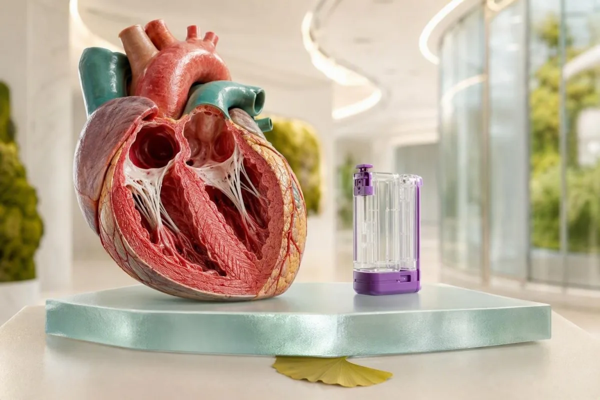
Chest pain is a symptom, not a diagnosis. The safest decision depends on your symptoms, ECG, examination and repeat blood tests—not one reassuring result.


- Call emergency services now for pressure-like chest pain lasting more than 15 minutes, especially with breathlessness, sweating, fainting, or pain spreading to the arm, jaw, back, or upper abdomen.
- High-sensitivity troponin above the laboratory 99th-percentile upper reference limit indicates heart-muscle injury, but does not alone prove a heart attack.
- Early normal troponin can be falsely reassuring when symptoms began less than 2-3 hours earlier; repeat sampling is often needed.
- A changing troponin value over 1-3 hours is usually more concerning than one stable, mildly raised result.
- D-dimer is useful only when pulmonary embolism risk is low or intermediate; a positive result does not diagnose a clot.
- Potassium below 3.0 mmol/L or above 6.0 mmol/L can destabilize heart rhythm and needs urgent clinician assessment, particularly with palpitations or weakness.
- NT-proBNP above 900 pg/mL in adults aged 50-75 with acute breathlessness supports heart failure, but kidney impairment and age can raise it.
- Do not drive yourself to hospital if chest pain is severe, recurrent, or paired with collapse, neurological symptoms, or shortness of breath.
Which chest-pain symptoms need emergency care before blood tests?
Call emergency services immediately for new central chest pressure, heaviness, squeezing, or burning that lasts more than 15 minutes or returns at rest—especially with breathlessness, cold sweating, nausea, fainting, or pain to the jaw, arm, back, or upper abdomen. A blood test for chest pain is never a safe reason to wait at home.
Chest pain can reflect a heart attack, pulmonary embolism, aortic emergency, severe pneumonia, collapsed lung, or a less dangerous cause such as reflux or chest-wall strain. The symptom pattern matters more than whether pain is “sharp” or “dull”; women, older adults, and people with diabetes may have nausea, fatigue, breathlessness, or upper-back discomfort rather than classic pressure.
I have seen a fit 47-year-old dismiss intermittent jaw tightness as stress because he had no dramatic chest pain. His first ECG was subtle, his repeat troponin rose, and angiography showed an acute coronary blockage. Dr. Thomas Klein’s practical rule is simple: new exertional symptoms or symptoms at rest deserve urgent assessment, not online reassurance.
Sudden tearing pain through the chest into the back, collapse, one-sided weakness, new speech difficulty, or a pulse difference between arms raises concern for an aortic catastrophe or stroke pathway. Blood work may support the assessment, but imaging and immediate clinical examination take priority; see our guide to blood tests for shortness of breath.
What chest pain blood work do emergency clinicians usually order?
Emergency chest pain blood work usually includes high-sensitivity troponin, a full blood count, electrolytes, kidney function, glucose, and sometimes D-dimer, BNP or blood gases. The exact panel follows the working diagnosis, not a fixed checklist.
High-sensitivity cardiac troponin I or T is the core blood test because it detects injury to heart-muscle cells. A complete blood count can identify anaemia or infection; a haemoglobin below 80 g/L can worsen cardiac oxygen supply, while a white-cell count alone cannot distinguish stress from infection. Kidney function affects both risk assessment and safe use of contrast imaging.
Clinicians add glucose because severe hyperglycaemia can accompany acute illness, and they check potassium, magnesium, sodium, and calcium because abnormal levels may provoke arrhythmias. Potassium above 6.0 mmol/L, calcium below 1.9 mmol/L, or magnesium below 0.5 mmol/L merits urgent ECG review even if chest discomfort seems mild. Our electrolyte urgent-care guide explains why symptoms change the response.
Kantesti is an AI blood test analyzer designed to organize uploaded laboratory results into clinical patterns, but it cannot replace emergency assessment for active chest pain. Our blood test biomarkers guide helps explain common analytes after a clinician has excluded an emergency, and our clinical use-case workflows describe appropriate follow-up boundaries.
Why can an early normal troponin fail to rule out a heart problem?
A normal high-sensitivity troponin drawn very early after chest pain begins may not rule out a heart attack because measurable release takes time. Most validated rapid pathways require symptoms to have started at least 3 hours before a single very-low result is used for rule-out.
Troponin is a structural protein released when heart-muscle cells are injured. With modern assays, concentrations often begin rising within 1-3 hours, but the exact timing varies with the size of injury, coronary flow, kidney function, and the assay used. The 2021 AHA/ACC chest-pain guideline recommends serial high-sensitivity troponin testing within 1-3 hours when acute coronary syndrome remains possible (Gulati et al., 2021).
A value below the assay’s limit of detection can be reassuring only in the right setting: low-risk history, non-ischaemic ECG, symptoms beginning at least 3 hours earlier, and no ongoing pain. A normal result at 45 minutes is a snapshot, not a verdict. This distinction is central to troponin I versus troponin T, since their numbers and reporting units are not interchangeable.
Do not compare a troponin result with a generic internet “normal range.” Laboratories set assay-specific 99th-percentile limits; one lab may report hs-cTnT in ng/L and another hs-cTnI in ng/L with a different cutoff. Kantesti AI reads the reported method and reference interval, while the emergency clinician decides whether the timing and symptoms make the result safe to act on.
Why does the troponin change matter more than one result?
A meaningful rise or fall in troponin over 1-3 hours suggests acute myocardial injury, whereas a stable elevation often reflects chronic injury. No universal delta number fits every assay, so clinicians use the local laboratory’s validated pathway.
A patient with chronic kidney disease may have hs-cTnT persistently above 14 ng/L without an acute heart attack. Conversely, a first value inside range that rises substantially on repeat testing can reveal a developing event. The Fourth Universal Definition of Myocardial Infarction separates myocardial injury from infarction by requiring a rise or fall plus evidence of ischaemia, such as symptoms, ECG changes, or imaging findings (Thygesen et al., 2018).
The absolute difference is often more informative at lower concentrations, while a relative change may help when the baseline is already high. In practice, I worry more about 8 ng/L becoming 27 ng/L in 2 hours than an isolated 32 ng/L that has remained unchanged for months. Recent hard exercise, tachyarrhythmia, severe hypertension, sepsis, and pulmonary embolism can also create a real rise without a blocked coronary artery.
Keep the exact collection times, not merely the result values. The blood-test timeline guide is useful for later review, but no timeline tool can determine whether active symptoms are dangerous.
Which high troponin results require urgent care, even if they are not a heart attack?
Any newly elevated troponin with current chest pain, breathlessness, fainting, or an abnormal ECG needs same-day emergency evaluation. Troponin identifies heart-muscle injury; it does not specify the cause.
A high troponin can occur with myocarditis, pulmonary embolism, rapid atrial fibrillation, heart failure, hypertensive crisis, sepsis, severe anaemia, kidney disease, or direct cardiac trauma. Values several times above the upper reference limit increase concern, but a small elevation can still be serious when it is rising or paired with symptoms. The safest interpretation is “heart injury until explained,” not “heart attack confirmed.”
After a marathon, troponin may transiently rise despite no coronary blockage; however, chest pressure, syncope, or persistent breathlessness after exercise should not be attributed to training. Creatine kinase can also rise massively after muscle work and is less specific for cardiac injury—our high CK warning guide covers the separate rhabdomyolysis risk.
Kantesti is an AI blood test interpretation platform that flags an out-of-range troponin alongside renal function, haemoglobin, and prior results, rather than treating it as an isolated number. A flagged result is a prompt to contact the ordering clinician; active symptoms mean emergency services first.
Can normal blood tests and a normal ECG still miss dangerous chest pain?
Yes. A single normal ECG and early normal blood tests do not exclude unstable angina, intermittent arrhythmia, aortic disease, or early coronary occlusion. Repeated ECGs and observation are often necessary when symptoms persist or recur.
An ECG records only a few seconds of cardiac electrical activity. Transient ischaemic changes may disappear between episodes, and some heart attacks initially have non-diagnostic ECGs. The 2023 ESC acute coronary syndrome guideline supports repeat ECGs when symptoms recur or clinical suspicion remains high (Byrne et al., 2023).
Unstable angina can cause genuine exertional or rest chest discomfort without troponin elevation because there is ischaemia without enough cell injury to release measurable troponin. That is why clinicians integrate age, diabetes, smoking, blood pressure, known coronary disease, symptom trajectory, and the physical examination. A 34-year-old can have acute coronary syndrome; a 74-year-old can have reflux—the history shifts probability but never settles it alone.
A later outpatient discussion may include cholesterol, HbA1c, lipoprotein(a), and blood pressure, but those are prevention tests rather than emergency rule-out tests. If you are comparing risk markers after discharge, read our piece on ApoB and ApoA1 ratio.
When does a D-dimer result make chest pain urgent?
A positive D-dimer does not diagnose pulmonary embolism, but chest pain with sudden shortness of breath, coughing blood, fainting, or a swollen painful leg needs urgent assessment regardless of the number. D-dimer is most useful for ruling out a clot in people with low or intermediate pre-test probability.
D-dimer measures fibrin breakdown and rises with clots, age, pregnancy, infection, surgery, cancer, trauma, and hospital admission. In patients older than 50 years, many pathways use an age-adjusted cutoff of age multiplied by 10 ng/mL FEU; for example, 72 years corresponds to 720 ng/mL FEU. This can reduce unnecessary scans without accepting a clinically meaningful loss of safety in selected low-risk patients.
A negative high-sensitivity D-dimer can help exclude pulmonary embolism only when clinical probability is low or intermediate. It should not delay imaging when probability is high, oxygen saturation is low, blood pressure is unstable, or there is unexplained syncope. The practical traps behind false-positive and false-negative results are covered in our D-dimer accuracy article.
Hormonal oestrogen exposure, recent surgery, a long journey, pregnancy, previous clot, cancer, and prolonged immobility change the threshold for concern. Tell the emergency team about these factors even if the D-dimer is “only mildly elevated”; the result has no safe standalone interpretation.
Can blood tests identify an aortic emergency causing chest pain?
No blood test can safely rule out aortic dissection in a person with abrupt severe chest or back pain, collapse, pulse asymmetry, or neurological symptoms. Emergency CT angiography or transoesophageal echocardiography is usually decisive when clinicians suspect it.
D-dimer may be elevated in acute aortic syndromes, but it is not reliable enough to be used casually as a home rule-out test. A normal D-dimer may have a role in carefully selected low-risk pathways within the first 24 hours, yet imaging remains necessary when clinical features are concerning. In my experience, the phrase “worst pain of my life” deserves attention, but so does sudden unexplained back pain in someone who looks pale or unwell.
Creatinine and eGFR are checked before contrast CT because kidney function influences imaging preparation, not because they diagnose dissection. An eGFR below 30 mL/min/1.73 m² requires a risk-benefit discussion, but suspected dissection is usually an emergency where lifesaving imaging outweighs kidney concerns. See GFR after dehydration for why one reduced result may not reflect baseline function.
Do not take leftover anticoagulants, aspirin, or pain medicines as a substitute for assessment when dissection is possible. The treatment for aortic dissection differs sharply from treatment for coronary clot, which is precisely why rapid diagnosis matters.
Which CBC and metabolic-panel results can worsen chest pain risk?
Severe anaemia, dangerous potassium abnormalities, marked glucose elevation, and acute kidney injury can worsen chest symptoms or trigger arrhythmias. These findings often change immediate treatment even when troponin is normal.
Haemoglobin below 80 g/L can reduce oxygen delivery enough to provoke angina in people with coronary disease; active bleeding, black stools, dizziness, or fast heartbeat make it urgent. Anaemia is not the same as a heart attack, but it can produce a type 2 myocardial infarction when oxygen supply cannot meet demand. The full blood count guide explains what the accompanying indices add.
Potassium below 3.0 mmol/L or above 6.0 mmol/L can cause dangerous rhythm disturbances, particularly when ECG changes, weakness, kidney disease, or potassium-altering medicines are present. A reported potassium of 6.2 mmol/L from a haemolysed sample may be falsely high, so clinicians often repeat it urgently while reviewing the ECG. Never assume a flagged result is a laboratory error without being told so.
Glucose of 11.1 mmol/L (200 mg/dL) or higher during acute illness is common and does not by itself diagnose diabetes, while glucose above 20 mmol/L (360 mg/dL) with dehydration, vomiting, or confusion needs urgent metabolic evaluation. Kantesti AI can organize these linked results after the acute episode, including potential potassium draw errors, but it does not triage active symptoms.
What do BNP and NT-proBNP mean when chest pain comes with breathlessness?
BNP and NT-proBNP help identify cardiac strain or heart failure in people with chest pain and breathlessness, but neither test diagnoses the cause alone. A low value makes acute heart failure less likely, whereas an elevated value needs clinical context and often echocardiography.
For acute presentations, NT-proBNP below 300 pg/mL makes acute heart failure unlikely in many adults. Age-based rule-in thresholds are often 450 pg/mL under age 50, 900 pg/mL at ages 50-75, and 1,800 pg/mL above 75, although laboratories and guidelines may vary. Kidney disease, atrial fibrillation, pulmonary embolism, and sepsis can raise the value without classic left-sided heart failure.
Obesity can suppress natriuretic peptide concentrations, so a seemingly modest result does not always exclude heart failure in a person with marked swelling, breathlessness lying flat, or rapid weight gain. I am particularly cautious when a patient reports needing three pillows for sleep or waking abruptly gasping—those practical details can matter as much as the number. Our NT-proBNP cutoff guide gives the broader context.
Kantesti is an AI-powered blood test analysis tool that can display NT-proBNP with eGFR, albumin, and sodium trends for clinician follow-up. Its clinical methodology and limitations are reviewed in our medical validation information.
Which chest pain blood-test results can be misleading?
Haemolysis, delayed processing, recent heavy exercise, and chronic kidney disease can distort or complicate chest pain blood work. A surprising result should be repeated or interpreted with the sample-quality note, symptoms, ECG, and prior values.
Haemolysis releases potassium from cellular elements during collection or transport, producing pseudohyperkalaemia. A haemolysis index on the lab report, a normal ECG, and a promptly repeated non-haemolysed sample can clarify the issue; however, clinicians treat a true potassium above 6.0 mmol/L as urgent until proven otherwise.
Strenuous exercise can raise CK, AST, creatinine, and occasionally troponin, especially after endurance events. That does not give athletes a free pass: chest pain, fainting, unusual breathlessness, or palpitations need assessment because myocarditis and coronary disease can occur in very fit people. The marathon runner blood-test guide separates expected training effects from red flags.
Biotin supplements can interfere with selected immunoassays, although the direction and extent depend on the specific test. Bring a medication and supplement list, including dose and last use, because it can prevent a misleading result or unnecessary repeat.
When is discharge after chest pain blood work reasonably safe?
Discharge is considered only after clinicians judge the overall short-term risk to be low using symptoms, examination, serial ECGs, serial troponin, vital signs, and sometimes a validated score. A low-risk label does not mean “nothing happened”; it means immediate hospital danger appears sufficiently low for outpatient care.
A commonly used HEART score combines History, ECG, Age, Risk factors, and Troponin. Scores of 0-3 are often associated with low short-term event risk when paired with serial high-sensitivity troponin protocols, but tools cannot overrule persistent symptoms, an abnormal examination, or clinician concern. The discharge plan should state exactly when to return and who will review follow-up tests.
Ask for the troponin assay name, values, collection times, ECG interpretation, and whether any finding needs repeat testing. Saving the original report is more useful than transcribing a single “normal” label; results before doctor review explains why portal release can be confusing.
Kantesti AI supports secure result organization and longitudinal comparison after discharge, not real-time emergency diagnosis. Our medical advisory board oversees clinical safety principles, including clear escalation advice when laboratory patterns and symptoms may signal urgent care.
What should you avoid doing while waiting with possible cardiac chest pain?
Do not drive yourself, exercise to “test” the pain, wait for a home blood test, or take someone else’s medicine when heart-related chest pain is possible. Sit or rest in a safe place, call emergency services, and unlock the door if you are alone.
If emergency services advise aspirin and you have no allergy, active bleeding, or previous instruction to avoid it, follow their local protocol rather than improvising a dose. Aspirin is not appropriate for every cause of chest pain, particularly suspected aortic dissection or significant gastrointestinal bleeding. Emergency dispatchers can tailor advice to your country and situation.
Do not use a smartwatch rhythm tracing or a normal oxygen reading to dismiss persistent chest pain. Consumer devices may detect useful clues, but they cannot exclude coronary ischemia, pulmonary embolism, or aortic disease. A normal pulse rate is also not a safety certificate.
In our review of patient reports, the dangerous delay is often caused by “I’ll see if it settles after an hour.” If the discomfort is new, recurrent, or associated with exertion, arrange urgent evaluation. For a calmer follow-up process after a clinician has assessed you, our AI report accuracy checklist helps verify source values and units.
Which blood tests matter after an emergency cause of chest pain is excluded?
After an emergency cause is excluded, lipid testing, HbA1c, kidney function, blood pressure review, and one-time lipoprotein(a) testing can clarify future cardiovascular risk. These prevention tests do not replace acute troponin testing during symptoms.
A fasting sample is not always required for a standard lipid panel, but triglycerides above 4.5 mmol/L (400 mg/dL) may prompt repeat fasting testing. LDL cholesterol drives long-term atherosclerotic risk, while HbA1c of 6.5% or higher supports diabetes when confirmed outside acute illness. The distinction matters because emergency stress can temporarily raise glucose.
Lipoprotein(a) is largely genetically determined and generally needs measuring once in adulthood; values above 125 nmol/L or 50 mg/dL are commonly considered risk-enhancing. Family history of early heart attack, stroke, or very high cholesterol makes this particularly useful. Read our lipoprotein(a) screening guide for unit differences and family implications.
As of September 2, 2026, prevention should be individualized rather than reduced to one cholesterol target. Dr. Thomas Klein recommends bringing family history, smoking status, pregnancy history, medicines, and prior lipid values to follow-up. Learn how Kantesti’s analysis methods handle trend context in our AI technology guide.
Frequently Asked Questions
Can a blood test tell if chest pain is a heart attack?
A high-sensitivity troponin blood test can detect heart-muscle injury, but it cannot diagnose a heart attack by itself. Clinicians diagnose myocardial infarction when a troponin rise or fall occurs together with evidence of ischemia, such as typical symptoms, ECG changes, or imaging findings. A value above the assay-specific 99th-percentile upper reference limit is abnormal, yet myocarditis, pulmonary embolism, severe anaemia, kidney disease, and fast arrhythmias can also raise troponin. New chest pain with an elevated troponin needs urgent medical assessment.
How soon does troponin rise after chest pain starts?
High-sensitivity troponin often begins to rise about 1-3 hours after acute heart-muscle injury, but the timing varies between people and assays. A single very-low result is generally used for rule-out only when symptoms began at least 3 hours earlier and the ECG and clinical risk are low. If blood is drawn 30-90 minutes after pain begins, a normal result may need repeating after 1-3 hours. Ongoing or severe symptoms require emergency assessment regardless of the first troponin result.
What troponin number is dangerous?
A dangerous troponin number is not universal because each assay has its own 99th-percentile upper reference limit and reporting method. For one common high-sensitivity troponin T assay, values above 14 ng/L are above the reference limit, but the trend over 1-3 hours and the patient’s symptoms are often more informative than a single value. A result several times above the upper reference limit with chest pain, breathlessness, sweating, or fainting is an emergency. Even a small rise from 8 ng/L to 27 ng/L can be clinically significant.
Can anxiety cause high troponin or chest pain?
Anxiety can cause chest tightness, fast breathing, palpitations, and tingling, but it should not be assumed to explain new chest pain before dangerous causes are excluded. Panic itself does not usually cause a substantial troponin elevation, although extreme tachycardia or severe blood-pressure elevation can occasionally contribute to myocardial injury. A troponin above the laboratory reference limit requires a medical explanation, especially if it changes on repeat testing. Chest pain with fainting, breathlessness, or exertional onset needs urgent assessment even in someone with known anxiety.
Is D-dimer high with a heart attack?
D-dimer can be elevated during a heart attack, but it is not a diagnostic blood test for coronary blockage. D-dimer measures fibrin breakdown and also rises with pulmonary embolism, age, pregnancy, surgery, infection, cancer, and inflammation. In adults over 50 years, some low-risk pulmonary embolism pathways use an age-adjusted cutoff of age multiplied by 10 ng/mL FEU, such as 700 ng/mL FEU at age 70. A positive D-dimer usually leads to further risk assessment or imaging rather than a diagnosis by itself.
Should I go to urgent care or the emergency department for chest pain?
The emergency department or emergency services are safer than routine urgent care for chest pressure lasting more than 15 minutes, pain at rest, pain with breathlessness or sweating, fainting, new neurological symptoms, or pain spreading to the jaw, arm, back, or upper abdomen. Emergency departments can perform serial ECGs, serial high-sensitivity troponin testing at 1-3 hour intervals, urgent imaging, and continuous monitoring. Urgent care may be appropriate only when symptoms are clearly mild, stable, non-exertional, and a clinician has judged emergency causes unlikely. If you are unsure, emergency evaluation is the safer choice.
Get AI-Powered Blood Test Analysis Today
Join over 2 million users worldwide who trust Kantesti for instant, accurate lab test analysis. Upload your blood test results and receive comprehensive interpretation of 15,000+ biomarkers in seconds.
📚 Referenced Research Publications
Klein, T., Mitchell, S., & Weber, H. (2026). Kantesti LTD. (2026). AI Blood Test Analyzer: 2.5M Tests Analyzed | Global Health Report 2026. Zenodo. https://doi.org/10.5281/zenodo.18175532. ResearchGate: https://www.researchgate.net/; Academia.edu: https://www.academia.edu/. Kantesti AI Medical Research.
Klein, T., Mitchell, S., & Weber, H. (2026). Kantesti LTD. (2026). RDW Blood Test: Complete Guide to RDW-CV, MCV & MCHC. Zenodo. https://doi.org/10.5281/zenodo.18202598. ResearchGate: https://www.researchgate.net/; Academia.edu: https://www.academia.edu/. Kantesti AI Medical Research.
📖 External Medical References
⚕️ Medical Disclaimer
This article is for educational purposes only and does not constitute medical advice. Always consult a qualified healthcare provider for diagnosis and treatment decisions.
E-E-A-T Trust Signals
Experience
Physician-led clinical review of lab interpretation workflows.
Expertise
Laboratory medicine focus on how biomarkers behave in clinical context.
Authoritativeness
Written by Dr. Thomas Klein with review by Dr. Sarah Mitchell and Prof. Dr. Hans Weber.
Trustworthiness
Evidence-based interpretation with clear follow-up pathways to reduce alarm.