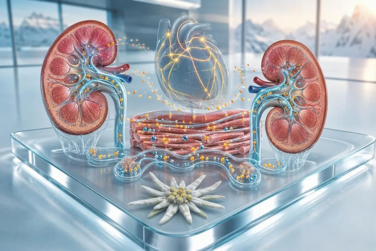
The most useful electrolyte interpretation asks which values moved together, how quickly they changed, and whether the kidneys, heart, or brain may be under strain.


- Emergency pattern means potassium of 6.5 mmol/L or higher, sodium below 120 mmol/L, or any electrolyte result paired with fainting, seizures, confusion, chest symptoms, or severe weakness.
- Low sodium plus low CO2 can signal a high-risk metabolic problem, especially with vomiting, diarrhoea, diabetes, kidney disease, or rapid breathing.
- High potassium plus reduced eGFR needs urgent clinical review because impaired kidneys cannot reliably remove potassium from the body.
- Low CO2 blood test usually reflects low bicarbonate; a value below 18 mmol/L deserves prompt interpretation with chloride, glucose, ketones, creatinine, and the anion gap.
- Potassium recheck is often sensible for a mild isolated rise, but it is not a reason to wait when potassium is 6.0 mmol/L or above or symptoms are present.
- Magnesium matters because magnesium below 0.5 mmol/L can make low potassium and low calcium difficult to correct and may contribute to rhythm instability.
- Corrected sodium should be considered when glucose is high; each 100 mg/dL glucose rise above 100 lowers measured sodium by roughly 1.6–2.4 mmol/L.
- Trend speed changes triage: a sodium fall from 139 to 124 mmol/L over 24 hours can be more dangerous than a stable sodium of 124 mmol/L in a closely monitored chronic case.
Which electrolyte combinations need urgent action?
Electrolyte panel results need urgent care when a dangerous number is paired with symptoms, a rapid change, kidney impairment, or evidence of acid-base disturbance. Potassium at 6.5 mmol/L or higher, sodium below 120 mmol/L, CO2 below 15 mmol/L, or calcium below 1.75 mmol/L generally warrants emergency assessment, particularly with confusion, collapse, seizures, palpitations, chest discomfort, marked weakness, or breathlessness. As of August 4, 2026, the combination matters more than a single red flag: Kantesti is an AI blood test analyzer that reads sodium, potassium, chloride, CO2, glucose, and kidney markers as one clinical pattern.
A typical adult serum sodium range is 135–145 mmol/L, potassium is 3.5–5.0 mmol/L, chloride is 98–107 mmol/L, and total CO2 is commonly 22–29 mmol/L; your laboratory's printed range takes priority. Dr. Thomas Klein's practical rule is simple: a value barely outside range may be routine, while two values pointing to the same physiological failure deserve attention. Our basic metabolic panel overview explains why creatinine and glucose belong beside these values.
In our review work, the combination that most often changes a calm outpatient plan into same-day triage is potassium 5.8–6.4 mmol/L with eGFR below 30 mL/min/1.73 m² or CO2 below 20 mmol/L. Acidosis shifts potassium from cells into the circulation, and reduced filtration limits its removal; that is a much different situation from potassium 5.3 mmol/L after a difficult sample collection.
A red laboratory flag is not itself a diagnosis. Kantesti analyses pattern relationships against the 15,000+ biomarker guide, but an AI interpretation never replaces an ECG, examination, repeat sample, or emergency clinician when a critical pattern appears.
Low sodium with low CO2: why this pair can be risky
Low sodium plus low CO2 can indicate water imbalance alongside metabolic acidosis or compensated respiratory alkalosis, and it requires faster assessment when sodium is below 125 mmol/L or CO2 is below 18 mmol/L. The immediate concern is not that the two values always share one cause; it is that diarrhoea, adrenal insufficiency, diabetic ketoacidosis, sepsis, kidney dysfunction, and some medicines can produce both.
Total CO2 on a chemistry panel is a close proxy for bicarbonate, not a measure of oxygen or carbon dioxide in the lungs. A CO2 value of 16 mmol/L with sodium 122 mmol/L should trigger a review of glucose, ketones, lactate, chloride, creatinine, medication exposure, and symptoms rather than advice to simply eat more salt; see our detailed discussion of low sodium warning signs.
The European hyponatraemia guideline advises immediate treatment in a monitored setting for severe neurological symptoms and uses 150 mL of 3% saline over 20 minutes as an initial clinician-directed approach (Spasovski et al., 2014). That is hospital care, not a home recipe: rapid overcorrection can injure the brain, and many specialists aim to limit sodium correction to roughly 8 mmol/L in 24 hours in people at high risk of osmotic demyelination.
One easily missed pattern is low sodium, low CO2, and normal-to-high chloride after several days of diarrhoea. Stool bicarbonate loss lowers CO2, while drinking large volumes of plain water can worsen sodium dilution; a diarrhoea-related lab review is useful when this follows gastrointestinal illness.
High potassium with kidney impairment: the ECG risk pattern
High potassium is more urgent when kidney filtration is reduced, CO2 is low, or potassium is rising quickly; potassium of 6.0 mmol/L or higher generally needs same-day clinical assessment and an ECG. The electrical effects on the heart are unpredictable enough that a person can feel well shortly before an abnormal rhythm develops.
Potassium of 5.1–5.4 mmol/L is often rechecked when it is isolated, kidney function is normal, and the laboratory reports haemolysis or a difficult collection. Potassium of 6.0–6.4 mmol/L, however, is not a routine repeat-and-wait result if eGFR is reduced, and potassium 6.5 mmol/L or higher is commonly managed as an emergency; read more about a slightly elevated potassium result.
The KDIGO potassium conference report identifies chronic kidney disease, diabetes, metabolic acidosis, and renin-angiotensin-aldosterone system medicines as overlapping drivers of hyperkalaemia (Clase et al., 2020). Kantesti AI highlights the combination of potassium, creatinine, eGFR, CO2, glucose, and medication entries because a potassium value alone does not distinguish impaired excretion from a collection artefact.
High potassium emergency signs include new palpitations, fainting, chest pressure, sudden profound weakness, or an unusually slow or irregular pulse. Do not take an extra diuretic, potassium binder, or bicarbonate from an old prescription without medical direction; the safe intervention depends on ECG findings, volume status, kidney function, and the cause.
Low CO2 blood test: use chloride and the anion gap
A low CO2 blood test should be interpreted with chloride and the anion gap, because CO2 below 22 mmol/L may represent bicarbonate loss, acid accumulation, or compensation for a breathing disorder. The basic calculation is anion gap = sodium − chloride − CO2; most modern laboratories consider roughly 8–12 mmol/L typical when potassium is excluded.
A high anion gap with CO2 below 18 mmol/L raises concern for ketoacidosis, lactic acidosis, advanced kidney failure, or certain toxin exposures. For example, glucose 420 mg/dL, CO2 12 mmol/L, anion gap 24 mmol/L, nausea, and deep rapid breathing is an emergency pattern that needs immediate hospital assessment, not a next-week appointment.
A normal anion gap with low CO2 and high chloride more often points toward bicarbonate loss through diarrhoea, renal tubular acidosis, or chloride-rich fluid administration. Albumin complicates the calculation: for every 1 g/dL albumin below 4.0 g/dL, clinicians commonly add about 2.5 mmol/L to the measured anion gap, which can reveal a hidden gap in malnutrition or serious illness.
Respiratory alkalosis can also lower panel CO2 because the kidneys excrete bicarbonate over time; panic-related hyperventilation is possible, but so are pulmonary embolism, severe infection, and liver disease. The high chloride and CO2 pattern is more informative than treating a single low CO2 flag as proof of one diagnosis.
Chloride tells you whether low CO2 is a loss or an acid load
Chloride moving opposite to CO2 often identifies the type of acid-base disturbance: high chloride with low CO2 suggests hyperchloraemic acidosis, while low chloride with high CO2 suggests vomiting, diuretic effect, or metabolic alkalosis. Chloride is usually 98–107 mmol/L, but its meaning is relational rather than independent.
A chloride of 112 mmol/L with CO2 17 mmol/L and a normal anion gap is commonly seen after bicarbonate loss or a chloride-heavy intravenous fluid load. This pattern may cause fatigue and faster breathing, yet the urgency depends on pH, kidney function, circulation, and cause—not merely on chloride being 5 mmol/L above range.
By contrast, chloride 88 mmol/L, CO2 34 mmol/L, and potassium 3.0 mmol/L after repeated vomiting is a recognisable alkalosis pattern. It can lead to weakness, cramps, light-headedness, and rhythm vulnerability, especially when magnesium is also low; our article on low chloride from vomiting or diuretics covers the practical questions to bring to a clinician.
Urine chloride, when ordered, can refine the story. A urine chloride below about 20 mmol/L in metabolic alkalosis often fits chloride depletion from vomiting or remote diuretic use, while a higher value can suggest ongoing diuretic effect or mineralocorticoid excess—one of those details that changes treatment.
Low potassium plus low magnesium: the correction-resistant pair
Low potassium with low magnesium is clinically meaningful because magnesium deficiency increases urinary potassium loss and can make potassium replacement fail until magnesium is addressed. Potassium below 3.0 mmol/L or magnesium below 0.5 mmol/L merits urgent assessment when symptoms, heart disease, or QT-prolonging medicines are present.
The usual adult potassium range is 3.5–5.0 mmol/L, while serum magnesium commonly runs 0.70–1.00 mmol/L. A potassium of 2.8 mmol/L with magnesium 0.48 mmol/L is more concerning than potassium 2.8 alone because both minerals influence cardiac repolarisation and muscle membrane stability.
I see this after prolonged diarrhoea, intensive endurance exercise, heavy alcohol exposure, poorly controlled diabetes, and use of loop or thiazide diuretics. Long-term proton-pump inhibitor use is another under-recognised contributor to low magnesium, which is why a low magnesium causes review can be relevant when cramping and palpitations accompany a low potassium result.
Severe weakness, inability to stand, fainting, new palpitations, or potassium below 2.5 mmol/L should prompt emergency care. Oral replacement is reasonable only in selected stable cases; intravenous replacement, infusion rate, ECG monitoring, and kidney-dose adjustments require a clinical setting.
Calcium, magnesium, and phosphate: neuromuscular red flags
Low calcium, magnesium, and phosphate together can cause tingling, cramps, confusion, weakness, and rhythm problems, particularly after malnutrition, alcohol-related illness, major surgery, or refeeding. Ionised calcium is the biologically active result; total calcium must be interpreted with albumin.
Total calcium is commonly 2.15–2.55 mmol/L, but albumin changes the measured total without necessarily changing ionised calcium. A rough correction adds 0.02 mmol/L calcium for each 1 g/L albumin below 40 g/L, although direct ionised calcium is more reliable in acute illness, major pH shifts, and critical care.
Phosphate below 0.32 mmol/L can impair muscle and respiratory function, while magnesium below 0.5 mmol/L may trigger tremor, tetany, or seizures. Kantesti AI is an AI biomarker interpretation platform that checks this mineral cluster against nutrition history, glucose treatment, kidney markers, and recent hospitalisation rather than suggesting that one low value has a single cause.
People restarting calories after several days of poor intake can develop a rapid intracellular shift of phosphate, potassium, and magnesium within 24–72 hours. That is why refeeding risk needs medical planning, and why our low phosphate symptom guide is especially relevant after fasting, eating disorders, chemotherapy, or prolonged illness.
Low sodium with high glucose: check the corrected sodium
High glucose can lower measured sodium by drawing water into the bloodstream, so sodium should be corrected before calling it true hyponatraemia. A commonly used estimate adds 1.6–2.4 mmol/L to sodium for each 100 mg/dL glucose above 100 mg/dL, although the exact factor varies at very high glucose levels.
For example, sodium 128 mmol/L with glucose 500 mg/dL may correct to approximately 134–138 mmol/L. That does not make the situation harmless: glucose above 300 mg/dL with thirst, vomiting, abdominal pain, drowsiness, deep breathing, or positive ketones can indicate hyperglycaemic crisis and needs urgent evaluation.
Measured serum osmolality helps distinguish dilutional hyponatraemia from hypertonic hyponatraemia. A sodium of 124 mmol/L with normal glucose and low serum osmolality is a different problem from sodium 124 with glucose 600 mg/dL; compare this with the fasting and post-meal values in our glucose range guide.
Do not drink litres of plain water to “flush out sugar” when sodium is low or symptoms are present. The right fluid, insulin plan, potassium monitoring, and rate of correction depend on whether the person has diabetic ketoacidosis, hyperosmolar state, dehydration, kidney impairment, or another process.
Sodium, potassium, CO2, and creatinine: the kidney stress cluster
A rising creatinine with high potassium and low CO2 suggests impaired kidney clearance plus acidosis and should be assessed promptly, particularly when eGFR is below 30 mL/min/1.73 m². This cluster can emerge with dehydration, acute kidney injury, advanced chronic kidney disease, urinary obstruction, or medicine-related reduction in kidney perfusion.
Creatinine is influenced by muscle mass and recent exercise, yet a rapid increase of 0.3 mg/dL within 48 hours or 1.5 times baseline within 7 days meets a commonly used acute kidney injury definition. When that rise occurs with potassium 5.8 mmol/L and CO2 18 mmol/L, the pattern is more urgent than any one result, and the chronic kidney disease staging guide provides useful background.
BUN or urea can add volume clues, but a high BUN-to-creatinine ratio is not proof of dehydration. Gastrointestinal bleeding, high protein intake, steroids, catabolism, and reduced kidney perfusion can all raise it; our BUN and creatinine ratio guide explains the limits of that shortcut.
The 2024 KDIGO guideline recommends evaluating both eGFR and urine albumin-creatinine ratio for kidney risk, not creatinine alone (KDIGO CKD Work Group, 2024). New ankle swelling, reduced urine output, breathlessness, vomiting, or an inability to keep fluids down should lower the threshold for urgent in-person care.
Medicine-related electrolyte patterns clinicians look for
Medication effects often explain electrolyte clusters: ACE inhibitors, ARBs, spironolactone, trimethoprim, and NSAIDs can raise potassium, while thiazide diuretics and several antidepressants can lower sodium. The highest-risk scenarios occur after a new prescription, dose increase, dehydration, or an acute illness that changes kidney perfusion.
A patient taking an ACE inhibitor plus spironolactone who develops creatinine 1.8 mg/dL, eGFR 32 mL/min/1.73 m², and potassium 5.9 mmol/L needs a prescribing clinician to review the combination urgently. Do not stop a heart or kidney medicine abruptly based on an app alert alone, but do contact the prescriber the same day; timing after blood-pressure medicine changes matters.
Thiazide-associated hyponatraemia can appear weeks or even months after initiation, particularly in older adults with lower body mass or high water intake. Sodium below 125 mmol/L, new unsteadiness, confusion, or falls is an urgent pattern even if the person initially describes only fatigue or “brain fog.”
Metformin rarely causes a low CO2 pattern by itself, but severe dehydration, sepsis, hypoxia, or substantial kidney dysfunction changes the risk calculation for lactic acidosis. Bring every prescription, over-the-counter pain medicine, herbal product, electrolyte powder, and salt substitute to the review; potassium-containing salt substitutes are easy to overlook.
When an abnormal electrolyte result may be a sample problem
A falsely high potassium result is common after cellular disruption during collection, but suspected sample error must be confirmed quickly when potassium is above 5.5 mmol/L or kidney function is impaired. Haemolysis can release intracellular potassium, and delayed processing, fist clenching, or a traumatic collection can exaggerate the value.
Laboratories often flag haemolysis, but the flag does not prove the potassium is false. A potassium of 6.2 mmol/L in a person with eGFR 22 mL/min/1.73 m² should still be treated as potentially real until an urgent repeat and ECG clarify it; this is why our potassium draw error explainer emphasises clinical context.
Pseudohyperkalaemia can also occur with extreme thrombocytosis or leukocytosis, because potassium leaks during clotting in a serum specimen. A clinician may request plasma potassium, a rapidly processed whole-sample measurement, or a repeat collection without prolonged tourniquet time when the result and clinical picture conflict.
Kantesti checks whether a dramatic change conflicts with prior reports, but it cannot see the collection event itself. A change from potassium 4.2 to 6.1 mmol/L within hours deserves a delta-check mindset, particularly when creatinine, CO2, symptoms, and the laboratory comment do not move in the same direction.
Which electrolyte imbalance symptoms mean emergency care?
Call emergency services now for seizures, fainting, new confusion, severe drowsiness, chest pain, a sustained irregular heartbeat, severe breathlessness, or rapidly worsening weakness with an abnormal electrolyte panel. Symptoms can outweigh a borderline number, because the speed of electrolyte change and the underlying illness are not fully visible on the report.
Brain symptoms are especially concerning with sodium below 125 mmol/L, calcium below 1.9 mmol/L, or severe glucose disturbance. A person with sodium 126 who is speaking normally and has a stable chronic history may be managed very differently from someone who fell from 138 to 126 over 24 hours and is now confused.
Heart-related symptoms deserve fast action with potassium above 5.5 mmol/L or below 3.0 mmol/L, particularly in people with known heart disease or medicines that affect rhythm. Our blood test and palpitations article explains why an ECG is necessary even when a person feels better by the time they are seen.
When I am unsure from a remote report, I advise a lower threshold for in-person evaluation in pregnancy, infancy, frail older age, established kidney or heart failure, diabetes using insulin, and active cancer treatment. The physicians on our Medical Advisory Board use the same principle: triage is based on the person and trajectory, not a traffic-light colour alone.
What to bring to a same-day electrolyte review
Bring the complete report, your previous electrolyte values, a medication list, and a brief timeline of symptoms to a same-day review; these details often identify the cause faster than repeating one number alone. The most useful timeline includes fluid intake, vomiting or diarrhoea, urine output, recent exercise, new supplements, and any medicine change in the previous 14 days.
Write down the exact test date and time, whether you were fasting, and whether you received intravenous fluids before collection. A sodium of 130 mmol/L after a marathon, large water intake, and nausea carries a different clinical question than sodium 130 after a new thiazide prescription and several quiet weeks at home.
Ask four focused questions: Is this result confirmed? Is the change acute? Does my ECG or acid-base status need checking? Which medicine, illness, or fluid pattern best explains it? A doctor-visit lab summary can keep those questions visible when anxiety makes it hard to remember them.
As Dr. Thomas Klein, I find that a two-line symptom timeline often prevents both underreaction and unnecessary panic. Kantesti AI can organise trends and prepare a pattern-based discussion, while our clinical workflow examples show how structured reports support—not replace—medical decision-making.
Using AI safely to understand electrolyte panel results
AI can help identify meaningful electrolyte patterns, but it cannot determine whether you have an ECG change, dehydration, altered mental status, or a time-critical cause of acidosis. Kantesti is an AI lab test interpretation service that flags interactions among kidney function, glucose, sodium, potassium, chloride, CO2, calcium, and magnesium for a clinician-ready discussion.
A high-quality interpretation starts by transcribing units and reference intervals correctly, then comparing the result with prior draws. For example, CO2 19 mmol/L may be chronic and stable in advanced kidney disease, while a decline from 26 to 19 in 48 hours after vomiting, infection, or a medication change can be clinically more meaningful.
Never use an interpretation tool to decide against emergency care when high potassium emergency signs, seizures, confusion, severe weakness, chest symptoms, or breathing difficulty are present. The sensible next step is to share the complete report with the clinician who knows your medications, examination findings, urine output, and baseline kidney function.
Our methods are reviewed against clinical standards and pattern-level safety checks; details are available in Kantesti's medical validation materials. In my experience, the best outcome comes when technology turns a dense panel into better questions, while a qualified clinician makes the treatment and triage decision.
Frequently Asked Questions
What electrolyte levels require emergency care?
Potassium of 6.5 mmol/L or higher, sodium below 120 mmol/L, total CO2 below 15 mmol/L, corrected calcium below 1.75 mmol/L, or phosphate below 0.32 mmol/L can require emergency evaluation. Emergency care is appropriate at any level when there are seizures, fainting, confusion, severe weakness, chest pain, palpitations, an irregular pulse, or significant breathlessness. A lower threshold is sensible for people with kidney disease, heart failure, diabetes using insulin, pregnancy, or a rapidly changing result. The laboratory's critical-value call and your clinician's advice should take precedence over general cutoffs.
What does low CO2 and low sodium on a blood test mean?
Low CO2 and low sodium can reflect a combination of water imbalance and acid-base disturbance, with possible causes including diarrhoea, diabetic ketoacidosis, adrenal insufficiency, kidney dysfunction, severe infection, and medication effects. CO2 below 22 mmol/L usually represents low bicarbonate on a chemistry panel, while sodium below 135 mmol/L is hyponatraemia. Sodium below 125 mmol/L or CO2 below 18 mmol/L deserves prompt clinical review, especially with vomiting, confusion, rapid breathing, high glucose, or reduced urine output. Clinicians usually interpret chloride, anion gap, creatinine, glucose, ketones, and serum osmolality alongside this pair.
Is high potassium always an emergency?
High potassium is not always an emergency, but potassium of 6.0 mmol/L or higher generally needs same-day clinical assessment and an ECG, while 6.5 mmol/L or higher is commonly treated as an emergency. A mild isolated result of 5.1–5.5 mmol/L may be caused by sample haemolysis or collection difficulty and can sometimes be repeated promptly. The result is more concerning when eGFR is below 30 mL/min/1.73 m², CO2 is below 20 mmol/L, creatinine is rising, or symptoms such as palpitations, fainting, chest discomfort, or weakness occur. Do not assume a haemolysis flag makes a high potassium value safe.
Can dehydration cause abnormal electrolytes and high creatinine?
Dehydration can raise sodium, urea or BUN, creatinine, and sometimes potassium, particularly when kidney blood flow falls during vomiting, diarrhoea, fever, or diuretic use. It can also lower potassium, magnesium, chloride, and CO2 when gastrointestinal losses are substantial, so there is no single dehydration pattern. A creatinine increase of 0.3 mg/dL within 48 hours or 1.5 times baseline within 7 days may meet acute kidney injury criteria and needs prompt medical assessment. Reduced urine output, dizziness on standing, confusion, severe thirst, or inability to keep fluids down increases urgency.
Why are potassium and magnesium checked together?
Potassium and magnesium are checked together because magnesium deficiency increases kidney potassium wasting and makes low potassium difficult to correct. Normal adult potassium is usually 3.5–5.0 mmol/L and serum magnesium is commonly 0.70–1.00 mmol/L, although laboratory ranges vary. Potassium below 3.0 mmol/L with magnesium below 0.5 mmol/L can increase the risk of muscle weakness and abnormal heart rhythms, particularly in people taking diuretics or QT-prolonging medicines. Clinicians often correct magnesium as part of the treatment plan rather than replacing potassium alone.
Should I drink electrolyte drinks if my sodium is low?
Do not self-treat low sodium with electrolyte drinks until the cause is clear, because sodium below 135 mmol/L can result from excess water, medication effects, heart failure, liver disease, adrenal disorders, kidney problems, or true salt loss. Sports drinks contain variable sodium and sugar amounts and are not a safe treatment for symptomatic hyponatraemia or sodium below 125 mmol/L. Drinking large volumes of plain water can worsen dilutional hyponatraemia, while concentrated salt intake can be unsafe in kidney or heart disease. A clinician may recommend a specific fluid plan after reviewing sodium, glucose, osmolality, urine studies, blood pressure, and symptoms.
Get AI-Powered Blood Test Analysis Today
Join over 2 million users worldwide who trust Kantesti for instant, accurate lab test analysis. Upload your blood test results and receive comprehensive interpretation of 15,000+ biomarkers in seconds.
📚 Referenced Research Publications
Klein, T., Mitchell, S., & Weber, H. (2026). Serum Proteins Guide: Globulins, Albumin & A/G Ratio Blood Test. Kantesti AI Medical Research.
Klein, T., Mitchell, S., & Weber, H. (2026). C3 C4 Complement Blood Test & ANA Titer Guide. Kantesti AI Medical Research.
📖 External Medical References
⚕️ Medical Disclaimer
This article is for educational purposes only and does not constitute medical advice. Always consult a qualified healthcare provider for diagnosis and treatment decisions.
E-E-A-T Trust Signals
Experience
Physician-led clinical review of lab interpretation workflows.
Expertise
Laboratory medicine focus on how biomarkers behave in clinical context.
Authoritativeness
Written by Dr. Thomas Klein with review by Dr. Sarah Mitchell and Prof. Dr. Hans Weber.
Trustworthiness
Evidence-based interpretation with clear follow-up pathways to reduce alarm.