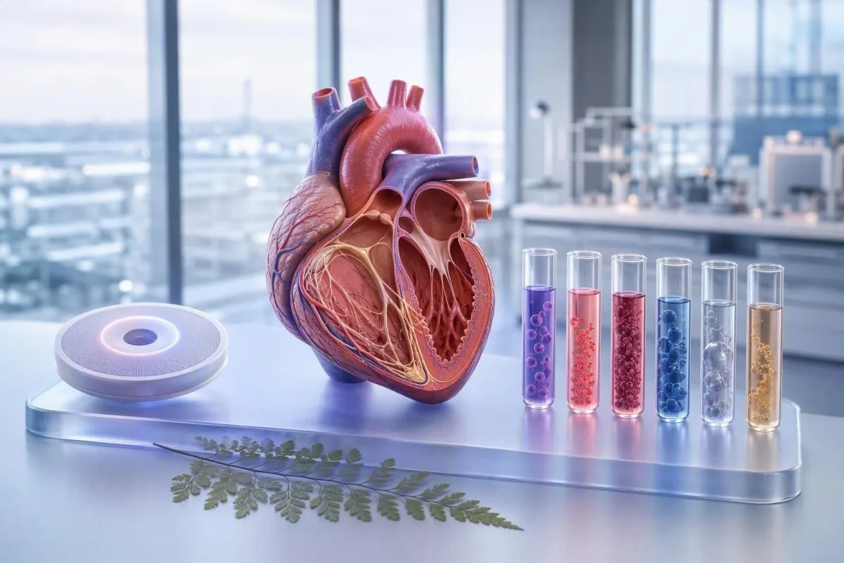
A targeted blood test for heart palpitations can uncover reversible triggers, but it cannot diagnose the rhythm itself. Here is how clinicians pair laboratory results with ECG-based assessment.


- First-line panel for palpitations commonly includes a CBC, ferritin, TSH with free T4, electrolytes, kidney function, and glucose or HbA1c.
- Hemoglobin below 12.0 g/dL in non-pregnant adult women or below 13.0 g/dL in adult men meets the WHO definition of anemia and can drive a compensatory fast pulse.
- Ferritin below 15 ng/mL indicates depleted iron stores in most adults, while many clinicians investigate symptoms when ferritin is below 30 ng/mL.
- TSH below 0.1 mIU/L with elevated free T4 strongly supports overt hyperthyroidism, a treatable cause of persistent sinus tachycardia and atrial fibrillation.
- Potassium below 3.5 mmol/L or magnesium below 0.70 mmol/L can increase cardiac electrical instability, especially with vomiting, diarrhoea, diuretics, or QT-prolonging medicines.
- Glucose below 70 mg/dL (3.9 mmol/L) can cause adrenaline-mediated pounding, shaking, and sweating; a normal HbA1c does not exclude a brief low-glucose episode.
- Normal blood work does not rule out arrhythmia. A 12-lead ECG, ambulatory monitor, or wearable rhythm strip is usually more useful when episodes are sudden, recurrent, or irregular.
- Emergency assessment is needed for palpitations with fainting, chest pressure, severe breathlessness, new weakness, or a sustained resting pulse above about 150 beats per minute.
What blood tests can and cannot show in palpitations
A blood test for heart palpitations can identify anemia, iron deficiency, thyroid excess, electrolyte disturbance, glucose shifts, kidney impairment, and systemic illness; it cannot tell whether the rhythm was atrial fibrillation, SVT, or an extra beat. In my practice, the most useful initial work-up is a focused panel plus a 12-lead ECG rather than an indiscriminate list of tests.
Palpitations describe an awareness of heartbeat—racing, skipping, fluttering, or thumping—not a diagnosis. A resting pulse of 95 beats per minute can feel alarming in someone whose usual rate is 55, while a pulse of 130 may be physiologically appropriate during fever, dehydration, or panic; the context changes the interpretation.
I am Dr. Thomas Klein, and after 15 years reviewing symptom-led panels, I have learned that a single abnormal flag rarely explains the whole story. Kantesti AI is an AI blood test analyzer that reads the CBC, ferritin, thyroid markers, glucose, and electrolytes as a pattern, including whether the finding matches the timing and character of the symptom.
Bring a short episode log to the appointment: start time, duration, pulse if available, activity, caffeine or alcohol exposure, medicines, menstrual bleeding, and whether the beat felt regular. This makes the blood test biomarkers guide far more clinically useful than a result viewed in isolation.
The practical first-line order
For new or recurrent palpitations, clinicians commonly request a CBC, ferritin with iron studies when indicated, urea/creatinine and electrolytes, TSH with free T4, and glucose or HbA1c. Pregnancy testing, troponin, CRP, drug levels, or plasma metanephrines are selective tests driven by symptoms and examination—not routine screening.
CBC results that point to anemia or blood loss
A complete blood count can find anemia that makes the heart beat faster to maintain oxygen delivery. Hemoglobin below 12.0 g/dL in non-pregnant adult women or 13.0 g/dL in adult men is anemia by WHO criteria, although symptoms depend heavily on how quickly the level fell.
A low hemoglobin accompanied by low MCV and a rising RDW often suggests evolving iron-restricted red-cell production. Palpitations are particularly common after rapid menstrual loss, a stomach illness with poor intake, or an endurance event; a gradual hemoglobin of 10.8 g/dL may be tolerated better than a sudden fall to 11.5 g/dL.
The reticulocyte count adds a detail that many standard explanations miss. A low reticulocyte response in anemia suggests underproduction, whereas a high count can follow recent loss or cellular breakdown; the CBC components explained guide helps patients see why red-cell count, hematocrit, MCV, and hemoglobin can appear to disagree.
Clinicians should not automatically blame a borderline hemoglobin for every flutter. A hemoglobin of 11.8 g/dL may contribute to exertional awareness of the pulse, but sudden irregular bursts at rest still need rhythm capture, especially in people over 50 or those with structural heart disease.
Iron deficiency can cause symptoms before severe anemia
Iron deficiency may contribute to palpitations even when hemoglobin remains within range, particularly when ferritin is below 30 ng/mL and exertional fatigue, restless legs, or heavy periods are present. Ferritin below 15 ng/mL is highly specific for depleted iron stores in an otherwise well adult.
Ferritin is an acute-phase reactant, so infection, obesity, liver inflammation, and recent strenuous exercise can raise it despite limited available iron. When ferritin is 45 ng/mL but transferrin saturation is below 20% and CRP is elevated, I consider iron restriction plausible rather than reassuring; our iron studies reference guide explains this common mismatch.
Dr. Thomas Klein's clinical rule of thumb is to ask about the source before recommending iron. Heavy menstrual bleeding, frequent donation, low intake, coeliac disease, acid-suppressing medicines, and gastrointestinal blood loss lead to very different follow-up plans; the age-specific context in ferritin levels for women is particularly useful.
Kantesti is an AI blood test interpretation platform that compares ferritin with MCV, RDW, hemoglobin, transferrin saturation, CRP, and prior values instead of calling one isolated number the answer. A rising RDW after starting iron can be expected because newly produced cells differ in size from older iron-restricted cells.
When iron studies need prompt review
New iron-deficiency anemia after menopause, in an adult male, or alongside black stools, unexplained weight loss, or persistent abdominal symptoms needs medical review for a source of loss. Iron tablets can darken stools, but they should never be used to explain away a new symptom without a clinician's assessment.
Palpitations thyroid blood test: TSH and free T4
A palpitations thyroid blood test should begin with TSH and, when TSH is outside range or suspicion is high, free T4. TSH below 0.1 mIU/L plus high free T4 usually indicates overt hyperthyroidism and warrants prompt clinical review because it increases the likelihood of sustained tachycardia and atrial fibrillation.
Most laboratories use a TSH interval around 0.4 to 4.0 mIU/L, but the assay, age, pregnancy status, and pituitary history matter. Low TSH with normal free T4 and free T3 is termed subclinical hyperthyroidism; it can still be relevant when TSH is persistently below 0.1 mIU/L, especially after age 65 or with osteoporosis or atrial fibrillation risk.
Hyperthyroid palpitations often come with heat intolerance, tremor, weight change, frequent stools, poor sleep, or a persistently fast pulse, though not everyone has the textbook picture. The American Thyroid Association guideline by Ross et al. recommends confirming biochemical hyperthyroidism and establishing the cause before definitive therapy (Ross et al., 2016); see our plain-language thyroid test glossary.
Biotin deserves a specific warning: doses of 5,000 to 10,000 mcg used in hair supplements can interfere with some immunoassays, producing a falsely low TSH and falsely high free T4. Kantesti AI asks about supplements and the laboratory method because a provocative result that conflicts with the clinical picture should be repeated after the prescriber's advised biotin pause; our AI methodology guide describes that context-first approach.
Electrolyte and kidney tests that affect cardiac rhythm
Potassium, magnesium, calcium, sodium, bicarbonate, creatinine, and eGFR are the core electrolyte and kidney tests for palpitations. Potassium below 3.5 mmol/L or above 5.5 mmol/L can alter cardiac electrical conduction, and risk rises when magnesium is also low.
A typical serum magnesium reference interval is 0.70 to 1.00 mmol/L, yet serum magnesium can look normal when total body stores are limited. I pay closer attention when a patient has diarrhoea, vomiting, long-term proton-pump inhibitor use, loop or thiazide diuretics, diabetes, or a prolonged QT interval on ECG—several small risks can add up.
Calcium must be read with albumin or measured as ionized calcium in selected cases. Total calcium of 2.10 mmol/L may reflect low albumin rather than physiologically low ionized calcium, while true hypocalcaemia may lengthen the QT interval and cause tingling or muscle spasm; the basic metabolic panel guide provides the practical units used across laboratories.
Reduced kidney clearance can both raise potassium and amplify the effects of prescribed medicines. An eGFR below 60 mL/min/1.73 m² lasting at least 3 months meets the definition of chronic kidney disease, but one low value during dehydration does not establish chronic disease.
Why potassium results sometimes mislead
A mildly high potassium result can be spurious if cellular elements break down during collection or the sample is delayed. With potassium above 6.0 mmol/L, symptoms, kidney function, medicines, and an urgent ECG matter more than speculation; laboratories may request a rapid repeat using a carefully handled specimen.
Glucose changes: why a normal HbA1c may miss an episode
Low glucose can trigger adrenaline release and produce a pounding heartbeat, tremor, sweating, hunger, and anxiety. Glucose below 70 mg/dL (3.9 mmol/L) is the standard alert threshold, but a laboratory draw hours later may be completely normal.
A fasting plasma glucose of 126 mg/dL (7.0 mmol/L) or higher on two separate diagnostic tests supports diabetes, while HbA1c of 6.5% or higher is another accepted diagnostic threshold. High glucose itself does not usually cause abrupt fluttering, but dehydration, illness, stimulant-containing products, and rapid swings during treatment can make the pulse feel forceful.
The symptom-timing question is more revealing than many people expect. Episodes that occur 2 to 4 hours after a high-carbohydrate meal, after missed meals, or overnight on glucose-lowering medication deserve paired finger-stick or continuous-monitor data at the time of symptoms; compare this with glucose ranges for women.
HbA1c can be misleadingly low after recent blood loss, hemolysis, or some hemoglobin variants, and it can read higher with iron deficiency. That is why clinicians sometimes rely on fasting glucose, symptom-time glucose, and the CBC together rather than treating HbA1c as a universal truth.
Magnesium, phosphate, and nutritional triggers
Magnesium and phosphate testing is most useful for palpitations when there is gastrointestinal loss, restrictive eating, alcohol excess, heavy endurance training, diuretic use, or poor nutritional intake. Phosphate below 0.80 mmol/L can contribute to weakness and impaired cellular energy production, while severe deficiency below 0.32 mmol/L needs urgent management.
Low magnesium is often blamed for every skipped beat, but evidence for empiric supplementation in people with normal levels and benign ectopy is mixed. In my experience, correcting a documented low value is sensible; escalating supplements without kidney-function awareness can cause diarrhoea and, in advanced kidney disease, magnesium accumulation.
Refeeding after prolonged undernutrition deserves special caution. Insulin release can shift phosphate, potassium, and magnesium into cells during the first 72 hours of nutritional rehabilitation, creating a riskier setting for rhythm disturbance than any single baseline test suggests; our low phosphate symptoms guide covers the warning signs.
A serum magnesium result should always be paired with the unit and reference interval printed by the laboratory. Kantesti AI does not infer deficiency from symptoms alone; it weighs magnesium against potassium, calcium, eGFR, medication exposures, and the result's collection context.
Medicines, stimulants, and the lab clues they leave behind
Caffeine, nicotine, decongestants, ADHD medicines, thyroid replacement, beta-agonist inhalers, energy products, and some supplements can provoke palpitations even when blood work is normal. Labs are useful here because they can reveal consequences—low potassium, suppressed TSH, reduced kidney clearance, or glucose variability—not because they detect every stimulant.
A frequent example is salbutamol or other beta-agonist use during a viral illness: the medicine can raise heart rate and shift potassium modestly into cells. A person taking a thiazide diuretic plus a laxative may develop a more meaningful potassium deficit, particularly if the bicarbonate is elevated from volume loss.
Do not stop prescribed rhythm, thyroid, psychiatric, or blood-pressure medicine abruptly because a number is flagged online. Instead, document every prescription, over-the-counter product, powder, tea, and pre-workout dose; our article on supplements changing blood tests explains why timing belongs in the laboratory history.
Inherited or acquired long-QT vulnerability is an ECG diagnosis, not a vitamin deficiency diagnosis. The combination that worries me is a QT-prolonging medicine, potassium below 3.5 mmol/L, magnesium below 0.70 mmol/L, and diarrhoea—each factor is manageable, but together they deserve same-day clinical advice.
When thyroid results need repeating rather than treatment
Borderline thyroid results often need confirmation because TSH changes more slowly than free T4 and can be temporarily altered by acute illness, pregnancy, medication changes, and assay interference. A mildly low TSH of 0.25 mIU/L without high free T4 is not proof that thyroid excess caused the palpitations.
TSH responds over weeks, not minutes. After changing levothyroxine, clinicians often wait about 6 to 8 weeks before judging the new steady state, unless severe symptoms or pregnancy requires earlier review; the TSH fluctuation guide explains why a small one-off shift is common.
Acute illness can lower T3 and sometimes lower or normalise TSH without primary thyroid disease, a pattern called non-thyroidal illness. Testing thyroid hormones during a hospital admission for infection or surgery can therefore create more uncertainty than clarity unless thyroid dysfunction is genuinely suspected.
The 2016 American Thyroid Association guideline recommends treating overt hyperthyroidism but individualising decisions in subclinical disease according to TSH degree, age, symptoms, cardiac risk, and bone risk (Ross et al., 2016). That nuance matters: treatment can settle sinus tachycardia, yet it will not necessarily eliminate unrelated premature beats.
Preparing blood work so a result answers the real question
Most blood tests for palpitations do not require fasting, but the timing of food, exercise, supplements, and the episode itself can alter what the result means. A sample drawn after a 20-km run may show transient hemoconcentration, higher creatinine, and a stress-related glucose rise that is not the patient's resting baseline.
For iron studies, a morning draw before iron tablets is often preferred because serum iron has diurnal variation and can rise after a dose. Ferritin is less affected by a single meal, while triglycerides and glucose can change substantially after eating; ask the ordering clinician whether fasting serves the specific question.
Avoid a major workout, dehydration, and new supplements immediately before a non-urgent baseline panel if possible. This is not about manufacturing a better number—it is about avoiding a misleading one; our guide to meaningful changes between visits explains biological variation and laboratory variation.
Kantesti is an AI-powered blood test analysis tool that can compare prior results and surface a genuine trend, such as ferritin falling from 52 to 18 ng/mL over 12 months, rather than overreacting to a 0.1 mmol/L potassium shift. As of August 1, 2026, trend context is still one of the most underused pieces of a palpitations evaluation.
Troponin, CRP, and tests that are not routine screening
Troponin is appropriate for suspected acute cardiac injury, not for uncomplicated intermittent palpitations. A high-sensitivity troponin result must be interpreted using the assay's 99th-percentile upper reference limit and the change over 1 to 3 hours, alongside symptoms and ECG findings.
An isolated low-level troponin elevation does not automatically mean a heart attack. Kidney disease, myocarditis, severe tachycardia, heart failure, pulmonary embolism, and even analytical issues can raise it; conversely, a normal early result may need repetition if chest pain began recently.
CRP and ESR can identify a broader inflammatory setting but are poor tests for explaining an isolated skipped beat. A CRP of 20 mg/L may support infection or tissue inflammation, yet it does not distinguish anxiety-related sinus tachycardia from supraventricular tachycardia; our troponin I versus T guide clarifies assay differences.
The 2022 European Society of Cardiology ventricular arrhythmia guideline places ECG assessment, clinical history, and structural evaluation ahead of broad biomarker testing for symptomatic rhythm concerns (Zeppenfeld et al., 2022). That ordering prevents a normal CRP from offering false reassurance.
Why normal labs may still require an ECG or monitor
Normal labs do not exclude atrial fibrillation, SVT, premature atrial contractions, premature ventricular contractions, or inherited electrical conditions. A 12-lead ECG is the next useful test when palpitations are recurrent, abrupt, irregular, exercise-related, or associated with dizziness, breathlessness, or chest discomfort.
The monitor choice should match episode frequency. Daily symptoms may be caught on a 24- to 48-hour Holter, weekly events often need a 7- to 14-day patch, and less frequent unexplained episodes may call for a longer event monitor; a normal resting ECG between episodes does not rule out intermittent arrhythmia.
Atrial fibrillation can feel like irregular fluttering, but many people have no sensation at all. The 2023 ACC/AHA/ACCP/HRS atrial fibrillation guideline requires clinician-confirmed ECG evidence for diagnosis rather than relying solely on a consumer device notification (Joglar et al., 2024); our women's heart blood-test guide also covers risk factors that can be missed.
I particularly encourage rhythm recording when a patient says, “It starts like a switch and stops like a switch.” That description is not diagnostic, but it often points us away from a metabolic trigger and toward a paroxysmal electrical rhythm that blood work cannot capture.
Palpitations red flags: when to seek urgent care
Palpitations need urgent emergency assessment when they occur with fainting, near-fainting, chest pressure, severe shortness of breath, confusion, new one-sided weakness, or a sustained very rapid heartbeat. A resting rate above roughly 150 beats per minute that does not settle, particularly with symptoms, should not be managed by waiting for outpatient blood work.
Risk is higher with known coronary disease, heart failure, cardiomyopathy, congenital heart disease, a prior arrhythmia, or a family history of sudden unexplained death before age 40. Pregnancy and the first months after delivery also change the threshold for assessment because anemia, thyroid disease, pulmonary embolism, and cardiomyopathy require tailored consideration.
Do not drive yourself if symptoms are severe or you feel likely to faint. A pulse reading alone cannot determine safety: a regular 140 beats per minute during fever differs from an irregular 140 with chest pressure, and both need the clinical setting to be understood.
For a non-emergency doctor visit, a concise lab-result visit summary can include episode timing, wearable strips, medicines, family history, and the actual laboratory report. That saves time and reduces the chance that an important pattern is lost in a long symptom list.
A sensible follow-up plan after heart palpitations blood work
The right follow-up depends on the pattern: correct a clear reversible abnormality, repeat a questionable result under better conditions, and record the rhythm when blood work is unrevealing. Most patients find that this three-part plan is more reassuring than repeatedly ordering broad panels.
If hemoglobin is 10.2 g/dL and ferritin is 7 ng/mL, the next question is the cause of iron deficiency and a monitored treatment plan—not whether the palpitations are “all anxiety.” If potassium is 3.2 mmol/L after gastroenteritis, prompt replacement advice, medicine review, and a recheck may be more useful than an extensive hormone panel.
If results are normal but episodes recur, ask specifically for ECG-based evaluation and whether echocardiography is indicated by examination, ECG, exertional symptoms, or family history. Kantesti AI interprets a palpitations-related panel by identifying clusters that justify faster follow-up, while making clear that AI interpretation is not a rhythm diagnosis.
Clinical oversight matters most at this handoff. Our medical validation standards describe how Kantesti uses source ranges, cross-marker checks, and clinician-informed safety language; our Medical Advisory Board reviews the boundaries between laboratory education and medical decision-making.
Research publications
Kantesti LTD. (2026). Diarrhea After Fasting, Black Specks in Stool & GI Guide 2026. Figshare. https://doi.org/10.6084/m9.figshare.31438111. Related record searches are available through ResearchGate and Academia.edu.
Kantesti LTD. (2026). Women's Health Guide: Ovulation, Menopause & Hormonal Symptoms. Figshare. https://doi.org/10.6084/m9.figshare.31830721. Related record searches are available through ResearchGate and Academia.edu.
Frequently Asked Questions
What blood tests should I ask for if I have heart palpitations?
A sensible first blood test for heart palpitations usually includes a CBC, ferritin or iron studies when iron deficiency is plausible, electrolytes with kidney function, TSH with free T4, and glucose or HbA1c. Potassium below 3.5 mmol/L, magnesium below 0.70 mmol/L, hemoglobin below 12.0 g/dL in most non-pregnant women, and TSH below 0.1 mIU/L are examples of findings that can materially change follow-up. The final selection should reflect your medicines, bleeding history, diet, symptoms, and examination. An ECG remains necessary when an arrhythmia is suspected.
Can low iron cause heart palpitations even if hemoglobin is normal?
Yes, low iron stores may contribute to exercise intolerance and awareness of heartbeat before anemia becomes obvious. Ferritin below 15 ng/mL usually indicates depleted stores, and many clinicians investigate symptoms at ferritin below 30 ng/mL when the broader pattern supports iron deficiency. Ferritin can be falsely normal or high during inflammation, liver disease, or recent strenuous exercise, so transferrin saturation and CRP may help. A clinician should identify why iron is low before treatment, especially in adult men and postmenopausal women.
Which thyroid blood test detects a thyroid cause of palpitations?
TSH is the usual screening test for a thyroid cause of palpitations, with free T4 added when TSH is abnormal or clinical suspicion is strong. TSH below 0.1 mIU/L with elevated free T4 supports overt hyperthyroidism, which can cause persistent tachycardia and raise atrial fibrillation risk. A mildly low TSH between 0.1 and 0.39 mIU/L needs context, repeat testing, and medication review rather than automatic treatment. High-dose biotin supplements can interfere with certain thyroid assays.
Can blood tests rule out atrial fibrillation?
No, normal blood tests cannot rule out atrial fibrillation because blood work does not record the heart rhythm. Thyroid, electrolyte, kidney, and anemia tests can identify triggers or contributors, but atrial fibrillation requires clinician-confirmed ECG evidence. A 12-lead ECG may detect persistent atrial fibrillation, while a 24-hour to 14-day ambulatory monitor is more useful for intermittent episodes. Seek urgent assessment if an irregular rapid heartbeat occurs with chest pressure, fainting, or severe breathlessness.
Can low potassium cause skipped beats?
Low potassium can increase susceptibility to extra beats and more significant rhythm disturbance, especially when potassium is below 3.5 mmol/L. The risk is greater when magnesium is below 0.70 mmol/L, kidney function is impaired, or the person takes QT-prolonging medicines, diuretics, laxatives, or certain inhalers. Potassium below 3.0 mmol/L, or any high potassium result with weakness, chest symptoms, or ECG changes, needs prompt medical assessment. A repeat result may be needed because collection and sample handling can occasionally create a falsely high potassium level.
When should palpitations send me to the emergency department?
Palpitations warrant emergency assessment when they occur with fainting or near-fainting, chest pain or pressure, severe breathlessness, confusion, new neurological symptoms, or a sustained resting pulse around 150 beats per minute or higher. People with known heart disease, cardiomyopathy, a prior arrhythmia, or a family history of sudden death before age 40 should use a lower threshold for urgent advice. Do not drive yourself if you are dizzy or feel likely to collapse. Routine blood work should never delay emergency evaluation for red-flag symptoms.
Get AI-Powered Blood Test Analysis Today
Join over 2 million users worldwide who trust Kantesti for instant, accurate lab test analysis. Upload your blood test results and receive comprehensive interpretation of 15,000+ biomarkers in seconds.
📚 Referenced Research Publications
Klein, T., Mitchell, S., & Weber, H. (2026). Diarrhea After Fasting, Black Specks in Stool & GI Guide 2026. Kantesti AI Medical Research.
Klein, T., Mitchell, S., & Weber, H. (2026). Women's Health Guide: Ovulation, Menopause & Hormonal Symptoms. Kantesti AI Medical Research.
📖 External Medical References
⚕️ Medical Disclaimer
This article is for educational purposes only and does not constitute medical advice. Always consult a qualified healthcare provider for diagnosis and treatment decisions.
E-E-A-T Trust Signals
Experience
Physician-led clinical review of lab interpretation workflows.
Expertise
Laboratory medicine focus on how biomarkers behave in clinical context.
Authoritativeness
Written by Dr. Thomas Klein with review by Dr. Sarah Mitchell and Prof. Dr. Hans Weber.
Trustworthiness
Evidence-based interpretation with clear follow-up pathways to reduce alarm.