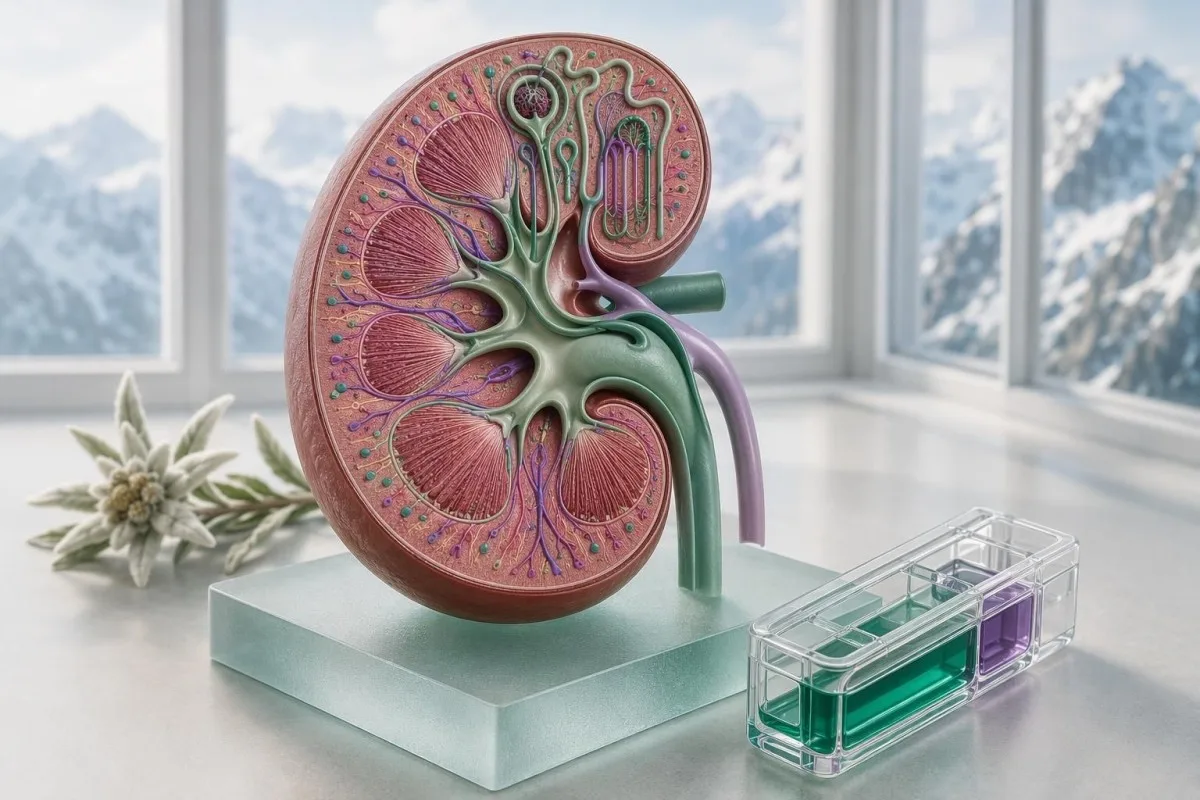
A BMP is most useful when you read its values as connected signals, not isolated flags. Sodium, CO2, glucose and kidney markers can reveal dehydration, medication effects, acid-base shifts or a need for prompt care.


- Sodium: Adult reference ranges are usually 135-145 mmol/L; sodium below 120 or above 160 mmol/L needs emergency assessment, especially with confusion or seizures.
- CO2: BMP CO2 reflects bicarbonate. A value below 18 mmol/L can indicate clinically meaningful metabolic acidosis and deserves prompt review.
- Glucose: Fasting glucose of 126 mg/dL (7.0 mmol/L) or higher meets a diabetes diagnostic threshold only when confirmed unless symptoms and unequivocal hyperglycaemia are present.
- Creatinine and eGFR: A single elevated creatinine can follow dehydration, creatine use or intense exercise; persistence and urine albumin determine kidney risk.
- Potassium: Potassium at or above 6.0 mmol/L, or below 2.5 mmol/L, can affect cardiac conduction and generally requires same-day clinical action.
- Patterns matter: Low CO2 plus high anion gap and high glucose is more concerning than any one result alone.
- Lab context: Recent vomiting, diarrhoea, fasting, IV fluids, exercise and medicines can shift several BMP values within hours.
- Follow-up: Compare the result with prior values, your symptoms, blood pressure, urine findings and the laboratory’s own reference interval.
What a basic metabolic panel actually measures
A basic metabolic panel (BMP) measures glucose, calcium, sodium, potassium, chloride, CO2, blood urea nitrogen (BUN) and creatinine; many laboratories also report eGFR. The fastest way to interpret a BMP is to ask whether its values point toward a fluid problem, an acid-base problem, altered glucose control or reduced filtration.
The BMP is not a general “health score.” It is a targeted chemistry snapshot, and the numbers change at different speeds: glucose can move within minutes, sodium over hours, and creatinine over days. Kantesti is an AI blood test analyzer that reads BMP values together with units, laboratory intervals and prior reports rather than treating a red flag as a diagnosis.
A normal BMP does not exclude early kidney disease, diabetes or hormonal disease. For example, albumin in urine may rise years before creatinine changes, which is why our biomarker reference guide distinguishes filtration markers from kidney-damage markers.
In my clinic, a “normal” result that has moved from creatinine 0.70 to 1.05 mg/dL over 6 months can deserve more attention than a stable 1.10 mg/dL result. Dr. Thomas Klein’s practical rule is simple: read the trend, then ask whether the physiology fits the person in front of you.
BUN is measured in mg/dL in the United States but urea is commonly reported in mmol/L elsewhere; they are related but not interchangeable numbers. A BUN-to-creatinine guide can prevent an unnecessary scare when reports from different countries are compared.
Check timing, fasting status and the sample before reading flags
BMP interpretation starts with the draw conditions because a non-fasting meal, prolonged tourniquet time, hard exercise or intravenous fluid can meaningfully shift results. Glucose is especially timing-sensitive, while creatinine and sodium require more clinical context.
A fasting glucose is typically interpreted after at least 8 hours without caloric intake; water is fine. A random glucose of 140 mg/dL after lunch may be unremarkable, whereas fasting 140 mg/dL is abnormal and should be confirmed.
Hemolysis during collection can falsely raise potassium because cellular contents leak into the sample. If potassium is unexpectedly 5.7 mmol/L in a well person with normal creatinine, I often check the laboratory comment and consider a repeat before assuming true hyperkalaemia; see why potassium samples fail.
Vigorous resistance exercise within 24-48 hours can raise creatinine slightly, and creatine supplements can raise it without necessarily reducing filtration. A comparison with cystatin C, urine albumin and a prior baseline is often more informative than stopping at one creatinine result.
Fasting changes some results less than patients expect: sodium and calcium should not be “corrected” by fasting, while glucose may be lower. Our detailed guide to fasting versus non-fasting tests explains which changes are real and which are noise.
Sodium results: water balance, not dietary salt alone
Adult serum sodium is commonly 135-145 mmol/L, and it primarily reflects the balance between body water and sodium rather than how much table salt you ate yesterday. Sodium below 130 mmol/L or above 150 mmol/L merits timely clinical review, particularly when symptoms are present.
Hyponatraemia below 135 mmol/L is common, but urgency depends on speed and symptoms. Headache, vomiting, new confusion, a seizure, severe unsteadiness or sodium below 120 mmol/L are emergency signals because brain cells can swell when sodium falls rapidly.
High glucose can lower measured sodium by drawing water into the bloodstream. A practical correction is to add about 1.6-2.4 mmol/L of sodium for every 100 mg/dL glucose above 100 mg/dL, although clinicians vary in the factor they use at very high glucose.
A sodium of 132 mmol/L after a marathon, with weight gain from heavy fluid intake, is a very different problem from sodium 132 mmol/L in someone taking a thiazide diuretic. For symptom thresholds and safer next steps, review low sodium warning signs.
Sodium above 155 mmol/L can signal a serious water deficit, impaired thirst, diabetes insipidus or limited access to fluids. Older adults and infants can become symptomatic before they describe thirst clearly, so a high sodium pattern should never be managed by guesswork alone.
CO2 on a BMP: the bicarbonate clue to acid-base balance
BMP CO2 usually represents serum bicarbonate, with a typical adult range of about 22-29 mmol/L. A low CO2 value below 18 mmol/L suggests metabolic acidosis until proven otherwise, while values above 32 mmol/L can reflect metabolic alkalosis or chronic respiratory compensation.
Low bicarbonate occurs when the body gains acid, loses bicarbonate or cannot excrete acid effectively. Diarrhoea, diabetic ketoacidosis, kidney failure, lactic acidosis and some medications are plausible causes, but the BMP alone cannot identify which one applies.
Calculate the anion gap when sodium, chloride and CO2 are available: sodium minus chloride minus CO2. With sodium 140, chloride 104 and CO2 18 mmol/L, the gap is 18; the laboratory’s interval matters because albumin, assay method and local calibration alter the expected result.
The combination of glucose above 250 mg/dL, CO2 below 18 mmol/L, nausea, abdominal pain, deep rapid breathing or confusion requires urgent assessment for ketoacidosis. Urine or capillary ketones and a blood gas clarify severity; do not wait for a routine appointment.
High chloride with low CO2 often produces a normal-gap acidosis after diarrhoea or large-volume saline, whereas high gap acidosis suggests added unmeasured acids. Our explanation of chloride and CO2 patterns explores that useful distinction.
Glucose results: separate screening thresholds from urgent levels
Fasting glucose of 70-99 mg/dL (3.9-5.5 mmol/L) is generally normal, 100-125 mg/dL indicates prediabetes, and 126 mg/dL or higher can diagnose diabetes when confirmed. A glucose result becomes urgent when it is very high with dehydration or ketones, or low enough to impair brain function.
The American Diabetes Association’s 2025 Standards use fasting plasma glucose of 126 mg/dL (7.0 mmol/L) or higher, A1c of 6.5% or higher, or a 2-hour glucose of 200 mg/dL or higher as diagnostic thresholds when confirmed in the absence of classic symptoms (American Diabetes Association, 2025). One unplanned BMP is a screening result, not the whole story.
A random glucose of 200 mg/dL (11.1 mmol/L) plus classic symptoms such as thirst, frequent urination and unintended weight loss can establish diabetes clinically. By contrast, glucose 62 mg/dL with sweating, shaking, confusion or inability to swallow safely needs immediate carbohydrate if the person is awake and emergency help if not.
Kantesti AI is an AI blood test interpretation platform that puts glucose beside CO2, sodium and prior A1c values, because glucose 280 mg/dL with CO2 26 mmol/L has a different immediate risk profile from glucose 280 mg/dL with CO2 14 mmol/L.
A glucose flag after eating does not mean you should skip a prescribed medicine or begin supplements on your own. For timing-specific thresholds, see our random glucose result guide and arrange confirmation with your clinician.
Creatinine, BUN and eGFR: what kidney clues can and cannot tell you
Creatinine and eGFR estimate filtration, while BUN is heavily influenced by hydration, protein intake and catabolic state. Chronic kidney disease requires an eGFR below 60 mL/min/1.73 m² or evidence of kidney damage for at least 3 months, not one abnormal BMP.
Many adult laboratories list creatinine around 0.6-1.1 mg/dL for women and 0.7-1.3 mg/dL for men, but muscle mass makes these broad intervals imperfect. A muscular 30-year-old may have creatinine 1.25 mg/dL with normal filtration, while an older adult with low muscle mass can have clinically reduced filtration at 0.95 mg/dL.
The 2024 KDIGO guideline classifies eGFR 60-89 mL/min/1.73 m² as mildly reduced only when kidney-damage evidence is also present; urine albumin-creatinine ratio is central to that decision (KDIGO, 2024). A urine ACR test is therefore often the next useful test.
BUN above 20 mg/dL with creatinine 1.0 mg/dL may reflect dehydration, gastrointestinal protein absorption, steroids or a high-protein diet rather than kidney disease. Conversely, rising creatinine with normal BUN can occur with medication effects or obstruction; no ratio can replace clinical assessment.
Kantesti is an AI-powered blood test analysis tool that compares creatinine and eGFR across dates, then prompts users to check urine findings and blood pressure. For stage-by-stage context, read our chronic kidney disease guide.
When a creatinine rise deserves fast action
An increase in creatinine of 0.3 mg/dL or more within 48 hours, or 1.5 times baseline within 7 days, meets a commonly used acute kidney injury criterion. Reduced urine output, new swelling, vomiting, severe illness or use of NSAIDs makes that change more urgent.
Potassium results: distinguish laboratory artifact from cardiac risk
Potassium is usually 3.5-5.0 mmol/L, and values below 2.5 mmol/L or at or above 6.0 mmol/L can disturb cardiac electrical activity. An unexpected potassium result should be checked against symptoms, kidney function, medicines and sample quality immediately.
High potassium is more likely to be genuine when creatinine is elevated, CO2 is low, or the person uses an ACE inhibitor, ARB, spironolactone, trimethoprim or potassium supplement. Muscle weakness, palpitations, fainting or chest discomfort alongside potassium 6.0 mmol/L warrants emergency assessment.
Pseudohyperkalaemia is a real laboratory phenomenon. Fist clenching, a difficult collection, delayed processing and hemolysis can release potassium from cellular elements after collection, which is why a repeat plasma sample and ECG may be safer than either dismissing or panicking over a single value.
Low potassium commonly follows vomiting, diarrhoea, diuretics or excess insulin effect. Potassium 2.8 mmol/L with CO2 35 mmol/L suggests a different mechanism from potassium 2.8 mmol/L with CO2 15 mmol/L; acid-base status helps direct the work-up.
Do not self-treat an elevated result with restrictive diets or a low result with high-dose supplements before advice, particularly if kidney function is uncertain. Our guide to slightly high potassium lays out when repeat testing and ECG assessment are appropriate.
Chloride and anion gap: the patterns hidden beside CO2
Chloride is commonly 98-106 mmol/L, but its clinical value often lies in its relationship with sodium and CO2. A raised chloride with low CO2 can indicate bicarbonate loss or saline-related acidosis, whereas a raised anion gap points toward unmeasured acids.
The anion gap is usually calculated as sodium minus chloride minus CO2; a typical value is roughly 8-12 mmol/L when potassium is excluded, although intervals differ by analyser. Low albumin lowers the gap, so an apparently normal gap can occasionally hide an acid accumulation in people with low albumin.
Chloride at 112 mmol/L and CO2 at 18 mmol/L after several days of diarrhoea usually fits bicarbonate loss better than a primary kidney filtration failure. That said, a urine test, medication review and repeat BMP may still be needed if the pattern persists.
A low anion gap is uncommon and often reflects low albumin, laboratory variation or elevated positively charged proteins; it is not normally an emergency by itself. Persistent low values can justify a broader protein and kidney review, especially if total protein is abnormal.
When a BMP changes sharply from one draw to the next, compare collection details before building a diagnosis. The mechanics of a meaningful laboratory delta check are useful when chloride or CO2 shifts by 6-8 mmol/L overnight.
Calcium on a BMP: useful screening, incomplete diagnosis
Total calcium is commonly about 8.5-10.2 mg/dL (2.12-2.55 mmol/L), but albumin changes its measured value. Calcium below 7.5 mg/dL or above 12.0 mg/dL merits prompt clinical assessment, especially with neurological, cardiac or dehydration symptoms.
Total calcium travels partly bound to albumin, so low albumin may make total calcium appear low even when biologically active ionised calcium is normal. The older correction formula adds 0.8 mg/dL for each 1 g/dL albumin below 4, but I use it cautiously because it performs poorly in critical illness and major acid-base shifts.
High calcium can follow primary hyperparathyroidism, dehydration, certain medicines, excess vitamin D, malignancy or prolonged immobility. A repeat calcium plus albumin, parathyroid hormone, phosphate and vitamin D is usually more revealing than assuming a calcium supplement is the sole cause.
Tingling around the mouth, muscle spasms, severe constipation, confusion, weakness or an abnormal heart rhythm changes the urgency. Calcium 13.2 mg/dL with thirst and confusion needs same-day care; calcium 10.4 mg/dL without symptoms often calls for confirmation rather than alarm.
For the next diagnostic branch, our guide to parathyroid hormone with normal calcium explains why hormone results must be interpreted alongside calcium, kidney function and vitamin D.
BMP combinations that warrant prompt follow-up
The BMP combinations most likely to require prompt follow-up are high glucose with low CO2, rising creatinine with high potassium, severe sodium abnormality with neurological symptoms, and calcium disturbance with weakness or rhythm symptoms. Together, these results identify physiology that one isolated flag can miss.
Glucose 320 mg/dL, CO2 16 mmol/L and an anion gap above 12 mmol/L should prompt urgent ketone assessment, particularly with nausea, abdominal pain or rapid breathing. This pattern may represent diabetic ketoacidosis even if the person has never been formally diagnosed with diabetes.
Creatinine rising from 0.9 to 1.5 mg/dL plus potassium 5.8 mmol/L after starting an ACE inhibitor, ARB, NSAID or diuretic combination deserves same-day clinician contact. The concern is not that one drug is always wrong; it is reduced renal potassium excretion in a susceptible patient.
Sodium 118 mmol/L with new confusion is an emergency, while sodium 132 mmol/L with no symptoms can often be investigated outpatient. The number and the tempo matter: a fall from 140 to 124 mmol/L in 24 hours is more dangerous than a stable 124 mmol/L in many chronic cases.
Kantesti AI flags linked abnormalities as follow-up patterns rather than implying a diagnosis from a colour code. If palpitations, fainting, confusion, seizure, severe shortness of breath or markedly reduced urine output occurs, seek emergency care; see our electrolytes and irregular heartbeat guide.
Medicines, illness and diet can shift a BMP within days
Diuretics, blood-pressure medicines, NSAIDs, steroids, metformin, laxatives and supplements can alter BMP results within days to weeks. Acute vomiting, diarrhoea, fever and reduced fluid intake can create a temporary pattern that resembles chronic disease.
Thiazide diuretics can lower sodium and potassium, while ACE inhibitors, ARBs and spironolactone can raise potassium and creatinine. A creatinine increase of up to roughly 30% after starting renin-angiotensin system blockade may be acceptable in some monitored patients, but the prescribing clinician must judge it against blood pressure, potassium and volume status.
Metformin does not typically raise creatinine, but worsening kidney function changes how safely it can be used. NSAIDs can reduce kidney blood flow, particularly during dehydration or when combined with a diuretic and an ACE inhibitor or ARB—the well-known “triple whammy” pattern.
Very high protein intake can raise BUN without proving kidney damage, and creatine may raise creatinine through assay-related physiology. Do not stop prescribed treatment based on an app interpretation alone; record every medicine, dose, supplement and illness before your review.
For people who ate before a renal panel, the direction and size of likely changes matter. Our renal-panel fasting explainer helps distinguish preparation effects from results that deserve a repeat.
When to repeat abnormal BMP results and what to ask for next
Mild, unexpected BMP abnormalities are often repeated within days to a few weeks, while critical values or symptoms need same-day assessment. The right next test depends on the pattern: urine albumin for kidney clues, A1c for glucose, ketones for acidosis, or magnesium and ECG for potassium-related risk.
Repeat a possible collection-related potassium elevation promptly, preferably with careful sample handling; do not wait months. Recheck a mildly low sodium or CO2 sooner if the value is new, falling, medication-related or accompanied by vomiting, diarrhoea, poor intake or confusion.
A1c reflects average glucose exposure over roughly 2-3 months, but it can mislead when red-cell turnover is altered by anaemia, recent bleeding or advanced kidney disease. Pairing fasting glucose with A1c is often more reliable than relying on either one in a borderline case.
Dr. Thomas Klein advises patients to bring three facts to the appointment: prior results, a complete medication list and the exact circumstances of the draw. These details can prevent a costly cascade after an avoidable sample or preparation problem.
The sensible question is not “How do I make this normal before a retest?” It is “What could have changed this value, and what result would alter care?” Our abnormal-test repeat guide offers a practical time-frame discussion.
Why trends beat single BMP flags in most outpatient care
A trend across two or more BMPs is usually more informative than a single borderline flag because each person has a physiological baseline. A creatinine rise of 25% or a sodium drift of 5 mmol/L can matter even when both values remain within a printed interval.
Kantesti AI compares result dates, units, reference intervals and related markers to identify changes that deserve clinician review. Kantesti is an AI lab test interpretation service designed to explain patterns in plain language, not to diagnose kidney disease or replace emergency care.
A stable eGFR of 58 mL/min/1.73 m² for 3 months is clinically different from an eGFR decline from 95 to 58 over a year. Trend interpretation should also account for lab method changes, creatine use, weight loss and shifts in muscle mass.
When I review a panel from a 52-year-old endurance athlete with creatinine 1.28 mg/dL after an event, the pre-race value, hydration status and urinalysis often settle the question. A single post-race result may be physiology, whereas persistent albuminuria is not something to wave away.
Patients can check whether an AI summary preserved the correct units, specimen date and fasting status using our AI lab accuracy checklist. The clinical approach behind trend review is described in our technology guide.
A practical BMP review checklist for your clinician visit
Bring the complete report, not only highlighted values, because chloride, CO2 and the laboratory interval may explain an apparently isolated sodium, glucose or kidney flag. Seek urgent help rather than a routine review for seizure, confusion, fainting, chest symptoms, severe weakness, deep rapid breathing or markedly reduced urine.
Write down whether the sample was fasting, your fluid intake, recent exercise, diarrhoea or vomiting, and every prescription, over-the-counter medicine and supplement. A 24-hour history is often enough to explain a BUN or glucose shift, but it should not be used to dismiss a persistently abnormal trend.
Ask four focused questions: Is this new? Could the sample or medicine explain it? What test would confirm the concern? What symptoms should make me seek care before the next appointment? Those questions usually get farther than asking whether a flagged number is “bad.”
As of July 17, 2026, no consumer interpretation should override the clinician responsible for your overall history and examination. Kantesti’s physician-governed approach is reviewed with our Medical Advisory Board, and our clinical standards are detailed in medical validation.
For readers who need a second clinical perspective, a timely review is reasonable when a result conflicts with how you feel or changes treatment. The purpose of basic metabolic panel results explained clearly is calmer, safer follow-up—not self-diagnosis or delay.
Research notes and clinical-review boundaries
A BMP is a high-value screening test, but it cannot by itself confirm the cause of an electrolyte, glucose or kidney abnormality. Safe interpretation combines the laboratory report with symptoms, examination, medicines, repeat testing and, when indicated, urine or blood-gas testing.
Klein, T. (2026). Serum Proteins Guide: Globulins, Albumin & A/G Ratio Blood Test. Zenodo. https://doi.org/10.5281/zenodo.18316300. Related academic profiles can be searched through ResearchGate and Academia.edu. Albumin context is especially relevant when a BMP calcium or anion gap seems discordant.
Klein, T. (2026). C3 C4 Complement Blood Test & ANA Titer Guide. Zenodo. https://doi.org/10.5281/zenodo.18353989. Complement testing is not part of a BMP, but it may be clinically relevant when kidney impairment occurs with blood or protein in urine and systemic immune features.
My own clinical threshold is deliberately conservative: if the symptoms and numbers disagree, repeat the test or broaden the assessment rather than forcing a neat explanation. This is particularly true for borderline CO2, creatinine and calcium results, where pre-analytic and biological variation are real.
Kantesti maintains clinical review processes for interpretation content, but the ordering clinician remains responsible for diagnosis and treatment. Our clinical team and methods explain how medical oversight is incorporated into educational lab-result support.
Frequently Asked Questions
What is included in a basic metabolic panel?
A basic metabolic panel usually includes sodium, potassium, chloride, CO2 (bicarbonate), glucose, calcium, BUN and creatinine; many reports also include eGFR. Typical adult sodium is 135-145 mmol/L, CO2 is about 22-29 mmol/L, and fasting glucose is 70-99 mg/dL. The BMP is used to assess fluid and electrolyte balance, glucose status and kidney-related filtration clues. Exact tests and reference intervals vary by laboratory and country.
Is a CO2 level of 18 on a BMP dangerous?
A BMP CO2 level of 18 mmol/L is below the common adult interval of 22-29 mmol/L and can indicate metabolic acidosis. It deserves prompt review when it is new or occurs with high glucose, a raised anion gap, kidney impairment, diarrhoea, rapid breathing, vomiting or confusion. CO2 of 18 mmol/L is not automatically an emergency in a well person, but glucose above 250 mg/dL or significant symptoms should trigger urgent assessment for ketones and acid-base disturbance. A repeat BMP or blood gas may be needed to confirm the cause.
What sodium level is considered dangerously low?
Serum sodium below 120 mmol/L is generally considered dangerously low because it can cause brain swelling, seizures, coma and death, particularly when the fall occurs over less than 48 hours. Sodium of 120-129 mmol/L also needs prompt medical review if there is headache, vomiting, confusion, unsteadiness or a recent medication change. A stable sodium of 132 mmol/L without symptoms is often investigated outpatient, but the cause still matters. People should not rapidly increase salt or restrict fluids without individual clinical advice.
Can dehydration raise creatinine and BUN?
Dehydration can raise BUN and creatinine by reducing kidney blood flow, and BUN often rises disproportionately because urea is reabsorbed more avidly during volume depletion. A BUN above 20 mg/dL with creatinine near baseline may fit dehydration, high protein intake or steroid use, but it does not prove any one cause. Persistent creatinine elevation, reduced urine output, swelling or potassium elevation requires timely evaluation for kidney injury. Comparing the result with a prior creatinine and checking urine albumin are often more useful than interpreting BUN alone.
Does a high glucose result on a BMP mean I have diabetes?
A high glucose result on a BMP does not always mean diabetes because meals, illness, stress medicines and timing can raise glucose temporarily. Fasting plasma glucose of 126 mg/dL (7.0 mmol/L) or higher meets a diabetes diagnostic threshold when confirmed on another day unless classic symptoms and unequivocal hyperglycaemia are present. Fasting glucose of 100-125 mg/dL indicates prediabetes, while a random glucose of 200 mg/dL or higher plus thirst, frequent urination and weight loss can support an immediate diagnosis. A1c, repeat fasting glucose and clinical context clarify the result.
When should I go to urgent care for abnormal BMP results?
Seek urgent or emergency care for abnormal BMP results accompanied by confusion, seizure, fainting, chest pain, severe weakness, palpitations, deep rapid breathing, persistent vomiting or markedly reduced urine output. Laboratory values that commonly need same-day action include potassium at or above 6.0 mmol/L or below 2.5 mmol/L, sodium below 120 mmol/L or at or above 160 mmol/L, and glucose above 300 mg/dL with ketones or dehydration symptoms. Calcium at or above 14.0 mg/dL or CO2 below 15 mmol/L also warrants urgent clinical assessment in most settings. The laboratory’s critical-value call and the treating clinician’s direction should always take priority.
Should I ask for a magnesium test if my potassium is low?
A BMP does not measure magnesium, and low magnesium can make low potassium difficult to correct. Ask your clinician about magnesium testing if potassium remains low, especially if you use diuretics, have ongoing diarrhea, or take medicines that affect electrolytes.
Get AI-Powered Blood Test Analysis Today
Join over 2 million users worldwide who trust Kantesti for instant, accurate lab test analysis. Upload your blood test results and receive comprehensive interpretation of 15,000+ biomarkers in seconds.
📚 Referenced Research Publications
Klein, T., Mitchell, S., & Weber, H. (2026). Serum Proteins Guide: Globulins, Albumin & A/G Ratio Blood Test. Kantesti AI Medical Research.
Klein, T., Mitchell, S., & Weber, H. (2026). C3 C4 Complement Blood Test & ANA Titer Guide. Kantesti AI Medical Research.
📖 External Medical References
Kidney Disease: Improving Global Outcomes (KDIGO) CKD Work Group (2024). KDIGO 2024 Clinical Practice Guideline for the Evaluation and Management of Chronic Kidney Disease. Kidney International.
American Diabetes Association Professional Practice Committee (2025). 2. Diagnosis and Classification of Diabetes: Standards of Care in Diabetes—2025. Diabetes Care.
⚕️ Medical Disclaimer
This article is for educational purposes only and does not constitute medical advice. Always consult a qualified healthcare provider for diagnosis and treatment decisions.
E-E-A-T Trust Signals
Experience
Physician-led clinical review of lab interpretation workflows.
Expertise
Laboratory medicine focus on how biomarkers behave in clinical context.
Authoritativeness
Written by Dr. Thomas Klein with review by Dr. Sarah Mitchell and Prof. Dr. Hans Weber.
Trustworthiness
Evidence-based interpretation with clear follow-up pathways to reduce alarm.