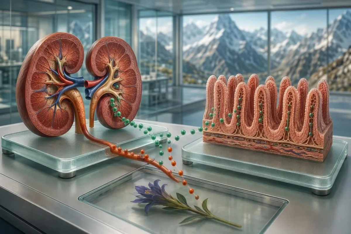
Low magnesium is usually caused by renal losses from medicines, reduced intestinal absorption, prolonged diarrhea, or heavy alcohol use—not simply a low-magnesium diet. The pattern of potassium, calcium, kidney function, medicines, and stool symptoms usually points toward the source.


- Hypomagnesemia means serum magnesium below 0.70 mmol/L or 1.7 mg/dL in most adult laboratories.
- Loop and thiazide diuretics can increase urinary magnesium loss, especially when potassium is also low.
- Proton-pump inhibitors can reduce intestinal magnesium absorption, usually after months to years of continuous use.
- Alcohol-related low magnesium often combines poor intake, diarrhea, vomiting, and renal magnesium wasting.
- Chronic diarrhea can lower magnesium quickly because digestive fluids contain magnesium and the intestine has less time to absorb it.
- Fractional excretion of magnesium above 2% during hypomagnesemia suggests inappropriate renal magnesium loss when kidney function is reasonably preserved.
- Severe symptoms such as fainting, seizure, sustained palpitations, or marked weakness need urgent clinical assessment.
- Low potassium or low calcium that will not correct can be a clue that magnesium deficiency is being missed.
What Low Magnesium Usually Means—and Why the Cause Comes First
Low magnesium most often results from medicines that waste magnesium through the kidneys, reduced gut absorption, ongoing gastrointestinal fluid loss, or heavy alcohol use. A serum magnesium below 0.70 mmol/L (1.7 mg/dL) is hypomagnesemia in most adult labs, but the cause determines whether the right next step is reviewing a medicine, treating diarrhea, addressing alcohol use, or checking renal losses. As of August 1, 2026, that distinction matters more than automatically taking a supplement.
Only about 1% of total body magnesium is in serum, so a normal serum result does not fully exclude depleted intracellular stores; conversely, a clearly low serum value deserves attention. In my clinical practice, the most revealing panels pair magnesium with potassium, corrected calcium, creatinine, bicarbonate, glucose, and a medication timeline rather than treating one number in isolation.
Dr. Thomas Klein sees a recurring pattern: a patient starts a diuretic or remains on a proton-pump inhibitor, develops low potassium months later, then magnesium finally gets measured. Kantesti is an AI blood test analyzer that reads magnesium alongside renal function, calcium, potassium, and longitudinal medication-related patterns rather than presenting an isolated flag.
Symptoms generally become more likely below 0.50 mmol/L (1.2 mg/dL), although rate of decline and coexisting hypokalemia matter as much as the result itself. Our biomarker reference guide can help readers identify the units used by their own laboratory before comparing values.
Which Medicines Are the Most Common Low Magnesium Causes?
Diuretics, proton-pump inhibitors, certain cancer medicines, calcineurin inhibitors, aminoglycoside antibiotics, amphotericin B, and some antivirals can cause low magnesium. They do so either by reducing intestinal uptake or by making the kidneys excrete magnesium when the body should conserve it.
Loop diuretics such as furosemide and bumetanide reduce magnesium reabsorption in the thick ascending limb, while thiazides can promote magnesium loss in the distal tubule. The risk rises with higher doses, older age, low dietary intake, diarrhea, and combined treatment with another magnesium-lowering drug; this is why diuretics and low magnesium often travel with potassium abnormalities.
Proton-pump inhibitors, including omeprazole and pantoprazole, appear to impair active intestinal magnesium transport through TRPM6 and TRPM7 pathways. The FDA has described cases after at least 3 months, although many occur after more than 1 year; in a subset, oral magnesium alone did not normalize the level until the PPI was stopped or changed under medical supervision.
Liamis et al. (2022) detail renal magnesium wasting from cisplatin, anti-EGFR therapies, tacrolimus, cyclosporine, aminoglycosides, foscarnet, and amphotericin B. Do not stop a prescribed medicine abruptly: bring a complete list—including over-the-counter acid remedies—to the clinician, then review long-term PPI monitoring and safer alternatives for your particular indication.
Why Diuretics Can Cause Low Magnesium and Low Potassium Together
Diuretics cause magnesium depletion by increasing sodium delivery and urine flow in kidney segments that normally reclaim magnesium. A low magnesium result with low potassium after a diuretic change is more suggestive of renal wasting than poor dietary intake alone.
In patients with normal or near-normal kidney function, fractional excretion of magnesium above 2% while serum magnesium is low supports inappropriate renal loss. A 24-hour urine magnesium above roughly 24 mg per day during hypomagnesemia can tell a similar story, although collection errors and reduced eGFR make either measure less reliable.
The clinical trap is assuming potassium replacement alone will fix the problem. Magnesium is required for renal potassium conservation, so persistent potassium below 3.5 mmol/L despite sensible replacement should prompt a magnesium check; potassium may simply continue leaking into urine until magnesium is restored.
I have seen this after a seemingly modest hydrochlorothiazide increase in a 68-year-old with a magnesium of 0.54 mmol/L and potassium of 3.0 mmol/L. The useful question was not whether the medicine was “bad,” but whether blood pressure control could be maintained with a revised regimen and repeat labs; see our guide to potassium after blood-pressure medicine changes.
How Alcohol Leads to Magnesium Depletion
Heavy alcohol use can lower magnesium through reduced food intake, vomiting, diarrhea, pancreatitis, and direct renal magnesium wasting. Acute withdrawal and hospital treatment can expose or worsen the deficiency because catecholamine surges and refeeding shift electrolytes into cells.
Alcohol-related hypomagnesemia is often a cluster rather than a single problem: magnesium may be low alongside phosphate below 0.80 mmol/L, potassium below 3.5 mmol/L, and low or low-normal calcium. In a person with heavy intake, this cluster should raise concern for poor nutrition, gastrointestinal loss, and renal tubular dysfunction at the same time.
A normal liver panel does not rule out alcohol-related magnesium loss. GGT, AST, ALT, mean corpuscular volume, albumin, and triglycerides can add context, but none diagnoses alcohol exposure or explains a low magnesium result by itself; Dr. Thomas Klein advises using the laboratory trend and an honest intake history together.
Stopping alcohol can improve urinary wasting, yet the first 72 hours may be medically risky for people with dependence because withdrawal can cause seizures, delirium, dehydration, and arrhythmias. For a practical view of what may shift during recovery, read about blood-marker trends after reducing alcohol; seek supervised care rather than attempting sudden withdrawal alone if dependence is possible.
When Diarrhea and Vomiting Are the Main Magnesium Losses
Persistent diarrhea is one of the fastest gastrointestinal causes of low magnesium because stool and digestive secretions contain magnesium while absorption time is reduced. Vomiting contributes indirectly through low intake, volume depletion, and hormonal kidney responses, though diarrhea is usually the larger direct magnesium loss.
Diarrhea lasting more than 14 days deserves a cause-focused review, particularly if it occurs with weight loss, nocturnal stools, fever, greasy stools, or a magnesium result below 0.70 mmol/L. Osmotic diarrhea from laxatives or magnesium-containing products can be confusing: excessive supplemental magnesium may cause diarrhea, then the stool loss can complicate the overall electrolyte picture.
Watery diarrhea plus low bicarbonate commonly points toward gastrointestinal bicarbonate loss, while vomiting more often produces low chloride and elevated bicarbonate. Neither pattern is perfect, but these accompanying electrolytes can keep a clinician from mislabeling every low magnesium result as dietary deficiency; our stool-pattern red flag guide explains which changes merit prompt review.
Infections, inflammatory bowel disease, microscopic colitis, bile acid diarrhea, thyroid excess, and medication side effects can all sustain losses. Rehydration fluids are helpful, but plain water alone may worsen dilutional symptoms in substantial fluid loss; the correct oral solution depends on the sodium, potassium, glucose, and kidney context.
Which Gut Conditions Reduce Magnesium Absorption?
Celiac disease, Crohn disease affecting the small bowel, pancreatic insufficiency, short-bowel states, and bariatric surgery can reduce magnesium absorption. These hypomagnesemia causes are more likely when diarrhea coexists with low iron, low vitamin D, low albumin, or unintended weight loss.
The distal small intestine and colon help absorb magnesium, including through regulated TRPM6 transport; disease or resection in these areas can matter disproportionately. In a patient with prior bowel surgery, a serum magnesium of 0.62 mmol/L may reflect chronic losses even when meals seem nutritionally adequate.
Greasy, floating, difficult-to-flush stool suggests fat malabsorption and should shift the work-up toward pancreatic, biliary, or small-bowel causes rather than simply escalating supplements. A fecal fat result is not a magnesium test, but it can be a useful fork in the diagnostic road when low magnesium accompanies weight loss.
Celiac testing has a nuance many patients miss: total IgA should accompany tissue-transglutaminase IgA because selective IgA deficiency can create a falsely reassuring result. If symptoms persist with anemia, low ferritin, or recurrent electrolyte loss, gastroenterology input is reasonable even after one negative screening panel.
How Clinicians Separate Kidney Magnesium Wasting From Gut Loss
Low magnesium with an inappropriately high urine magnesium level points to kidney wasting, whereas low urine magnesium suggests the kidneys are appropriately conserving magnesium during poor intake or gut loss. The comparison is most useful when the laboratory sample and urine collection are obtained before large replacement doses.
A clinician may order serum magnesium, creatinine, potassium, calcium, phosphate, urine magnesium, and urine creatinine together. Kantesti is an AI blood test interpretation platform that can organize these related results over time, but urinary indices and treatment decisions still need clinician review because eGFR and recent supplements alter interpretation.
Reduced kidney function usually causes magnesium retention, not loss, but exceptions exist: tubulointerstitial injury, recovery after acute kidney injury, uncontrolled diabetes with osmotic diuresis, and certain medicines can all waste magnesium. New glycosuria, high glucose, or a changing creatinine may therefore be part of the explanation rather than unrelated findings.
Urine albumin-creatinine ratio does not measure magnesium wasting, yet albuminuria changes the renal risk conversation and the safety of supplementation. Readers with diabetes or hypertension can review urine ACR preparation before assuming a single urine result answers every kidney question.
Diabetes, Hormones, and Other Metabolic Causes of Low Magnesium
Poorly controlled diabetes can cause low magnesium by osmotic diuresis, and hyperaldosteronism can contribute through renal electrolyte wasting. These causes become more plausible when magnesium is low with high glucose, frequent urination, hypertension, or persistent low potassium.
When glucose exceeds the kidney’s reabsorptive capacity, usually around 10 mmol/L (180 mg/dL) though variable by person, glucose enters urine and carries water and electrolytes with it. Magnesium may fall in parallel with sodium, potassium, and phosphate, especially during dehydration or treatment of severe hyperglycemia.
Primary aldosteronism classically produces hypertension with low potassium, but magnesium can also be low because distal sodium transport increases urinary losses. A potassium of 3.2 mmol/L in a person taking no diuretic and with difficult-to-control blood pressure merits a directed endocrine review rather than a supplement-only approach.
Inherited renal tubulopathies such as Gitelman syndrome are uncommon but memorable: low magnesium, low potassium, metabolic alkalosis, and low urinary calcium are a classic combination. If these abnormalities began young, recur despite treatment, or affect relatives, the chronic kidney disease guide helps frame why renal assessment and specialist input matter.
Why Low Magnesium Can Appear During Hospital Care or Refeeding
Low magnesium can emerge during refeeding, diabetic ketoacidosis treatment, prolonged hospitalization, and recovery from critical illness because magnesium shifts into cells or is lost in urine. A previously normal result does not guarantee safety once calories, insulin, fluids, or diuretics are introduced.
Refeeding syndrome is most concerning after prolonged inadequate intake, significant weight loss, alcohol dependence, or severe illness. Insulin-driven cellular uptake may lower phosphate, potassium, and magnesium within 24 to 72 hours of restarting calories; phosphate is often the earliest dramatic abnormality, but magnesium can be equally clinically significant.
In diabetic ketoacidosis, measured serum magnesium can be normal before treatment despite whole-body deficit because dehydration concentrates serum values. Once insulin and fluids begin, repeat electrolytes—not just the admission panel—guide replacement; a falling magnesium with muscle twitching or rhythm changes requires active management.
This is one setting where “eat more magnesium” is not a sufficient answer. Hospital teams use serial electrolytes, cardiac monitoring when indicated, thiamine, and measured replacement; our refeeding laboratory guide explains why all three minerals are checked together.
Low Magnesium Symptoms and the Electrolyte Patterns That Matter
Low magnesium symptoms include tremor, muscle cramps, weakness, tingling, fatigue, seizures, and heart-rhythm symptoms, but mild deficiency is often silent. The most useful warning pattern is magnesium below 0.50 mmol/L with low potassium, low calcium, or ECG symptoms.
Magnesium deficiency increases neuromuscular excitability, which can present as eyelid twitching, tremor, carpopedal spasm, or generalized weakness. These signs are not specific—anxiety, thyroid disease, stimulant use, low calcium, and overexertion can look similar—so symptoms should guide testing, not self-diagnosis.
Low magnesium can suppress parathyroid hormone release and create resistance to its action, producing low calcium that may not normalize until magnesium is replaced. This is a valuable clinical clue: hypocalcemia plus hypomagnesemia is more concerning than either mildly abnormal result alone, especially if tingling or spasms are present.
Severe hypomagnesemia increases the risk of ventricular arrhythmias, particularly with low potassium or medicines that prolong the QT interval. New palpitations, near-fainting, or a rapid irregular heartbeat should not be managed through supplements at home; use our palpitations testing guide to prepare for—but not delay—clinical assessment.
How to Test Magnesium Without Overreading One Result
Serum magnesium is the standard first test, with most adult reference ranges around 0.70 to 0.95 mmol/L or 1.7 to 2.3 mg/dL. Values below 0.50 mmol/L (1.2 mg/dL) generally warrant timely assessment for symptoms, ECG risk, and the cause of loss.
Reference intervals vary: some European laboratories use a lower limit near 0.66 mmol/L, while many clinicians view 0.75 mmol/L as a more physiologically comfortable threshold in high-risk patients. The number is not a diagnosis; a stable result of 0.69 mmol/L in an asymptomatic person differs from a fall from 0.86 to 0.69 mmol/L after chemotherapy or diarrhea.
RBC magnesium and ionized magnesium are marketed in some settings, but neither has universally standardized clinical cutoffs or broad guideline support for routine diagnosis. I generally start with a repeat serum magnesium, medication review, and paired electrolytes, then add urine testing when the cause remains unclear.
Kantesti AI interprets magnesium trends by comparing the laboratory’s own range, result trajectory, and coexisting electrolyte changes rather than treating a red flag as a diagnosis. For the technical guardrails behind this approach, see clinical validation standards.
A low result after a difficult draw is usually real, unlike falsely high magnesium caused by hemolysis, because cellular magnesium can leak into the specimen. Tell the laboratory or clinician about recent intravenous magnesium, laxative use, severe exercise, diarrhea, and the exact time of the last dose before repeating the test.
When Low Magnesium Needs Urgent Care Rather Than Routine Follow-Up
Low magnesium needs urgent assessment when it is severely low, causes seizure or sustained palpitations, accompanies fainting, or occurs with major potassium or calcium abnormalities. A result below 0.40 mmol/L (1.0 mg/dL) deserves same-day clinical advice even if symptoms seem modest.
Call emergency services for seizure, collapse, chest pain, severe breathlessness, or a persistent fast or irregular heartbeat. Emergency teams assess the ECG, repeat magnesium and potassium, check glucose and renal function, and determine whether intravenous magnesium is safer than oral replacement.
People at higher risk include those taking QT-prolonging medicines, digoxin, loop diuretics, cisplatin, tacrolimus, or multiple laxatives; risk also rises after severe diarrhea or alcohol withdrawal. A magnesium result of 0.47 mmol/L may be managed differently in an otherwise well outpatient than in a person with QT prolongation and potassium of 2.9 mmol/L.
Do not take repeated large doses while waiting for care if you have kidney disease, because reduced excretion can turn replacement into high magnesium. The low phosphate warning-sign guide is also relevant when weakness follows malnutrition, alcohol use, or refeeding.
How Treatment Changes Once the Cause Is Clear
The safest treatment for low magnesium corrects the source of loss and replaces magnesium at a dose matched to symptoms, kidney function, and severity. Mild stable deficiency is often managed orally, while severe symptomatic deficiency or poor absorption may require monitored intravenous replacement.
For uncomplicated outpatient deficiency, clinicians often use oral magnesium in divided doses because absorption falls and diarrhea rises with larger single doses. Magnesium citrate, chloride, lactate, and glycinate are often better tolerated than oxide, while oxide contains more elemental magnesium per tablet but can be less bioavailable; the practical trade-off is individual.
A typical non-prescription elemental dose might range from 100 to 300 mg daily, but this is not a universal prescription. People with eGFR below 30 mL/min/1.73 m², heart block, myasthenia gravis, or active kidney injury should ask a clinician before supplementation, and anyone with ongoing diarrhea needs the cause treated first.
The evidence for magnesium solely for nighttime cramps is mixed, so I avoid letting a common symptom obscure medication-related or gastrointestinal loss. Our magnesium dose and form guide covers interactions with levothyroxine, tetracyclines, and bisphosphonates, while our medical advisory board oversees the clinical review principles used in Kantesti content.
A Practical Recheck Plan After Low Magnesium Is Found
Most stable outpatient magnesium abnormalities should be rechecked after the suspected cause is addressed, often within 1 to 2 weeks for a meaningful low result or sooner when medicines change and symptoms exist. The same laboratory, similar timing, and documented supplement use make the trend far easier to interpret.
Record the exact magnesium result and units, recent stool frequency, alcohol intake, medicine changes, supplement dose, and whether the sample preceded or followed treatment. A single result may be noisy; two comparable results can distinguish a transient diarrheal loss from ongoing renal wasting with far more confidence.
Kantesti is an AI-powered blood test analysis tool that helps users compare magnesium, potassium, calcium, creatinine, and phosphate across reports in about 60 seconds, while preserving the need for a clinician to diagnose and prescribe. A trend is especially useful when magnesium remains below 0.70 mmol/L despite replacement, because that persistence argues for an unaddressed loss source.
Recheck earlier—within 24 to 72 hours—when severe diarrhea continues, intravenous replacement was given, potassium is low, kidney function changes, or a high-risk medicine is continued. For a sensible way to compare like with like, use our longitudinal lab-trend guide and bring the full sequence to your clinician.
Questions That Help Your Clinician Find the Real Source
The highest-yield questions ask whether magnesium is being lost through the kidneys or gut, whether a medicine changed, and whether potassium or calcium are also abnormal. These questions shorten the path from a flagged result to a targeted plan.
Ask: “Could my diuretic, acid suppressant, antibiotic, transplant medicine, cancer treatment, or laxative be contributing?” Also ask whether urine magnesium or fractional excretion would change management; that test is most useful when the answer determines whether to pursue renal loss or gastrointestinal disease.
Ask whether low magnesium explains potassium or calcium that remains abnormal after treatment, and whether an ECG is warranted. Patients with vomiting, diarrhea, greasy stool, weight loss, reduced appetite, or heavy alcohol intake should say so plainly—these details often explain more than an additional vitamin panel.
Kantesti supports multilingual result interpretation across 127+ countries, but it does not replace urgent care or a prescribing clinician. For transparent clinical methods and limitations, our AI technology guide explains how our AI lab test interpretation service handles context, ranges, and follow-up prompts.
Frequently Asked Questions
What is the most common cause of low magnesium?
The most common low magnesium causes in clinical practice are medicines, chronic diarrhea or malabsorption, heavy alcohol use, and urinary loss from diabetes or kidney tubule problems. Serum magnesium below 0.70 mmol/L or 1.7 mg/dL is considered low in most adult laboratories. Diuretics and long-term proton-pump inhibitors are particularly common medication contributors. The most useful next step is reviewing potassium, calcium, creatinine, stool symptoms, alcohol intake, and all medicines together.
Can diuretics cause low magnesium?
Yes, loop diuretics and thiazide diuretics can cause low magnesium by increasing urinary magnesium loss. The risk is higher when potassium is below 3.5 mmol/L, the diuretic dose has increased, diarrhea is present, or more than one magnesium-lowering medicine is used. A fractional magnesium excretion above 2% during hypomagnesemia can support renal wasting when kidney function is reasonably preserved. Do not stop a blood-pressure or heart-failure medicine without speaking to the prescribing clinician.
Can alcohol cause low magnesium even if liver tests are normal?
Yes, alcohol can cause low magnesium even with normal AST, ALT, and GGT results. Heavy alcohol use may reduce food intake, cause vomiting or diarrhea, increase urinary magnesium loss, and contribute to low phosphate and potassium. Magnesium below 0.50 mmol/L or 1.2 mg/dL is more likely to produce symptoms, especially during withdrawal or refeeding. Anyone at risk of alcohol withdrawal should seek supervised medical advice because sudden cessation can be dangerous.
What low magnesium symptoms need urgent care?
Low magnesium symptoms that need urgent care include seizure, fainting, chest pain, persistent palpitations, severe weakness, sustained muscle spasm, or significant shortness of breath. A magnesium value below 0.40 mmol/L or 1.0 mg/dL warrants same-day clinical advice even without obvious symptoms. Urgency rises if potassium is below 3.0 mmol/L, calcium is low, kidney function is changing, or the person takes a QT-prolonging medicine. Emergency assessment may include an ECG and intravenous replacement.
Can a normal magnesium blood test still miss deficiency?
A normal serum magnesium result can miss some body magnesium depletion because only about 1% of total body magnesium circulates in serum. Serum magnesium remains the standard first-line test because it is widely available and identifies clinically meaningful deficiency when low. In a person with persistent low potassium, low calcium, chronic diarrhea, or a high-risk medicine, clinicians may repeat serum testing and add urine magnesium studies. RBC magnesium testing has less standardized cutoffs and is not routinely preferred in major clinical practice.
How quickly should magnesium be rechecked after treatment?
Stable outpatient low magnesium is commonly rechecked within 1 to 2 weeks after addressing the likely cause, although timing depends on the starting value and treatment. Rechecking within 24 to 72 hours is more appropriate after intravenous replacement, ongoing severe diarrhea, low potassium, changing kidney function, or continuation of a high-risk medicine. Use the same laboratory when possible and record the last supplement dose before the draw. Persistent magnesium below 0.70 mmol/L despite replacement should trigger a search for continuing gut or kidney losses.
Get AI-Powered Blood Test Analysis Today
Join over 2 million users worldwide who trust Kantesti for instant, accurate lab test analysis. Upload your blood test results and receive comprehensive interpretation of 15,000+ biomarkers in seconds.
📚 Referenced Research Publications
Klein, T., Mitchell, S., & Weber, H. (2026). RDW Blood Test: Complete Guide to RDW-CV, MCV & MCHC. Kantesti AI Medical Research.
Klein, T., Mitchell, S., & Weber, H. (2026). BUN/Creatinine Ratio Explained: Kidney Function Test Guide. Kantesti AI Medical Research.
📖 External Medical References
⚕️ Medical Disclaimer
This article is for educational purposes only and does not constitute medical advice. Always consult a qualified healthcare provider for diagnosis and treatment decisions.
E-E-A-T Trust Signals
Experience
Physician-led clinical review of lab interpretation workflows.
Expertise
Laboratory medicine focus on how biomarkers behave in clinical context.
Authoritativeness
Written by Dr. Thomas Klein with review by Dr. Sarah Mitchell and Prof. Dr. Hans Weber.
Trustworthiness
Evidence-based interpretation with clear follow-up pathways to reduce alarm.