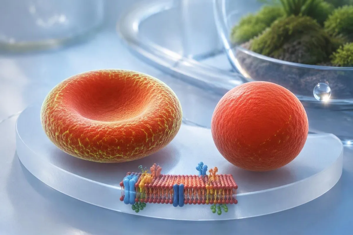
Reduced fluorescence is a clue to a red-cell membrane disorder—not a diagnosis by itself. The laboratory method, transfusion history, and accompanying evidence of hemolysis determine what that clue means.


- Low EMA binding supports hereditary spherocytosis when the clinical history, smear, and hemolysis markers agree; one abnormal result is not definitive.
- Fluorescence reduction is measured against laboratory controls. Some laboratories use decision limits around 15–20%, but validated cutoffs differ.
- Report format matters: 80% fluorescence relative to controls can mean a 20% reduction, not an 80% reduction.
- Recent transfusion can dilute affected cells with donor cells. Some laboratories request approximately 3 months after the last transfusion, but the testing laboratory sets the interval.
- Diagnostic performance reached 93% sensitivity and 98% specificity in a study of 150 patients with hereditary spherocytosis; these figures do not make every positive result conclusive.
- Other membrane disorders can also reduce EMA fluorescence, including hereditary pyropoikilocytosis and congenital dyserythropoietic anemia type II.
- Additional testing may include a direct antiglobulin test, osmotic gradient ektacytometry, selected fragility tests, or a red-cell membrane gene panel.
- Urgent symptoms such as breathlessness at rest, fainting, or rapidly worsening pallor need prompt assessment regardless of the EMA percentage.
Does a low EMA binding result mean hereditary spherocytosis?
A low EMA binding result supports hereditary spherocytosis, but does not establish the diagnosis on its own. Reduced fluorescence becomes convincing when it accompanies compatible red-cell morphology, evidence of hemolysis, and a fitting personal or family history; transfusion and other membrane disorders can complicate that interpretation.
The EMA binding test measures a membrane-associated fluorescence signal, not the percentage of cells that are spherocytes. A result showing a 20% reduction therefore does not mean that 20% of your red cells are abnormal, nor does it grade the condition as 20% severe.
Consider an illustrative adult with hemoglobin of 11.2 g/dL, reticulocytes of 6%, recurrent jaundice, and a parent who had a splenectomy: reduced EMA binding fits that story much better than it fits an otherwise well person with an isolated borderline flag. An accompanying high MCHC result adds another clue, although analyzer artifacts can produce that finding too.
Kantesti is an AI blood test analyzer that can help organize the accompanying CBC and hemolysis results, but cannot turn one fluorescence measurement into a confirmed membrane-disorder diagnosis. I’m Thomas Klein, writing as Chief Medical Officer at Kantesti Ltd; my interpretive starting point is to separate evidence supporting a diagnosis from evidence that actually settles it.
What does the eosin-5-maleimide test actually measure?
The eosin-5-maleimide test measures fluorescent dye binding to red-cell membrane proteins, predominantly band 3 and associated membrane components. Flow cytometry compares stained patient cells with control cells; reduced binding can reflect a membrane defect even when the affected gene does not encode band 3 itself.
Band 3, also called the anion exchanger AE1, helps connect the membrane to the supporting protein skeleton. Changes involving spectrin or ankyrin can disturb that architecture indirectly, so a low signal does not identify which of several membrane proteins is responsible.
Many flow cytometers excite EMA-stained cells using a 488 nm laser, then collect the emitted fluorescence through an appropriate optical filter. Instrument settings, staining conditions, and control selection influence the reported signal; an absolute fluorescence value from one laboratory is not automatically comparable with another laboratory’s value.
EMA fluorescence and LDH answer different questions: the first concerns membrane labeling, while the second can support increased cell turnover but has many other causes. Our explanation of what LDH measures helps prevent a common mistake—assuming that a high turnover marker proves a particular membrane defect.
How should you read the EMA percentage and laboratory cutoff?
An EMA percentage must be read according to the laboratory’s reporting convention: some reports show fluorescence remaining, while others show percentage reduction. There is no single universal cutoff, and a low numerical value can mean different things depending on which quantity the report describes.
For a mean-based method, percentage reduction is calculated as 100 × (control mean − patient mean) ÷ control mean. If the control signal is 1,000 arbitrary units and the patient signal is 800, fluorescence is reduced by 20%, while fluorescence remaining is 80%; neither figure is a concentration in mg/dL.
Some laboratories use decision limits around 15–20% reduction, while others validate lower limits or separate borderline intervals. Cell size matters because smaller red cells may carry less total dye signal; an adult MCV below approximately 80 fL should prompt consideration of microcytosis and whether the controls were suitably matched, as discussed in our borderline MCV interpretation.
The most useful request is not simply 'What is the normal number?' but 'Was this clearly abnormal using your validated method, and were the controls appropriate?' Marked size variation can complicate the average signal, making the RDW and cell-size relationship relevant when iron deficiency, reticulocytosis, or two cell populations coexist.
How accurate is EMA for hereditary spherocytosis diagnosis?
The EMA binding test is a useful, relatively sensitive screening investigation, but neither a positive nor a negative result is infallible. In a comparison involving 150 patients with hereditary spherocytosis, EMA achieved 93% sensitivity and 98% specificity under that study’s conditions (Bianchi et al., 2012).
Sensitivity of 93% means that approximately 7% of affected participants were not detected by EMA in that study—not that a negative result leaves every patient with a 7% chance of disease. The probability after testing depends on the original clinical suspicion, the laboratory method, and whether the patient resembles the study population.
Specificity also needs context: with hypothetical 1% disease prevalence, 93% sensitivity, and 98% specificity, the positive predictive value would be approximately 32%. At 50% prevalence it would be approximately 98%; these calculated examples explain why indiscriminate screening creates more uncertain positives than targeted hematology testing.
A patient can have compensated hemolysis and a near-normal hemoglobin because marrow production keeps pace with cell removal. That is why a apparently reassuring CBC should not end the assessment when reticulocytes and bilirubin are persistently abnormal; our hemoglobin and hematocrit comparison explains another reason the headline CBC values may not tell the whole story.
Why can a recent transfusion mask an abnormal EMA result?
Recent red-cell transfusion can make an abnormal EMA result appear less abnormal or normal because donor cells are measured alongside the patient’s cells. The laboratory may detect separate populations, but an averaged fluorescence value can conceal the affected population rather than exclude hereditary spherocytosis.
The impact depends on how much was transfused, when it was transfused, and the patient’s own cell survival. Two units do not produce the same donor-cell fraction in every person; in severe hemolysis, donor cells may make up a disproportionately large share because the patient’s affected cells disappear faster.
Some testing services ask clinicians to wait approximately 3 months after the last transfusion, but this is a laboratory-specific precaution, not a universal clearance rule. Give the laboratory the exact transfusion dates and ask whether it accepts the sample now, can review distinct populations, or recommends postponement.
A pre-transfusion sample is particularly valuable when it was collected and stored within the laboratory’s validated conditions. Similar masking occurs in other red-cell investigations, including G6PD activity testing, but the mechanism and retesting plan are test-specific; do not apply one assay’s waiting period to every other assay.
Which other red-cell disorders can reduce EMA fluorescence?
Reduced EMA fluorescence is not exclusive to hereditary spherocytosis; hereditary pyropoikilocytosis, some hereditary elliptocytosis cases, Southeast Asian ovalocytosis, and congenital dyserythropoietic anemia type II can also produce reduced binding. Smear morphology, inheritance, and evidence of ineffective production help distinguish these possibilities.
Congenital dyserythropoietic anemia type II can resemble spherocytosis but involves abnormal red-cell development, making the relationship between anemia and reticulocyte production especially informative. A relatively inadequate marrow response, characteristic specialist findings, and SEC23B testing may redirect the diagnosis; EMA alone cannot make that distinction.
Acquired immune hemolysis can produce spherocytes on a smear, although EMA binding is commonly preserved. A direct Coombs test therefore answers a different question—whether immunoglobulin or complement is attached to the cells—and must not be replaced by EMA simply because both are used during an anemia workup.
PNH testing examines a different membrane-protein problem and generally uses FLAER and lineage-appropriate markers, not EMA. Our explanation of PNH flow cytometry clarifies why ordering 'a flow test' is insufficient: the target, cell populations, and clinical indication determine which disorder the assay can detect.
Which accompanying blood results make low EMA more convincing?
Low EMA is more persuasive when the CBC, reticulocytes, bilirubin, haptoglobin, and smear form a coherent hemolysis pattern. No single companion marker is specific, and normal hemoglobin does not rule out ongoing red-cell destruction when marrow compensation is adequate.
An MCHC above approximately 36 g/dL can support spherocytosis, but the local reference interval and analytical flags matter. Cold agglutination, sample interference, or calculation problems can also raise MCHC; the next step may be an analyzer or smear review rather than another inherited-disease test.
Low haptoglobin plus increased indirect bilirubin supports hemolysis more strongly than either result alone. However, reduced liver synthesis can lower haptoglobin, and an adult total bilirubin reference interval is often approximately 0.2–1.2 mg/dL, with laboratory variation; our low haptoglobin differential explains why the combination still needs clinical interpretation.
Kantesti is an AI blood test interpretation platform that can place these accompanying values together, while specialist assessment establishes their cause. For a reticulocyte result of 6%, the absolute count and degree of anemia matter more than the percentage alone; our reticulocyte turnover guide explains how a seemingly high percentage can overstate marrow compensation.
How does EMA interpretation differ in babies and children?
EMA testing can support hereditary spherocytosis diagnosis in children, including infants, but age-related cell size, recent transfusion, and neonatal clinical findings need special attention. A baby may have significant jaundice without a classic adult smear or a clearly elevated MCHC.
Newborn red cells are normally larger than adult cells, so adult control populations may not be ideal for every laboratory method. An adult MCV interval around 80–100 fL should not be imposed on a neonatal CBC; age-matched or size-appropriate controls and the laboratory’s pediatric validation are more useful than a universal numerical rule.
Parvovirus B19 can temporarily suppress red-cell production and cause an aplastic crisis in someone with hereditary spherocytosis. A falling hemoglobin with unexpectedly low reticulocytes deserves prompt assessment; a positive IgG result alone does not prove a current crisis, as explained in our parvovirus antibody interpretation.
An illustrative 8-year-old with longstanding jaundice, a convincing family history, and a low EMA result needs a different conversation from an infant tested immediately after transfusion. Families should bring neonatal records and earlier CBCs when available; our guidance on children’s AI result interpretation explains why age-specific medical review cannot be replaced by an adult reference-range checker.
When does family history or genetic testing help?
Family history increases diagnostic confidence, while genetic testing is most useful for atypical, severe, or unresolved cases. The main hereditary spherocytosis genes include ANK1, SPTB, SPTA1, SLC4A1, and EPB42, but a negative family history does not exclude an inherited membrane disorder.
Ask about jaundice, anemia, early gallstones, splenectomy, and neonatal treatment, not just whether anyone remembers a formal diagnosis. A relative may have compensated hemolysis and never have been labeled anemic; a three-generation history can reveal a pattern that a single yes-or-no question misses.
For a parent with a confirmed heterozygous autosomal-dominant disease-causing variant, each pregnancy generally has a 50% chance of inheriting that variant. This does not predict identical severity: relatives carrying the same variant can have different hemoglobin levels, transfusion needs, or complications.
A membrane gene panel and hemoglobin electrophoresis investigate different mechanisms, although both may be appropriate in a complex anemia workup. Our hemoglobin pattern guide explains the latter; for gene panels, a variant of uncertain significance is not a confirmed diagnosis, and a negative panel does not eliminate every membrane defect.
Which additional tests can resolve an uncertain EMA result?
Specialist smear review, a direct antiglobulin test, osmotic gradient ektacytometry, selected fragility tests, and genetic testing can resolve discordant EMA findings. The appropriate sequence depends on whether the unresolved question concerns immune hemolysis, membrane mechanics, ineffective production, or a specific inherited defect.
Osmotic gradient ektacytometry measures how cells deform across changing osmotic conditions, generating a characteristic curve rather than another fluorescence percentage. It can help separate membrane disorders with overlapping EMA results, although transfused donor cells and access to specialist laboratories remain practical limitations.
Traditional osmotic fragility testing can miss mild disease: in the 150-patient comparison, sensitivity was 68% with fresh samples and 81% after incubation (Bianchi et al., 2012). This is why a normal fragility result should not automatically cancel a convincing EMA and clinical pattern; combining complementary evidence is usually more informative than collecting interchangeable 'confirmation' tests.
Kantesti can explain why these investigations answer different questions, but an uploaded PDF does not provide the raw flow histogram, gating decisions, or ektacytometry curve unless those materials are actually included. Our clinical methodology standards are relevant here: technical interpretation must acknowledge missing source information rather than infer an unreported specialist result.
Does a lower EMA result mean more severe disease?
The degree of EMA reduction does not reliably determine disease severity or select treatment. Severity assessment depends on hemoglobin, symptoms, transfusion requirements, growth in children, hemolytic complications, and quality of life—not on whether fluorescence reduction is 18% rather than 28%.
Splenectomy is not a response to a low EMA number; it is a major clinical decision that requires a secure diagnosis and assessment of benefits and long-term risks. The British guideline update emphasizes disease severity and individual circumstances rather than a fluorescence threshold (Bolton-Maggs et al., 2012).
Correct classification matters before splenectomy because some related disorders—particularly hereditary stomatocytosis syndromes—carry substantial thrombotic concerns after spleen removal. EMA is not a reliable stand-alone classifier for these disorders, so unusual morphology, dehydration-related findings, or an atypical family history should trigger specialist reconsideration.
Chronic hemolysis increases the risk of pigment gallstones, but abdominal symptoms still need their own assessment. Fever with persistent right-upper-abdominal pain, vomiting, or worsening jaundice is not explained away by an established membrane diagnosis; our discussion of gallbladder blood-test limitations shows why reassuring laboratory values cannot exclude every biliary complication.
When should you seek specialist or urgent medical review?
Hematology review is appropriate when low EMA is unexplained, borderline, inconsistent with other findings, or affected by transfusion. Breathlessness at rest, fainting, chest pain, rapidly worsening pallor, or a sudden functional decline requires urgent assessment regardless of the fluorescence result.
A sudden hemoglobin fall with a low reticulocyte response raises a different concern from stable compensated hemolysis. The illustrative child whose hemoglobin drops from 11 to 7 g/dL over several days needs prompt assessment for reduced production or another acute process; no particular EMA percentage determines whether that decline is safe.
Ongoing hemolysis can make HbA1c falsely low because red cells have less time to accumulate glucose-related modification. HbA1c usually reflects roughly 2–3 months of glycemic exposure, but transfusion adds another source of distortion; our A1c after transfusion guide explains why glucose-based assessment or another appropriate marker may be needed.
Stable patients should ask for a plan with three clear elements: the current working diagnosis, what remains uncertain, and the next investigation or review date. Repeat EMA is useful when it addresses a specific concern about sample conditions or donor-cell interference; repeated testing without a stated question can produce more conflicting numbers without improving diagnostic confidence.
How can you prepare your EMA report for a useful review?
Bring the complete EMA report, transfusion dates, CBC, reticulocyte count, bilirubin, haptoglobin, DAT result, and smear comments to the review. The laboratory’s cutoff and interpretive comment are often more useful than an isolated percentage copied into a message.
Kantesti is an AI-powered blood test analysis tool that can help explain accompanying laboratory reports, but it cannot reconstruct a missing histogram or substitute for a hematologist’s membrane-disorder assessment. Our AI technology explanation describes the interpretation workflow; check extracted values and units against the original report before acting on any explanation.
Kantesti AI should be used to clarify the questions for your clinician—not to convert 'suggestive of hereditary spherocytosis' into 'confirmed hereditary spherocytosis.' Keep fluorescence remaining and fluorescence reduction distinct, retain the local reference interval, and tell the reviewing clinician whether the sample preceded or followed transfusion.
My practical checklist as Thomas Klein is to establish sample validity, pattern consistency, and diagnostic consequences before interpreting one low result as definitive. Our Medical Advisory Board provides information about medical expertise behind the service; for an individual patient, specialist review is particularly valuable when the diagnosis would change family counseling, transfusion planning, or a discussion about splenectomy.
Research publications and the evidence behind EMA interpretation
The evidence supporting EMA interpretation comes from membrane-disorder guidelines and assay studies, not from general iron or urinalysis resources. As of October 17, 2026, this article distinguishes those diagnostic references from the two companion publications listed below, which address related laboratory context rather than EMA validation.
The ICSH laboratory guideline recommends interpreting hereditary red-cell membrane investigations through the clinical history, morphology, and complementary testing framework (King et al., 2015). A newer page date does not create a new diagnostic standard; the directly relevant external references below identify the publications on which the major assay and workup statements are based.
The companion iron studies publication helps distinguish iron deficiency or altered iron handling from a membrane defect. The accompanying urinalysis publication discusses urinary findings, including urobilinogen; neither an iron pattern nor urinary urobilinogen establishes hereditary spherocytosis, and the two supplied Zenodo DOIs should not be presented as evidence of EMA diagnostic accuracy.
Kantesti’s medical content standards describe the distinction between patient education and individual medical advice. Formal title-first citations are supplied below because publication authorship and dates were not provided; ResearchGate and Academia.edu links are clearly identified as title searches, not verified publication records or proof of peer review.
Frequently Asked Questions
Does a low EMA binding test confirm hereditary spherocytosis?
A low EMA binding test supports hereditary spherocytosis but does not confirm it by itself. In a study of 150 affected patients, EMA achieved 93% sensitivity and 98% specificity, but individual diagnostic confidence still depends on the clinical setting. Other membrane disorders and cell-size differences can also reduce fluorescence. A hematologist should reconcile the result with the smear, hemolysis markers, family history, and transfusion history.
What percentage is considered a low EMA binding result?
The abnormal EMA threshold is laboratory-specific rather than universal. Some laboratories use decision limits around 15–20% fluorescence reduction, while others validate lower thresholds or define a borderline interval. A report showing 80% fluorescence remaining corresponds to a 20% reduction if the control is defined as 100%. Check the reporting convention and laboratory comment before comparing your number with an online cutoff.
Can I have a normal EMA test and still have spherocytosis?
A normal EMA test does not exclude every mild or atypical case of hereditary spherocytosis. The reported sensitivity of 93% in the Bianchi et al. comparison means that some affected patients had results the assay did not classify as abnormal. Recent transfusion can also mask reduced fluorescence. Persistent evidence of hemolysis or a strong family history can justify specialist review and complementary testing despite a normal result.
How long after a blood transfusion should EMA testing be done?
The testing laboratory should determine when EMA testing is reliable after transfusion. Some services request approximately 3 months after the last red-cell transfusion, but this is not a universal rule or a guarantee that donor-cell interference has disappeared. Transfusion volume, the patient’s cell survival, and the ability to examine separate populations all matter. Provide exact transfusion dates and ask whether a valid pre-transfusion sample is available.
Is the EMA binding test the same as a Coombs test?
The EMA binding test and the direct Coombs test investigate different red-cell problems. EMA measures dye binding to membrane proteins, whereas a direct Coombs test detects immunoglobulin or complement attached to red cells. Spherocytes can occur in both inherited membrane disease and immune hemolysis, so these 2 tests may be complementary. Neither result should be used as a substitute for the other when distinguishing those causes.
Does a very low EMA result mean I need my spleen removed?
A very low EMA result does not by itself justify splenectomy. A fluorescence reduction of 28% is not a treatment threshold and does not reliably indicate more severe disease than an 18% reduction. Decisions depend on the confirmed diagnosis, anemia, symptoms, transfusion requirements, and complications. Specialist classification is necessary because spleen removal can be particularly problematic in some other inherited membrane disorders.
Do I need to fast before an eosin-5-maleimide test?
Fasting is not generally required for an eosin-5-maleimide test alone. If the appointment includes other tests, follow the collection instructions for the full panel rather than assuming the EMA assay requires an 8–12-hour fast. Sample anticoagulant, transport conditions, and time to analysis are more relevant to EMA reliability. Tell the laboratory about recent transfusion and follow its specific preparation instructions.
Get AI-Powered Blood Test Analysis Today
Join over 2 million users worldwide who trust Kantesti for instant, accurate lab test analysis. Upload your blood test results and receive comprehensive interpretation of 15,000+ biomarkers in seconds.
📚 Referenced Research Publications
Klein, T., Mitchell, S., & Weber, H. (2026). Urobilinogen in Urine Test: Complete Urinalysis Guide 2026. Kantesti AI Medical Research.
Klein, T., Mitchell, S., & Weber, H. (2026). Iron Studies Guide: TIBC, Iron Saturation & Binding Capacity. Kantesti AI Medical Research.
📖 External Medical References
Bolton-Maggs PHB et al. (2012). Guidelines for the diagnosis and management of hereditary spherocytosis—2011 update. British Journal of Haematology.
Bianchi P et al. (2012). Diagnostic power of laboratory tests for hereditary spherocytosis: a comparison study in 150 patients grouped according to molecular and clinical characteristics. Haematologica.
King MJ et al. (2015). ICSH guidelines for the laboratory diagnosis of nonimmune hereditary red cell membrane disorders. International Journal of Laboratory Hematology.
⚕️ Medical Disclaimer
This article is for educational purposes only and does not constitute medical advice. Always consult a qualified healthcare provider for diagnosis and treatment decisions.
E-E-A-T Trust Signals
Experience
Physician-led clinical review of lab interpretation workflows.
Expertise
Laboratory medicine focus on how biomarkers behave in clinical context.
Authoritativeness
Written by Dr. Thomas Klein with review by Dr. Sarah Mitchell and Prof. Dr. Hans Weber.
Trustworthiness
Evidence-based interpretation with clear follow-up pathways to reduce alarm.