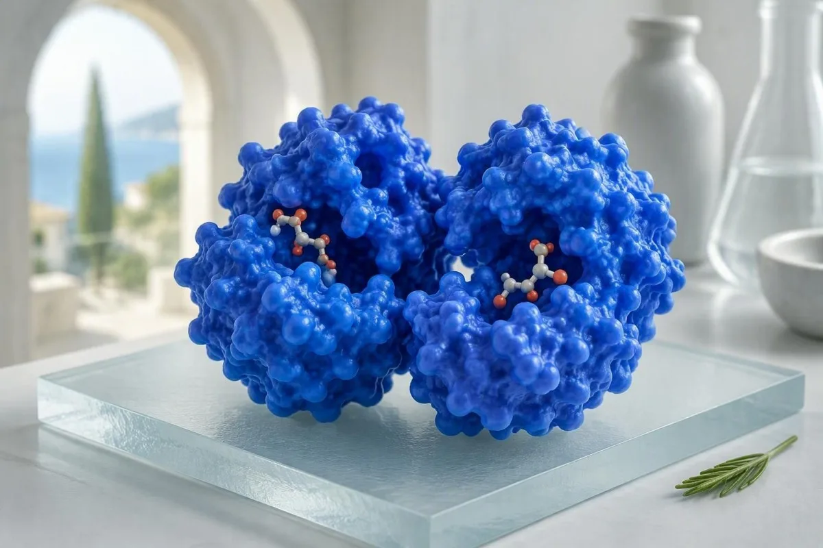
LDH is a useful clue to cell stress or cell breakdown, not a diagnosis. The number becomes meaningful only when it is read beside your symptoms, sample quality, CBC, liver tests, and recent activity.


- LDH stands for lactate dehydrogenase, an enzyme found in nearly all body tissues that rises when cells release their contents.
- Typical adult ranges are about 140-280 U/L, but the laboratory-specific upper limit is the only range that should guide interpretation.
- A high LDH is nonspecific: exercise, sample hemolysis, liver injury, red-cell breakdown, severe infection, and some cancers can all raise it.
- A hemolyzed sample can falsely elevate LDH because red cells contain abundant LDH; a clean redraw often answers the question.
- LDH above 2-3 times the laboratory upper limit deserves prompt clinical context, especially with anemia, jaundice, fever, chest symptoms, or weight loss.
- LDH plus high reticulocytes, bilirubin, and low haptoglobin supports hemolysis more than LDH alone.
- LDH plus elevated AST and ALT points clinicians toward liver or muscle injury; CK helps separate muscle from liver patterns.
- LDH trends matter more than one result when monitoring a known condition, provided the same laboratory method is used.
LDH means lactate dehydrogenase in a blood test
LDH stands for lactate dehydrogenase, an enzyme that helps cells convert lactate and pyruvate as they make energy. A raised LDH blood result means cells have released more enzyme into the sample; it does not identify which organ is responsible.
The LDH abbreviation is sometimes written as LD, especially on older UK and European laboratory reports. The test measures enzyme activity in units per litre, usually U/L or IU/L, rather than the physical amount of enzyme. That distinction matters because two laboratories can obtain slightly different results from the same specimen.
LDH catalyses the reversible conversion of pyruvate to lactate while regenerating NAD+ for energy metabolism. Cells in red blood cells, skeletal muscle, liver, lung, kidney, placenta, and lymphatic tissue all contain it. This broad distribution is exactly why a high result is a signal to investigate, not a shortcut to a diagnosis.
As of September 17, 2026, I still see patients interpret LDH as a “cancer test” after reading a portal flag. That is too blunt. Dr. Thomas Klein's practical rule is simpler: ask whether the result is real, whether it is new, and whether the rest of the panel gives it a tissue source. Our biomarker reference guide helps place LDH beside the tests that answer those questions.
What the lactate dehydrogenase test actually measures
A lactate dehydrogenase test measures LDH enzyme activity released into serum or plasma, not your lactate level and not oxygen delivery directly. LDH can rise within hours of cell disruption, while the clinical explanation may be temporary or serious.
LDH is often ordered when a clinician suspects tissue turnover, red-cell destruction, liver injury, muscle injury, or a complication of a known disease. It is also used as a trend marker in selected lymphomas and germ-cell tumours, but it cannot screen for cancer in someone without a compatible clinical picture.
Do not confuse LDH with lactate. Lactate is a small metabolic molecule measured in mmol/L and can rise during shock, seizures, severe asthma, or intense exertion; LDH is an enzyme measured in U/L. If your report says lactate, use our guide to high lactate beyond sepsis rather than applying LDH advice.
Kantesti is an AI blood test analyzer that reads LDH with its neighbouring markers, including bilirubin, AST, ALT, CK, hemoglobin, reticulocytes, and sample comments. In our review workflow, an isolated LDH of 310 U/L has a very different meaning from 310 U/L combined with falling hemoglobin and an elevated indirect bilirubin.
LDH normal ranges: use your laboratory's upper limit
Most adult laboratories report an LDH reference interval near 140-280 U/L, although a range of 120-246 U/L or 135-225 U/L is also common. A result is high only relative to the method and reference interval printed on your own report.
There is no universal “danger level” for LDH. A value of 420 U/L may be a mild 1.5-fold elevation in one laboratory and nearly twice the upper limit in another. Children, newborns, pregnancy, and intense physical activity can also produce values above adult ranges without the same implication.
Clinicians often describe LDH as a multiple of the upper limit of normal (ULN). LDH at 2 × ULN means more diagnostically than simply saying 400 U/L because it travels better across methods. A persistently rising value above 3 × ULN, particularly with symptoms or abnormal blood counts, warrants timely medical review.
One useful safeguard is checking whether the report says “hemolysed,” “unsuitable,” or “repeat recommended.” A high LDH paired with a high potassium on a visibly compromised specimen can be a collection artifact; our hemolysis redraw guide explains why repeating the sample is usually safer than guessing.
Why a high LDH result is so nonspecific
LDH is nonspecific because nearly every major tissue contains it, so one raised value cannot distinguish red-cell turnover from liver, muscle, lung, kidney, or lymphatic causes. The test answers “is there increased enzyme release?” rather than “where is the problem?”
This is one of those areas where context matters more than the number. A 26-year-old who completes a hard hill race can have elevated LDH and CK for 24-72 hours; the same LDH value in a person with fever, night sweats, and enlarged lymph nodes has a completely different pre-test probability.
LDH may increase with pneumonia, severe viral illness, liver inflammation, pulmonary embolism, renal injury, hemolysis, large bruises, seizures, and advanced malignancy. It can also remain normal in early or localised disease. Therefore, both a normal and high result have limits.
A result that is only slightly above range is particularly vulnerable to overinterpretation. I advise patients to write down recent exertion, alcohol intake, injections, illness, and new medicines before a repeat draw; our blood-test timeline guide shows why timing changes laboratory meaning.
Sample hemolysis is a common reason for falsely high LDH
In-vitro hemolysis can falsely raise LDH because red blood cells contain much higher LDH activity than serum. A difficult collection, vigorous tube mixing, temperature extremes, or delayed processing can disrupt cells after they leave the body.
When I review a panel showing high LDH, potassium, AST, and a laboratory hemolysis index, I first question the sample rather than diagnose disease. Lippi and colleagues describe hemolysis as a major source of pre-analytical laboratory error, with LDH among the analytes particularly susceptible to interference (Lippi et al., 2008).
True in-vivo hemolysis is different: red cells are breaking down within the body, often producing anemia, increased reticulocytes, indirect bilirubin, and reduced haptoglobin. The laboratory cannot settle that distinction from LDH alone. A peripheral smear may show useful clues such as spherocytes or schistocytes.
Kantesti AI flags the pattern of LDH with potassium and specimen-quality comments as a potential redraw scenario rather than treating every high value as biological. For a more detailed comparison, see our guide to low haptoglobin causes.
Exercise, muscle injury, and LDH: the CK comparison
Hard exercise can increase LDH for one to three days, especially when it causes unfamiliar eccentric muscle work. Creatine kinase, or CK, is generally more useful than LDH for identifying a skeletal-muscle source.
A marathon runner with an AST of 89 U/L, CK of 1,400 U/L, and modestly raised LDH does not automatically have liver disease. Muscle contains AST and LDH, and CK helps establish the source. Rest, hydration, and a repeat sample after 48-72 hours are often more informative than an immediate imaging scan when symptoms are absent.
Muscle pain, weakness, dark urine, reduced urine output, or CK values several thousand U/L change the urgency. These features can signal significant muscle breakdown and deserve same-day medical evaluation. LDH itself is not the best severity marker in that setting.
Avoid an unusually hard workout for 24-48 hours before a planned LDH retest unless your clinician has asked otherwise. Our high CK warning guide separates benign post-training rises from patterns that need urgent care.
The LDH pattern clinicians use when hemolysis is possible
LDH supports hemolysis when it rises alongside reticulocytes and indirect bilirubin while haptoglobin falls. A low hemoglobin result alone does not prove hemolysis because iron deficiency, bleeding, kidney disease, and marrow conditions can also cause anemia.
A typical hemolysis panel includes CBC, reticulocyte count, LDH, total and direct bilirubin, haptoglobin, and a blood-film review. In intravascular hemolysis, haptoglobin can become very low because it binds free hemoglobin; in liver disease it may also be low because hepatic production falls. That is why no single marker should be used as proof.
Reticulocyte counts are best interpreted as an absolute count or reticulocyte production index, not only as a percentage. A reticulocyte percentage of 3% may be inadequate in someone with severe anemia because the denominator has shrunk. Our explanation of reticulocyte testing covers this frequently missed calculation.
LDH can be impressively high in megaloblastic anemia because fragile precursor cells break down in the marrow before they circulate. If MCV is elevated and B12 is borderline, clinicians often check methylmalonic acid and folate rather than assuming immune hemolysis; our borderline B12 guide explains the next steps.
How clinicians separate liver, lung, and other LDH sources
LDH with high ALT and AST suggests hepatocellular or muscle injury, while LDH with a normal liver panel often redirects the work-up. ALP, GGT, bilirubin, CK, CBC, kidney function, symptoms, and imaging determine the next step.
A disproportionate rise in LDH compared with ALT and AST can occur in ischemic liver injury, but the diagnosis depends on the clinical event and often involves enzyme levels in the thousands. Mild isolated LDH elevation is not a reliable way to diagnose fatty liver. For that, clinicians use metabolic risk, liver enzymes, fibrosis scores, and sometimes imaging.
In respiratory illness, LDH may reflect widespread tissue stress but cannot diagnose pneumonia, pulmonary embolism, or interstitial lung disease. New breathlessness, chest pain, blue lips, fainting, or oxygen saturation below 92% needs urgent assessment rather than an online LDH interpretation.
Kantesti is an AI lab test interpretation service that identifies liver-pattern combinations rather than labelling LDH as a liver test. Readers with ALT, AST, GGT, or bilirubin changes can review our practical liver test abbreviation guide before discussing the full panel with their clinician.
LDH isoenzymes: useful historically, limited today
LDH has five isoenzymes formed from LDHA and LDHB subunits, but routine LDH isoenzyme testing is now uncommon. Modern imaging, troponin, CK, haptoglobin, flow cytometry, and targeted disease tests usually locate the source more accurately.
LDH-1 is relatively abundant in red cells and heart, whereas LDH-5 is more associated with liver and skeletal muscle. The old “LDH flip,” where LDH-1 exceeds LDH-2, was once used in suspected myocardial infarction. It has been superseded by high-sensitivity troponin because troponin is markedly more cardiac-specific.
Isoenzyme fractions can still be requested in unusual cases, but they are vulnerable to hemolysis and rarely resolve a routine isolated elevation. A normal troponin does not require an LDH isoenzyme panel to rule out a heart attack in modern practice. Symptoms and serial troponin protocols drive acute cardiac care.
If your report lists LDH-1 through LDH-5, the fraction pattern deserves specialist interpretation alongside sample quality. Our deeper article on LDH isoenzyme patterns explains why the historical tissue map is less definitive than many websites suggest.
LDH and cancer: a prognostic marker, not a screening test
LDH can help assess disease burden or prognosis in selected cancers, but it does not diagnose cancer and should not be used for population screening. Many non-cancer conditions cause the same elevation, including hemolysis and exercise.
In aggressive non-Hodgkin lymphoma, LDH above the laboratory upper limit is one component of the International Prognostic Index. Shipp and colleagues incorporated LDH into that score in 1993, alongside age, stage, performance status, extranodal involvement, and stage-related burden; it was never intended to stand alone (Shipp et al., 1993).
For people being monitored after a diagnosed malignancy, the trajectory matters: a sustained rise on comparable samples may prompt a clinician to review symptoms, imaging, or disease-specific markers. A one-off isolated elevation, especially from a hemolyzed tube, should not be read as recurrence.
I have had patients arrive frightened by LDH of 290 U/L found during a routine panel, with no symptoms and a documented hemolyzed specimen. The responsible approach is repeat testing and clinical review, not ordering broad tumour-marker panels. If cancer risk runs in your family, our preventive testing guide explains evidence-based first steps.
When to repeat an LDH test and how to prepare
A mildly elevated isolated LDH is commonly repeated within days to a few weeks after checking for collection error, recent exercise, and intercurrent illness. The exact interval depends on the level, symptoms, and abnormalities elsewhere on the panel.
For a stable person with LDH below roughly 1.5 × ULN, no alarming symptoms, and a possible pre-analytical issue, a clinician may repeat LDH with CBC, bilirubin, haptoglobin, AST, ALT, and CK. Use the same laboratory where possible; changing assays can create apparent trends that are methodological rather than biological.
Preparation is modest: maintain usual hydration, avoid strenuous exercise for 24-48 hours, and disclose alcohol, supplements, injections, and new medications. Fasting is not routinely required for LDH. Do not stop prescribed medication merely to “improve” a laboratory result.
Kantesti's trend analysis can compare dated reports and detect when an apparent change crosses a laboratory reference limit rather than a meaningful personal baseline. For the technical safeguards behind that approach, read our clinical validation framework.
When high LDH needs urgent medical assessment
High LDH needs urgent assessment when it occurs with serious symptoms, not because of a single universal LDH cutoff. Seek emergency care for chest pain, new severe shortness of breath, confusion, fainting, dark urine with marked muscle pain, jaundice with fever, or rapidly worsening weakness.
A high LDH with falling hemoglobin, jaundice, and breathlessness can reflect significant hemolysis and needs same-day assessment. High LDH with severe muscle pain and cola-coloured urine can accompany rhabdomyolysis, where clinicians check CK, potassium, creatinine, urine findings, and heart rhythm promptly.
People receiving chemotherapy or treatment for high-burden blood cancers need immediate guidance if LDH rises alongside potassium, phosphate, uric acid, or creatinine. That combination can occur in tumour lysis syndrome, a metabolic emergency that requires supervised care. Our tumour lysis laboratory guide explains the pattern.
A portal alert is not an emergency diagnosis. Still, never wait for an automated explanation if symptoms are acute or escalating. In my experience, patients do best when they bring the full report, the collection date, and a concise symptom timeline to the treating team.
Questions to ask after an elevated LDH result
The most useful question after high LDH is “what pattern accompanies it?” Ask whether the sample was hemolyzed, whether the result is new, and which targeted tests can identify a red-cell, liver, muscle, or systemic source.
Ask your clinician: “What is my LDH as a multiple of this lab's ULN?” and “Were potassium or AST also affected by hemolysis?” These questions are more useful than asking whether the result is “good” or “bad.” A laboratory flag only compares you with a reference population.
Also ask whether a CBC, reticulocyte count, bilirubin fractionation, haptoglobin, CK, liver panel, renal panel, or smear is appropriate for your symptoms. A low haptoglobin with normal hemoglobin does not automatically prove disease, and a normal LDH does not eliminate every cause of fatigue.
Kantesti is an AI-powered blood test analysis tool used across 127+ countries to turn a report into a structured discussion list, not to replace clinical examination. Our AI interpretation technology guide describes how source ranges, units, and adjacent biomarkers are checked before an explanation is generated.
How Kantesti interprets LDH in clinical context
Kantesti AI interprets LDH by checking its laboratory range, trend, specimen comments, and related biomarker patterns before suggesting a follow-up conversation. An isolated number cannot establish a diagnosis, and our interpretation clearly separates a possible artifact from a clinically coherent pattern.
For example, LDH of 360 U/L with a hemolysis warning, potassium of 5.7 mmol/L, and otherwise normal CBC is handled differently from LDH of 360 U/L with hemoglobin 92 g/L, reticulocytosis, indirect bilirubin elevation, and low haptoglobin. The latter combination merits prompt clinician-led assessment for hemolysis or another cause of accelerated cell turnover.
Our medical review process includes physicians and laboratory-aware safety rules; readers can meet the clinicians on the Medical Advisory Board. Dr. Thomas Klein's advice remains deliberately unglamorous: preserve the original PDF, check units, note the collection conditions, and share the complete panel rather than a cropped abnormal flag.
Kantesti has evaluated its interpretation engine through a pre-registered synthetic-case benchmark, while recognising that technical benchmarking is not the same as diagnosis at the bedside. The methodological details are available in our LDH and hematology research guide, and every meaningful abnormal result should be confirmed through appropriate clinical care.
The practical bottom line on an LDH blood result
LDH is best treated as a sensitive but nonspecific marker of cell stress or cell breakdown. A repeat sample and companion tests usually provide more clinical value than trying to diagnose the cause from LDH alone.
If LDH is mildly high and you feel well, first check the reference interval, sample comment, recent exercise, and whether other results are abnormal. If it remains elevated on a clean repeat, the next test should be chosen by pattern rather than by anxiety-driven broad testing.
If LDH is markedly elevated or accompanies anemia, jaundice, dark urine, severe pain, chest symptoms, fever, or unexplained weight loss, contact a clinician promptly. That is not because LDH identifies one disease; it is because the combined pattern may identify a time-sensitive problem.
Kantesti supports clearer laboratory conversations, but it does not replace diagnosis, examination, imaging, or urgent care. For a transparent account of our technical standards and clinical oversight, review our medical validation approach.
Frequently Asked Questions
What does LDH stand for in a blood test?
LDH stands for lactate dehydrogenase, an enzyme involved in converting lactate and pyruvate during cellular energy metabolism. Laboratories measure LDH activity in U/L or IU/L, with many adult reference intervals falling near 140-280 U/L. LDH enters the sample when cells are disrupted, but it cannot identify the affected tissue by itself. A high result should be interpreted with the laboratory range, symptoms, and related tests such as CBC, bilirubin, AST, ALT, CK, and haptoglobin.
Is an LDH of 300 U/L high?
An LDH of 300 U/L is mildly high in a laboratory with an upper limit of 280 U/L, but it can be normal in a laboratory whose upper limit is 330 U/L. The clinically useful calculation is the result divided by that laboratory's upper limit of normal. A value of 300 U/L with a hemolyzed sample or a hard workout in the preceding 48 hours is often handled differently from the same value with anemia, jaundice, or abnormal liver tests. A repeat sample may be appropriate when the elevation is isolated.
Can stress or exercise raise LDH?
Strenuous or unfamiliar exercise can raise LDH because skeletal muscle releases enzymes during recovery, often for 24-72 hours. CK usually rises more clearly than LDH when muscle is the principal source. Emotional stress alone is not a well-established explanation for a substantial LDH elevation, although illness, poor sleep, dehydration, and exercise that accompany stress can affect results indirectly. Avoiding vigorous exercise for 24-48 hours before a planned repeat test can reduce this ambiguity.
Does a high LDH mean cancer?
A high LDH does not mean cancer because exercise, hemolysis, liver injury, infection, anemia, lung disease, and muscle injury can all elevate it. LDH is used as a prognostic or monitoring marker in some diagnosed cancers, including aggressive lymphomas, but it is not a screening test. In the International Prognostic Index for non-Hodgkin lymphoma, LDH above the laboratory upper limit is only one of five clinical factors. An isolated LDH elevation should be confirmed and interpreted clinically before cancer testing is considered.
What blood tests are checked with LDH for hemolysis?
A hemolysis evaluation commonly includes a CBC, absolute reticulocyte count, LDH, total and direct bilirubin, haptoglobin, and a peripheral blood-film review. The pattern most supportive of hemolysis is elevated LDH and indirect bilirubin with reticulocytosis and low haptoglobin, often alongside falling hemoglobin. Haptoglobin can also be low in liver disease, and LDH can be falsely high in a hemolyzed collection tube. Doctors therefore interpret the complete pattern rather than relying on any one value.
When should I worry about a high LDH result?
A high LDH needs prompt medical assessment when it accompanies chest pain, severe shortness of breath, fainting, confusion, jaundice with fever, dark urine, marked muscle pain, rapidly worsening weakness, or a falling hemoglobin level. There is no single LDH number that defines an emergency; values above 3 times the laboratory upper limit are more concerning, but symptoms and companion results decide urgency. A mildly elevated isolated result below 1.5 times the upper limit is often repeated after checking sample quality and recent exercise. Do not delay urgent care for acute symptoms while waiting for a repeat test.
Get AI-Powered Blood Test Analysis Today
Join over 2 million users worldwide who trust Kantesti for instant, accurate lab test analysis. Upload your blood test results and receive comprehensive interpretation of 15,000+ biomarkers in seconds.
📚 Referenced Research Publications
Klein, T., Mitchell, S., & Weber, H. (2026). A Pre-Registered, Rubric-Based Automated Technical Benchmark of the Kantesti Blood-Test Interpretation Engine on 100,000 Synthetic Test Cases. Kantesti AI Medical Research.
Klein, T., Mitchell, S., & Weber, H. (2026). Clinical Validation Framework v2.0 (Medical Validation Page). Kantesti AI Medical Research.
📖 External Medical References
⚕️ Medical Disclaimer
This article is for educational purposes only and does not constitute medical advice. Always consult a qualified healthcare provider for diagnosis and treatment decisions.
E-E-A-T Trust Signals
Experience
Physician-led clinical review of lab interpretation workflows.
Expertise
Laboratory medicine focus on how biomarkers behave in clinical context.
Authoritativeness
Written by Dr. Thomas Klein with review by Dr. Sarah Mitchell and Prof. Dr. Hans Weber.
Trustworthiness
Evidence-based interpretation with clear follow-up pathways to reduce alarm.