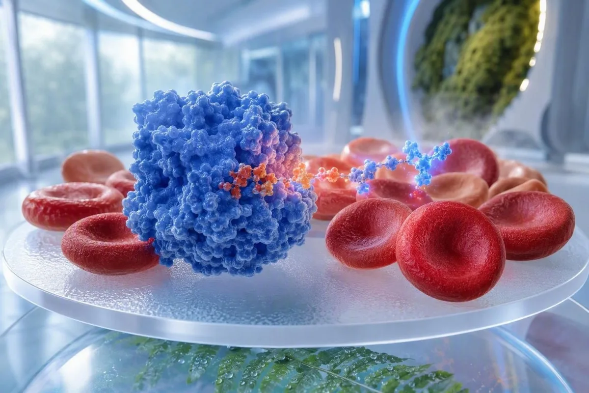
Low G6PD enzyme activity means red cells may be less able to handle oxidative stress, but a normal result during or soon after hemolysis can be falsely reassuring. The safest next step is usually a quantitative repeat test after recovery, with medication decisions guided by the prescribing clinician.


- Low G6PD enzyme activity can increase the risk of red-cell breakdown after certain medicines, infections, fava beans, or chemical exposures.
- False-normal timing is common during acute hemolysis because the most enzyme-deficient older red cells are removed first, leaving younger cells with relatively higher activity.
- Repeat testing is commonly considered about 2 to 3 months after an acute hemolytic episode or a transfusion, unless a specialist advises a different schedule.
- Quantitative testing reports activity in units per gram of hemoglobin or as a percentage of an adjusted male median; the laboratory's own reference interval matters.
- Intermediate results in the roughly 30% to 70% activity range deserve particular care in females because X-chromosome inactivation can create mixed red-cell populations.
- Urgent symptoms include dark tea-colored urine, jaundice, breathlessness, faintness, severe weakness, or rapidly worsening pallor after a new medicine or illness.
- Rasburicase and methylene blue require special caution in confirmed G6PD deficiency; prescribers should review alternatives before use.
- Fava beans and naphthalene mothballs are classic non-prescription triggers, but medication lists differ by dose, illness severity, and the variant involved.
What low G6PD enzyme activity means on lab test results
A low G6PD result means your red cells have reduced protection against oxidative stress and may break down faster when challenged by a trigger. As of August 7, 2026, the result should be treated as a medication-safety finding rather than a diagnosis made from one number alone.
G6PD, or glucose-6-phosphate dehydrogenase, helps red cells produce NADPH, which keeps glutathione in its protective reduced form. Red cells cannot make new enzyme proteins once mature, so activity naturally declines as a cell approaches its usual 120-day lifespan.
A result below the reporting laboratory's lower limit supports G6PD deficiency, but the number is not universally comparable between laboratories. Many quantitative assays report approximately 5.5 to 20.5 U/g hemoglobin in adults, yet the analytical method, hemoglobin correction, and local population determine what is truly low.
Kantesti is an AI blood test analyzer that reads a G6PD result alongside hemoglobin, bilirubin, LDH, reticulocytes, and the date of any acute illness rather than treating a single flag as definitive. Our clinical team regularly sees concern caused by a result that was technically normal but collected at exactly the wrong time.
Dr. Thomas Klein, MD, has found that patients often assume low activity means they are unwell every day. Most people with common variants feel entirely well between exposures; the practical issue is documenting the result clearly and knowing which situations merit a call to a clinician. For context on lab flags, see out-of-range result meanings.
How G6PD test results are measured and classified
A quantitative G6PD assay measures enzyme activity in a whole-blood sample and is more informative than a simple screen when a result may influence treatment. Activity is commonly expressed as U/g Hb or as a percentage of an adjusted male median, not as a universal concentration.
The preferred diagnostic sample is typically EDTA-anticoagulated whole blood, with enzyme activity normalized to the hemoglobin concentration. A sample with very low hemoglobin can produce a misleadingly high U/g Hb ratio because there is less hemoglobin in the denominator.
Many services use activity below 30% of the adjusted male median as deficient, about 30% to 70% as intermediate, and above 70% as normal; these operational bands are not interchangeable with every laboratory's reference interval. The report's assay name and lower reference limit should travel with the result.
A fluorescent spot screen can identify many markedly deficient samples, but it may miss intermediate activity, especially in heterozygous females or during reticulocytosis. A quantitative assay is generally the better choice when tafenoquine, primaquine, dapsone, or a similar oxidative medicine is being considered.
Reticulocytes are young red cells and usually contain more G6PD activity than older cells, which is why they matter so much here. Our reticulocyte and hematology guide explains why a count above about 100 × 10⁹/L during anemia changes how enzyme tests are interpreted.
Why a G6PD deficiency test can look normal after hemolysis
A G6PD test can appear normal during or soon after hemolysis because the oldest, most enzyme-deficient red cells are preferentially cleared first. The remaining circulation becomes enriched with reticulocytes and younger cells that have relatively higher measured activity.
This is a selection effect, not a cure. If a person loses a large fraction of vulnerable cells over 24 to 72 hours, the assay may mainly measure newly released cells whose G6PD content has not yet declined.
A falsely reassuring result is most likely when anemia, jaundice, dark urine, high LDH, low haptoglobin, or a rising reticulocyte count were present near the collection date. Even a hemoglobin fall from 145 g/L to 105 g/L can materially change the circulating cell-age mix.
Recent transfusion creates a second problem: donor red cells may have normal enzyme activity and can mask a recipient's deficiency for weeks. Save the date and number of units transfused; this often matters more than a borderline value on the PDF.
This broader lesson applies to many lab test results after acute illness. Comparing the current draw with the pre-illness baseline in a between-visit blood test review can show whether the enzyme result was obtained during a biologically unstable window.
How clinicians recognize active hemolysis around a G6PD result
Active hemolysis is suggested by falling hemoglobin plus increased reticulocytes, raised LDH, indirect bilirubin elevation, and low haptoglobin; no single marker proves it. The combined pattern determines whether a G6PD result should be deferred or repeated.
A haptoglobin result below about 0.3 g/L supports intravascular hemolysis in the right setting, but low haptoglobin can also reflect reduced liver production. LDH may rise several-fold above the upper reference limit, although liver injury, muscle injury, and sample handling can also increase it.
Unconjugated bilirubin often rises when red-cell clearance accelerates, while urine bilirubin is usually negative because unconjugated bilirubin is not water soluble. A positive urine bilirubin result instead points clinicians toward conjugated bilirubin and a different diagnostic branch.
When I review a panel with hemoglobin at 96 g/L, reticulocytes at 180 × 10⁹/L, LDH at 780 IU/L, and undetectable haptoglobin, I do not accept a normal enzyme activity result at face value. Thomas Klein, MD, would usually ask whether the sample was taken after a trigger rather than label the patient non-deficient.
A peripheral cell sample slide can show bite cells or blister cells after oxidative stress, though their absence does not exclude G6PD-related hemolysis. Read more about when cellular morphology changes demand prompt review in our guide to urgent schistocyte findings and haptoglobin interpretation.
When to repeat a G6PD test after hemolysis or transfusion
Repeat quantitative G6PD testing is often most reliable about 2 to 3 months after hemolysis has fully settled and at least 3 months after a transfusion, because reticulocytes and donor cells can obscure the baseline. A hematologist may adjust that timing when treatment cannot wait.
Do not use the calendar alone. Before repeat testing, clinicians look for stable hemoglobin, a reticulocyte count returning toward its personal range, bilirubin and LDH settling, and no ongoing exposure to a suspected trigger.
After an uncomplicated episode, a 12-week interval generally permits most transiently young-cell enrichment to fade. After several units of red-cell transfusion, the interval may need to reflect donor-cell survival, ongoing anemia, and the urgency of the medication decision.
If a medication must be started urgently, the prescriber may use a combination of prior records, molecular testing, specialist advice, and a risk-benefit decision rather than waiting for one perfect assay. This is particularly relevant in malaria treatment, where delaying therapy can itself be dangerous.
Kantesti AI can display the sequence of hemoglobin, bilirubin, LDH, and reticulocytes across multiple reports so that the repeat date is clinically explainable. For a practical recovery timeline after an admission, see changing labs after hospital discharge and our lab trend analysis guide.
Medicines that require clinician-guided caution with G6PD deficiency
Confirmed or strongly suspected G6PD deficiency warrants a prescriber or pharmacist review before oxidative medicines are used; the risk varies by dose, variant, age, infection, and baseline anemia. Do not stop a prescribed medicine solely from an online avoidance list.
Rasburicase is contraindicated in known G6PD deficiency because it can generate substantial oxidative stress, and methylene blue may be ineffective or harmful when NADPH generation is impaired. These are hospital-level decisions where an alternative should be selected promptly.
Primaquine and tafenoquine require validated quantitative G6PD assessment before use because they can cause dose-related hemolysis in susceptible people. Dapsone, nitrofurantoin, sulfonamide-containing medicines, and some antimalarials also deserve individualized review rather than blanket assumptions.
Luzzatto and Seneca described G6PD deficiency as a continuing pharmacogenetic issue precisely because drug risk is conditional, not binary (Luzzatto and Seneca, 2014). A viral illness, fever, kidney impairment, or a 2-week course of treatment may shift a previously tolerated exposure into a clinically relevant one.
Bring the laboratory report—not just a remembered diagnosis—to the prescriber. Medication and supplement preparation can also alter other labs, so our article on supplements before blood tests is useful when a repeat draw is planned.
Foods and household exposures linked with G6PD-related hemolysis
Fava beans are the best-established food trigger for G6PD-related hemolysis, while naphthalene-containing mothballs are a recognized household exposure to avoid. The reaction can occur within hours to several days and does not require an enormous serving in a highly susceptible person.
Favism is not simply a food allergy. Vicine and convicine metabolites can create oxidative stress in vulnerable red cells, and susceptibility differs substantially among G6PD variants and between individuals.
Naphthalene may be present in some mothballs and deodorizer products; accidental exposure is more concerning in infants and small children because a relatively small dose represents more exposure per kilogram. Store products in original packaging and seek poison-service advice after a suspected ingestion or heavy inhalation exposure.
Ordinary diets do not need to become restrictive by default. Garlic, legumes other than fava beans, blueberries, vitamin C foods, and most culinary herbs are often included on unreliable lists without consistent clinical evidence of harm at food amounts.
Kantesti is an AI-powered blood test analysis tool that keeps a documented enzyme result with the date, symptoms, and potential trigger, which can make a future medication conversation less chaotic. The evidence on supplements is uneven; our review of glutathione supplements and lab values explains why a product label is not the same as proven clinical protection.
Why G6PD results differ in females, newborns, and pregnancy
G6PD test interpretation is less straightforward in many females because random X-chromosome inactivation can leave a mixture of normal and deficient red cells. Newborns also have higher reticulocyte proportions, so adult cutoffs should not be applied without a pediatric reference interval.
A female can carry one altered G6PD allele and still have clinically meaningful intermediate activity, even if a qualitative screen is reported normal. Quantitative enzyme testing is preferable when a female patient may need primaquine, tafenoquine, or another treatment with known oxidative potential.
Newborn screening is especially valuable in regions with higher prevalence because severe neonatal jaundice can develop before a family knows there is an enzyme variant. A bilirubin rise in the first 24 hours of life always needs pediatric assessment regardless of G6PD status.
Pregnancy itself does not create G6PD deficiency, but anemia during pregnancy has many overlapping causes, including iron deficiency, folate deficiency, and dilution from increased plasma volume. A hemoglobin below 110 g/L deserves obstetric interpretation rather than automatic attribution to enzyme activity.
For pediatric context, review age-based blood test ranges. If iron deficiency is also suspected, the more useful question is often whether ferritin, transferrin saturation, and the red-cell indices tell the same story.
When genetic testing helps after a low or uncertain G6PD result
Genetic testing can clarify an uncertain G6PD phenotype, especially after hemolysis, transfusion, or an intermediate female result, but it does not replace enzyme activity testing in every clinical setting. The best test depends on whether the immediate question is drug safety, family counseling, or variant identification.
G6PD is located on the X chromosome, so many hemizygous males have a more uniform enzyme phenotype, while heterozygous females may have wide activity variation from lyonization. More than 200 variants are known, and genotype does not always predict the degree of hemolysis in a particular infection.
Molecular testing is useful when the enzyme result was taken during an unreliable period, when a specific familial variant is known, or when repeated activity tests conflict with the clinical history. It is not a shortcut for interpreting symptoms without a CBC and hemolysis markers.
The global distribution is uneven: the meta-analysis by Nkhoma et al. estimated that G6PD deficiency affects hundreds of millions of people worldwide, with higher frequencies in historical malaria-endemic areas (Nkhoma et al., 2009). That geography informs testing strategies but should never be used to dismiss testing in someone from another background.
Record relatives with neonatal jaundice, unexplained dark urine after illness, or a known enzyme variant. Our guide to family laboratory patterns can help families preserve useful details without assuming every relative has the same activity level.
Symptoms after a trigger that need same-day medical assessment
Dark brown urine, yellow eyes or skin, severe fatigue, shortness of breath, chest discomfort, fainting, or rapidly increasing pallor after a trigger require same-day clinical assessment. These symptoms can signal clinically significant hemolysis, but they can also occur with dehydration, liver disease, urinary problems, or other urgent conditions.
A sudden drop in oxygen-carrying capacity can produce a racing heart or breathlessness before a person recognizes jaundice. In a previously healthy adult, hemoglobin falling by 20 to 30 g/L over a few days is clinically meaningful and should not be managed by hydration alone.
Emergency clinicians commonly check CBC, reticulocytes, bilirubin fractions, LDH, haptoglobin, creatinine, electrolytes, urinalysis, and sometimes a direct antiglobulin test. The direct antiglobulin test helps separate immune-mediated hemolysis from non-immune causes, although results must be read in context.
A laboratory sample can itself become hemolyzed during collection and falsely elevate potassium or LDH without causing symptoms in the patient. That is why symptoms, urine color, and serial hemoglobin are more informative than an isolated laboratory hemolysis index.
If you have yellowing with dark urine, our explanation of bilirubin warning signs may help organize the history for urgent care. Do not wait for an app interpretation if you feel faint, confused, or breathless at rest.
How to read G6PD lab test results with the rest of the panel
The most useful G6PD interpretation combines enzyme activity, collection timing, hemoglobin, reticulocytes, bilirubin, LDH, haptoglobin, and transfusion history. A normal activity value has limited reassurance when the surrounding panel shows active red-cell turnover.
Start with the assay and units. A result of 4.2 U/g Hb may be low in a laboratory with a lower reference limit of 5.5 U/g Hb, while the same number cannot be interpreted safely without that laboratory's method and reference interval.
Then look at whether the CBC suggests anemia: hemoglobin below 130 g/L in many adult males or below 120 g/L in many non-pregnant adult females is commonly flagged, although local ranges vary. MCV, RDW, iron status, and B12 status can identify a second cause of anemia that is unrelated to G6PD.
Kantesti's AI lab test interpretation service uses pattern checks to identify when a seemingly reassuring G6PD result sits beside high reticulocytes or low haptoglobin and should be discussed with a clinician. That logic is reviewed against our medical validation standards and does not replace confirmatory laboratory testing.
For a source-checking workflow before acting on any automated summary, read our AI medical report safety guide. The clinician's question is usually not “is it red or green?” but “was this value biologically reliable on that date?”
Questions to take to your clinician after low G6PD enzyme activity
After a low or uncertain G6PD result, ask whether the assay was quantitative, whether hemolysis or transfusion could have distorted it, and whether repeat testing or genetic confirmation would change care. Bring the original report, not a cropped screenshot, because units and reference intervals are decisive.
Useful questions include: Was my test collected during anemia or within 12 weeks of transfusion? What was my reticulocyte count? Is my result below 30%, intermediate, or normal by this laboratory's method? These questions narrow the uncertainty quickly.
Also ask which medicines are relevant for your own care rather than requesting an indiscriminate “never take” list. A medication that carries risk at 100 mg daily may have a different risk profile at a single low dose, and infection itself can be the dominant hemolytic trigger.
Keep a photograph or PDF of the full report, dates of symptoms, and any medication started in the preceding 14 days. This becomes especially helpful if an emergency department or travel clinic needs to make a fast prescribing decision.
A blood test second opinion is reasonable when the result conflicts with past hemolysis, a family history, or a drug decision with meaningful consequences. Good follow-up is not overtesting; it is selecting the test at the right time.
G6PD precautions before travel, procedures, and new prescriptions
Before travel, surgery, or a new prescription, tell the clinician about confirmed or suspected G6PD deficiency and provide the actual result if available. This allows medicine selection before an urgent situation forces a hurried decision.
Travel medicine deserves early planning because antimalarial choices vary by destination, species risk, and G6PD activity. Tafenoquine cannot be safely treated as a routine travel tablet when quantitative activity has not been verified.
For procedures, the anesthetic team should know about the diagnosis, but G6PD deficiency does not automatically make routine anesthesia unsafe. The relevant issue is the complete medication plan, concurrent infection, kidney function, and whether a hemolytic event has occurred recently.
Kantesti AI can help users organize chronological reports for a clinician, but it cannot authorize a medicine or replace a pharmacist's interaction check. The Medical Advisory Board supports our clinician-led approach to safety language around potentially consequential lab findings.
A small medical alert card or phone note should state “G6PD deficiency—verify medicines,” the test date, and whether the result was quantitative. Avoid writing a long unverified drug list; the clinician needs accurate data, not an alarming label.
A practical next-step plan after G6PD test results
The safest plan after G6PD lab test results is to document the exact activity and test date, identify any recent hemolysis or transfusion, review medicines with a clinician, and repeat a quantitative assay after recovery if timing was unreliable. Most patients do not need daily treatment; they need a reliable record and a trigger-aware plan.
If your activity is clearly low on a stable outpatient sample, ask the clinician to add it to your medical record and discuss the medicines most relevant to you. If the result was normal during a suspected episode, do not assume the question is closed—arrange a repeat at roughly 8 to 12 weeks when appropriate.
If you develop dark urine, jaundice, breathlessness, marked weakness, or dizziness after illness, fava beans, naphthalene exposure, or a new medicine, seek same-day medical advice. Bring the trigger details, because the first 24 hours of history often determine which tests are prioritized.
In my experience, the most avoidable harm comes from two opposite mistakes: calling every borderline result dangerous, or ignoring a normal result obtained during obvious hemolysis. Luzzatto et al. emphasize that both phenotype and clinical context drive management of this inherited enzyme disorder (Luzzatto et al., 2016).
Kantesti's AI biomarker interpretation platform can preserve trends and prompt questions, while a physician confirms diagnosis and treatment decisions. Our technology guide explains how context-based pattern review is designed to support—not replace—clinical judgment.
Frequently Asked Questions
Can G6PD test results be normal during hemolysis?
Yes. G6PD activity can be falsely normal during or shortly after hemolysis because the oldest, most enzyme-deficient red cells are cleared first and younger reticulocytes have relatively higher enzyme activity. This effect is most likely when hemoglobin is falling, reticulocytes are elevated, or bilirubin and LDH are rising. A quantitative repeat test is often considered about 2 to 3 months after recovery, provided there has been no recent transfusion.
How low does G6PD enzyme activity need to be to indicate deficiency?
Many laboratories consider activity below about 30% of the adjusted male median to be deficient, but the exact cutoff depends on the assay and laboratory reference interval. Intermediate activity around 30% to 70% can be clinically relevant, particularly in heterozygous females. A result in U/g Hb must always be interpreted against the reference range printed on that specific report.
How long after a blood transfusion should I wait for a G6PD test?
A G6PD test is commonly delayed for about 3 months after a red-cell transfusion because donor cells with normal activity can mask the recipient's enzyme deficiency. The correct interval can be longer or shorter depending on how many units were given, ongoing anemia, and whether an urgent medication decision is needed. A hematologist can advise on enzyme testing versus genetic testing when waiting is not practical.
Which medicines are unsafe with G6PD deficiency?
Rasburicase is contraindicated in known G6PD deficiency, and methylene blue can be ineffective or harmful because it depends on NADPH generation. Primaquine and tafenoquine require validated quantitative G6PD assessment before prescribing, while dapsone, nitrofurantoin, and some sulfonamide-containing medicines require individualized clinician review. Do not stop prescribed treatment without speaking to the prescriber, because dose, infection severity, and personal enzyme activity affect risk.
Do females need quantitative G6PD testing?
Females often benefit from quantitative G6PD testing because random X-chromosome inactivation can create a mixed red-cell population with intermediate activity. A qualitative screen may miss clinically relevant activity in the roughly 30% to 70% range. Quantitative testing is particularly appropriate before primaquine or tafenoquine and when there is a personal or family history of hemolysis.
Can fava beans cause hemolysis in G6PD deficiency?
Yes. Fava beans can trigger acute hemolysis in susceptible people with G6PD deficiency because vicine and convicine metabolites increase oxidative stress inside red cells. Symptoms may begin within hours to several days and can include dark urine, jaundice, fatigue, pallor, or breathlessness. The reaction varies by G6PD variant and serving exposure, so a person with known deficiency should discuss avoidance and emergency symptoms with a clinician.
Get AI-Powered Blood Test Analysis Today
Join over 2 million users worldwide who trust Kantesti for instant, accurate lab test analysis. Upload your blood test results and receive comprehensive interpretation of 15,000+ biomarkers in seconds.
📚 Referenced Research Publications
Klein, T., Mitchell, S., & Weber, H. (2026). Multilingual AI Assisted Clinical Decision Support for Early Hantavirus Triage: Design, Engineering Validation, and Real-World Deployment Across 50,000 Interpreted Blood Test Reports. Kantesti AI Medical Research.
Klein, T., Mitchell, S., & Weber, H. (2026). A Pre-Registered, Rubric-Based Automated Technical Benchmark of the Kantesti Blood-Test Interpretation Engine on 100,000 Synthetic Test Cases. Kantesti AI Medical Research.
📖 External Medical References
⚕️ Medical Disclaimer
This article is for educational purposes only and does not constitute medical advice. Always consult a qualified healthcare provider for diagnosis and treatment decisions.
E-E-A-T Trust Signals
Experience
Physician-led clinical review of lab interpretation workflows.
Expertise
Laboratory medicine focus on how biomarkers behave in clinical context.
Authoritativeness
Written by Dr. Thomas Klein with review by Dr. Sarah Mitchell and Prof. Dr. Hans Weber.
Trustworthiness
Evidence-based interpretation with clear follow-up pathways to reduce alarm.