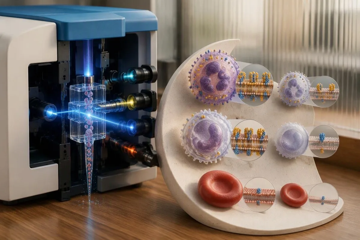
PNH flow cytometry detects cells missing protective surface proteins: FLAER checks their membrane anchors, while CD59 testing assesses a key complement defense. Different cell populations can have different clone percentages, and a small clone alone does not establish classical PNH or a need for treatment.


- PNH flow cytometry identifies GPI-deficient populations using multiple markers; an isolated abnormal marker is not enough for reliable diagnosis.
- FLAER test assesses GPI anchors on white cells, usually alongside CD24 on granulocytes and CD14 on monocytes.
- CD55 CD59 testing evaluates complement-protective proteins; CD59 is particularly useful for identifying partially and completely deficient red cells.
- PNH clone size is the percentage of abnormal cells within a specific lineage, not the percentage of all cells in the body.
- High-sensitivity testing can reach approximately 0.01% in a validated assay, but detection and accurate quantification are different capabilities.
- Red-cell percentages can fall after transfusion or hemolysis, so a 3% red-cell clone can coexist with a much larger granulocyte clone.
- Small clones may accompany aplastic anemia without clinically significant hemolysis; even a genuine 0.5% clone does not automatically require complement treatment.
- LDH interpretation uses the laboratory’s upper limit: 480 U/L with an upper limit of 240 U/L equals 2 times that limit, but LDH alone cannot diagnose PNH.
- Urgent symptoms such as sudden breathlessness, severe abdominal pain, or a new neurological deficit require immediate assessment regardless of clone percentage.
What does PNH flow cytometry actually detect?
PNH flow cytometry detects populations of cells missing GPI-anchored surface proteins, usually by examining granulocytes, monocytes, and red cells separately. FLAER checks the anchor on white cells; CD59 helps assess red-cell protection. A result such as 0.5% describes a population within one lineage—not, by itself, classical PNH.
A PNH clone is a group of cells descended from an altered hematopoietic stem cell, and the laboratory identifies its characteristic surface phenotype rather than simply measuring anemia. Most diagnostic approaches examine at least 2 cell lineages because agreement across populations helps distinguish a genuine clone from technical noise. A routine complete blood count cannot provide this information.
As Thomas Klein, MD, I would separate 3 questions when reading a report: is the abnormal population real, is it causing clinical problems, and does it require treatment? Those questions need different evidence. A tiny reproducible population may answer the first question while leaving the other 2 unanswered—particularly when the patient has bone marrow failure.
Kantesti is an AI blood test analyzer that can help explain a reported 0.5% clone alongside its accompanying CBC and chemistry results, without replacing specialist flow-cytometry interpretation. Our biomarker terminology guide helps distinguish surface markers from ordinary blood chemistry measurements. The original laboratory comments remain essential, especially when they describe results below a quantification limit.
Why do PNH cells lose their protective surface proteins?
PNH usually arises from an acquired PIGA mutation in a hematopoietic stem cell, impairing production of glycosylphosphatidylinositol, or GPI, anchors. Without these anchors, several proteins cannot attach properly to the cell membrane. The same stem-cell population can therefore produce abnormal cells in 3 routinely tested lineages: granulocytes, monocytes, and red cells.
CD55 and CD59 perform different complement-control tasks: CD55 accelerates breakdown of complement convertases, while CD59 inhibits formation of the membrane attack complex. Losing both defenses makes affected red cells vulnerable to complement-mediated destruction. Brodsky’s 2014 review in Blood explains this acquired stem-cell mechanism and why the clinical consequences extend beyond the red-cell percentage alone (Brodsky, 2014).
A PIGA change is usually somatic, meaning acquired rather than inherited through the family. The gene sits on the X chromosome, but that does not make an ordinary acquired PNH clone an inherited X-linked disorder. For a patient with a 12% granulocyte clone, testing relatives is generally not the next step; confirming the phenotype and assessing clinical consequences are more useful.
PNH and enzyme-related hemolysis need different tests, even when both produce anemia and dark urine. For example, G6PD activity testing measures an intracellular enzyme rather than GPI anchors; reticulocytosis can complicate that assay’s interpretation. A normal G6PD result does not exclude PNH, and CD59 deficiency does not establish G6PD deficiency. They answer 2 separate biological questions.
Why do doctors request FLAER and CD59 together?
FLAER and CD59 provide complementary evidence of GPI deficiency, rather than repeating the same measurement. FLAER is a fluorescent, non-lytic aerolysin reagent that binds GPI anchors on suitable white cells. CD59 testing examines a specific anchored protein, especially on red cells, helping identify both partial and complete deficiency.
The FLAER test is performed on the laboratory sample, not administered to the patient. A common granulocyte combination uses FLAER plus CD24; a monocyte combination uses FLAER plus CD14, with other markers defining the correct cell population. Demonstrating concordant loss of 2 GPI-associated signals reduces the chance that a single unusual antigen pattern will be mistaken for a PNH clone.
Red cells require a different approach because FLAER is not suitable for routine red-cell PNH analysis. Laboratories commonly identify erythrocytes with a lineage marker such as CD235a and assess CD59, sometimes alongside CD55. Type II cells show partial deficiency; type III cells show complete deficiency. Their percentages should be reported distinctly when the assay can resolve both populations reliably.
A normal complement concentration does not exclude PNH: the issue is missing cell-surface protection, not necessarily a low amount of circulating complement. Our C3 and C4 explanation covers those separate measurements. Ordering C3 and C4 cannot substitute for the 2-marker, lineage-specific approach described in the 2010 flow-cytometry guideline (Borowitz et al., 2010).
What does a clone percentage mean for each cell type?
PNH clone size is the proportion of GPI-deficient cells within the population being analyzed. A 20% granulocyte clone means approximately 20 of every 100 analyzed granulocytes have the relevant deficient phenotype. It does not mean 20% of all white cells, all marrow cells, or all cells in the body.
Granulocyte and monocyte clone sizes often resemble one another, but they do not have to match exactly. Differences can reflect biological production, cell survival, gating, or the smaller number of monocytes acquired. A report showing 18% granulocytes and 16% monocytes may be broadly concordant; a report showing 18% and 0.02% deserves closer review of both biology and assay quality.
Percentages can also be confused with ordinary differential-count percentages. A monocyte count of 8% on a CBC describes monocytes among white cells; an 8% PNH monocyte clone describes deficient cells among the analyzed monocytes. Our monocyte count versus percentage discussion explains the denominator problem. Those 2 measurements cannot be substituted for one another or added together.
The granulocyte clone often provides a better estimate of clone burden than the red-cell clone, because affected granulocytes are not selectively removed by complement-mediated red-cell destruction. Still, it is an estimate from peripheral blood, not a direct census of stem cells. When percentages differ across 3 lineages, I would first ask which population was measured well—not average the numbers.
Why can the red-cell clone be much smaller?
The red-cell PNH clone can look smaller because deficient red cells are destroyed or diluted by transfused donor cells. For example, a patient may have a 65% granulocyte clone but only a 7% red-cell clone. That mismatch can be clinically plausible and does not automatically mean either result is wrong.
A red-cell transfusion adds cells with normal GPI-linked protection, increasing the denominator without necessarily changing the patient’s stem-cell clone. The measured red-cell percentage can remain distorted for weeks to months, depending on transfusion volume and cell survival. The laboratory needs the transfusion date; even 2 recently transfused units can materially change interpretation in a patient with substantial anemia.
Complement inhibition can move the red-cell percentage in the opposite direction: protected PNH red cells survive longer, so their measured fraction may rise despite improved disease control. A change from 10% to 35% red cells after treatment is therefore not sufficient evidence of stem-cell clone expansion. Compare granulocyte results, hemoglobin, LDH, and treatment timing before drawing that conclusion.
Transfusion also affects other erythrocyte-based measurements, including A1c after transfusion, although the mechanism and clinical question differ. The useful habit is the same: label the result with its recent treatment context. Do not postpone clinically necessary PNH testing solely to wait 3 months after transfusion; a specialist can often interpret the white-cell analysis immediately.
What makes a PNH flow-cytometry result reliable?
Reliable PNH testing requires correct cell identification, multiple deficiency markers, adequate event counts, and validated sample handling. A white-cell population negative for just 1 marker is not convincing on its own. Borowitz and colleagues’ 2010 guideline emphasizes lineage-specific analysis and complementary markers rather than reliance on a single absent antigen.
A fresh EDTA sample is commonly used, but transport limits are laboratory-specific. Many services prefer analysis within 24–48 hours; other intervals are acceptable only when the method has been validated for them. Aging, poor viability, debris, and altered marker expression can create misleading negative events. Ask the receiving laboratory about its deadline rather than applying a universal shipping rule.
A high sample hemolysis index concerns release of intracellular material into the specimen and can distort chemistry tests such as LDH. It is not itself proof of hemolysis inside the patient. In a report containing both a 0.03% suspected clone and unexpectedly high LDH, sample quality and clinical hemolysis need to be assessed as 2 separate issues.
Kantesti can explain the distinction between a clone measurement and accompanying chemistry abnormalities, but we do not independently validate a laboratory’s raw cytometry gates. Our clinical validation framework should not be mistaken for certification of every external PNH assay. A discrepant result across 2 lineages is a reason to consult the testing laboratory, not to let software settle it.
How small a PNH clone can flow cytometry detect?
A validated high-sensitivity PNH assay can detect populations around 0.01%, but the achievable limit depends on cell lineage and sample quality. Detection is not the same as precise quantification. “No clone detected” means no qualifying population was found above that assay’s detection limit—not proof that every cell is normal.
Event counts explain why sensitivity is not just a marketing label. At 0.01%, 500,000 analyzed cells contain about 50 deficient cells, before exclusions or losses; 10,000 analyzed cells would contain only about 1. A tiny apparent population based on a handful of events has much greater counting uncertainty. The laboratory’s detection and quantification criteria must account for that difference.
Some reporting schemes call populations below 0.1% “rare GPI-deficient cells” and those around 0.1%–1% “minor populations,” but terminology is not identical everywhere. These are reporting categories, not treatment thresholds. A result reported as “detected below quantification” should not be interpreted with the same numerical confidence as a well-resolved 12% clone across granulocytes and monocytes.
Kantesti is an AI blood test interpretation platform that helps distinguish a qualitative statement such as “detected” from a quantitative claim such as 0.08%. Our qualitative versus quantitative results guide explains why these statements carry different information. For serial monitoring, retain the laboratory’s sensitivity, lineage, and wording—not just the percentage copied into a spreadsheet.
Does a small PNH clone mean classical PNH?
A small PNH clone does not, by itself, establish classical PNH. Small populations can accompany aplastic anemia and other marrow-failure settings without substantial complement-mediated hemolysis. Babushok’s 2021 review distinguishes subclinical clones from symptomatic PNH and emphasizes that clinical significance depends on more than finding GPI-deficient cells.
Consider a hypothetical patient with hemoglobin 8.5 g/dL, platelets 38 × 10^9/L, neutrophils 0.7 × 10^9/L, and a 0.6% granulocyte clone. If LDH is normal and reticulocyte production is low, marrow failure is more immediately explanatory than hemolysis from that tiny clone. The clone still matters, but it does not explain every abnormality or dictate complement treatment.
Aplastic anemia requires its own assessment, including the severity of cytopenias and marrow findings. Our aplastic anemia CBC patterns guide explains why low counts across 3 lineages deserve prompt attention. PNH flow cytometry cannot diagnose aplastic anemia alone, and a negative PNH result does not exclude it. These tests provide complementary, rather than competing, information.
Small clones are not automatically irrelevant or permanently stable. Their size may increase, decrease, or remain similar, and the marrow disorder may determine the patient’s immediate risk. Babushok discusses generally small clones in marrow-failure settings versus larger clones in classical PNH, often above 50%; those typical distributions overlap and 50% is not a diagnostic boundary (Babushok, 2021).
Which findings turn a clone into clinically significant PNH?
Clinically significant PNH requires the clone to be interpreted alongside hemolysis, symptoms, thrombosis, and marrow function. Doctors generally distinguish classical PNH, PNH associated with a marrow disorder, and subclinical clones. These 3 categories describe clinical behavior—not simply low, medium, and high percentages on a flow-cytometry report.
PNH commonly causes direct-antiglobulin-test-negative hemolysis before complement treatment, but that pattern is not unique to PNH. LDH, indirect bilirubin, haptoglobin, and reticulocytes help establish whether red-cell destruction is occurring. Our direct Coombs test explanation covers a different mechanism: antibodies or complement detected on red cells. A positive result can require a broader explanation rather than immediately excluding coexisting PNH.
LDH should be expressed relative to the local upper limit because reference intervals and methods differ. LDH 480 U/L with an upper limit of 240 U/L equals 2.0 times the upper limit, whereas 480 U/L against 300 U/L equals 1.6 times. Our LDH result interpretation explains non-PNH causes, including liver, muscle, and specimen-related abnormalities.
Dark urine is neither required nor confined to nighttime. A patient with a 55% granulocyte clone, recurrent abdominal discomfort, falling hemoglobin, and convincing intravascular hemolysis needs a different discussion from someone with a 0.2% clone and stable counts. Brodsky’s clinical review describes this broader presentation, including fatigue, smooth-muscle symptoms, thrombosis, and renal consequences (Brodsky, 2014).
When does a PNH clone justify treatment?
Complement treatment is considered for clinically consequential PNH, not prescribed solely because a clone exceeds a particular percentage. Relevant findings include symptomatic hemolysis, anemia attributable to PNH, transfusion requirements, thrombosis, and organ complications. A stable 0.5% clone without hemolysis usually calls for clinical follow-up rather than automatic complement inhibition.
Anemia in PNH may reflect hemolysis, marrow failure, or both, and complement inhibitors do not correct all 3 possibilities equally. Hemoglobin 8 g/dL with an absolute reticulocyte count of 30 × 10^9/L suggests an inadequate production response for substantial anemia, although the whole picture matters. Our reticulocyte response guide explains why cell production must be assessed alongside destruction.
C5 inhibitors such as eculizumab and ravulizumab reduce terminal-complement-mediated intravascular hemolysis; other medicines target earlier complement steps in selected patients. Persistently low hemoglobin with lower LDH can reflect extravascular hemolysis or marrow failure rather than treatment failure from clone growth. Haptoglobin alone cannot resolve that distinction; our low haptoglobin causes discussion includes liver-related and other confounders.
When explaining a 45% clone through Kantesti, I would frame the report as a starting point for hematology discussion, not a prescription trigger. Drug choice, infection-prevention requirements, vaccination timing, and any antimicrobial precautions depend on the treatment and local guidance. Complement inhibition carries serious infection risks, particularly with meningococci for terminal inhibitors; patients need a specific prevention and urgent-fever plan.
Can clone size predict clot risk or urgency?
Larger PNH clones generally raise concern for thrombosis, but clone percentage cannot safely rule a clot in or out. Hemolysis, prior thrombosis, symptoms, and other clinical factors also matter. A small clone is not permission to ignore severe abdominal pain, sudden breathlessness, a swollen leg, or a new neurological deficit.
PNH-associated thrombosis can occur in hepatic, portal, mesenteric, or cerebral venous circulation, as well as more familiar locations. An unexplained clot in an unusual site can justify PNH testing, particularly with cytopenias or hemolysis. An ordinary provoked leg clot without supporting features does not make PNH screening mandatory; the clinical pretest probability determines whether specialized testing adds useful information.
Normal PT or aPTT does not exclude thrombosis, and neither test measures the presence of a PNH clone. Our normal coagulation test limitations guide explains that distinction. For a patient with a known 60% clone and sudden severe abdominal pain, waiting for another flow result or a routine clotting panel can delay the imaging and assessment actually needed.
A newly identified clone plus suspected thrombosis needs urgent specialist coordination because anticoagulation, complement therapy, and cytopenias may interact. A platelet count of 25 × 10^9/L makes the bleeding-risk discussion particularly complex, but it does not mean a clot is impossible. This is not a situation for self-starting aspirin or changing anticoagulant doses based on an online explanation.
When should PNH flow cytometry be repeated?
Repeat PNH flow cytometry is used to confirm uncertain findings and follow clinically relevant clone changes, especially in marrow-failure settings. A stable small clone is often reassessed at roughly 6–12 months, depending on the hematologist’s plan. New hemolysis, worsening counts, or suspected thrombosis can justify earlier reassessment rather than waiting for that interval.
A change from 0.04% to 0.07% may reflect rare-event counting variation, particularly near a laboratory’s quantification limit. It should not automatically be called clinical progression. Conversely, a reproducible rise from 0.4% to 8% across granulocytes and monocytes deserves review even if the patient feels well. Compare the assay method, sensitivity, and acquired cell numbers before interpreting the direction.
Using the same experienced laboratory improves comparability, but a changed service is not automatically unreliable. Obtain both reports and check lineage definitions, type II/type III red-cell reporting, and detection limits. Our between-visit result differences guide explains why some numerical changes are analytical or biological variation. Cytometry rare-event uncertainty needs its own assessment rather than a generic chemistry change formula.
Kantesti’s trend tools can help organize dated CBC and chemistry results alongside reported clone percentages, but a 3-lineage clone trend needs specialist interpretation. Attach transfusion dates, complement-treatment changes, LDH upper limits, and any new symptoms to each time point. A rising red-cell fraction with falling LDH after treatment can represent better cell survival—the opposite of the conclusion a percentage-only graph might suggest.
What should you ask your hematologist about the report?
Ask whether the clone is confirmed, which lineages were tested, and whether there is evidence of clinically significant hemolysis or marrow failure. A useful report discussion separates analytical certainty from clinical severity. For a result around 0.01%, the laboratory’s detection and quantification limits may matter more than an extra decimal place.
Bring the complete report rather than a cropped percentage. Ask 4 practical questions: were granulocytes and monocytes concordant, was the red-cell result affected by transfusion, were type II and type III cells distinguished, and when should testing be repeated? Mention dark urine, abdominal symptoms, transfusion history, and previous clots—even if the report describes the population as “small.”
Kantesti is an AI-powered blood test analysis tool that can organize a 3-lineage report with related blood results and explain terminology; it cannot reconstruct raw flow data that the laboratory has not provided. Our AI technology explanation describes the interpretation workflow. Decimal accuracy in a PDF or photo still needs checking against the source report, especially when 0.01% and 0.1% imply different event burdens.
I’m Thomas Klein, MD, Chief Medical Officer; my approach is to avoid turning 1 abnormal line into a diagnosis without its clinical context. Our medical advisory team supports that separation of explanation from personal treatment decisions. As of October 15, 2026, this article uses identifiable PNH literature from 2010, 2014, and 2021 rather than inventing a new “2026 cutoff.”
Which publications support PNH interpretation and further reading?
PNH assay and treatment statements should be supported by condition-specific medical literature, not general AI publications. The 3 external references below address flow-cytometry standards, PNH biology, and clone significance. Our global health report provides separate platform background; it does not establish the diagnostic accuracy of a FLAER or CD59 assay.
Borowitz et al. (2010), Brodsky (2014), and Babushok (2021) support different parts of this explanation: assay methodology, disease mechanism, and the clinical meaning of finding a clone. The 2 Zenodo records listed here are background reading, not substitutes for those medical references. Our RDW and red-cell indices article adds CBC context, but RDW cannot detect or quantify a PNH clone.
Kantesti LTD. (2026). AI blood test analyzer: 2.5M tests analyzed | Global health report 2026. Zenodo. DOI record. A ResearchGate publication search and an Academia.edu publication search are discovery links, not verified repository copies; neither the title’s test count nor its DOI demonstrates PNH-specific clinical validation.
Kantesti LTD. (n.d.). RDW blood test: Complete guide to RDW-CV, MCV & MCHC. Zenodo. DOI record. A ResearchGate publication search and an Academia.edu publication search help locate possible listings; the publication year and full record metadata should be confirmed before reuse in a formal bibliography.
Frequently Asked Questions
What is a PNH flow cytometry test?
PNH flow cytometry detects populations of cells missing GPI-anchored surface proteins associated with paroxysmal nocturnal hemoglobinuria. Laboratories usually examine granulocytes, monocytes, and red cells separately using multiple markers. A validated high-sensitivity assay may detect a population around 0.01%, but the actual limit depends on the lineage and sample. Finding a clone does not, by itself, establish classical PNH or a need for treatment.
Why are FLAER and CD59 both checked for PNH?
FLAER checks GPI anchors on suitable white cells, while CD59 testing assesses a specific complement-protective protein, especially on red cells. Laboratories commonly pair FLAER with CD24 on granulocytes or CD14 on monocytes to demonstrate concordant deficiency of 2 signals. CD59 can help separate partially deficient type II red cells from completely deficient type III red cells. FLAER is a laboratory reagent and is not administered to the patient.
Is a small PNH clone dangerous or does it need treatment?
A small PNH clone does not automatically require complement treatment or establish classical PNH. A 0.5% clone may occur with aplastic anemia without clinically significant hemolysis, and the marrow disorder may explain the patient’s symptoms and low counts. Hemolysis, thrombosis, organ complications, and clone trends determine clinical significance alongside the percentage. Severe new symptoms still require prompt assessment because a small clone cannot rule out other serious illness.
Why is my red-cell PNH clone smaller than my granulocyte clone?
The red-cell PNH clone can be smaller because deficient red cells are selectively destroyed and transfused donor cells dilute the measured percentage. A 60% granulocyte clone and a 5% red-cell clone can therefore coexist without a laboratory error. Complement treatment may later increase the red-cell fraction by improving survival rather than expanding the stem-cell clone. Transfusion dates, treatment changes, and LDH trends are necessary for interpretation.
Does a negative FLAER test rule out PNH completely?
A negative FLAER-based assay means no qualifying deficient population was detected above the laboratory’s validated limit in the analyzed cells. It does not prove that the body contains absolutely zero GPI-deficient cells. An assay with 0.01% sensitivity provides a different level of exclusion from one with 1% sensitivity, and poor cell numbers can reduce performance. Persistent unexplained hemolysis or marrow-failure findings warrant discussion with hematology about assay quality and whether repeat testing is appropriate.
How often should a small PNH clone be monitored?
A stable small PNH clone is often monitored at approximately 6–12-month intervals, but the schedule depends on the marrow disorder, symptoms, and hematologist’s assessment. New hemolysis, worsening blood counts, or suspected thrombosis may require earlier review. A change from 0.04% to 0.07% near the quantification limit may represent counting variation rather than progression. Follow-up should compare the same lineages and document transfusions, treatment changes, and the laboratory’s detection limits.
Get AI-Powered Blood Test Analysis Today
Join over 2 million users worldwide who trust Kantesti for instant, accurate lab test analysis. Upload your blood test results and receive comprehensive interpretation of 15,000+ biomarkers in seconds.
📚 Referenced Research Publications
Klein, T., Mitchell, S., & Weber, H. (2026). AI Blood Test Analyzer: 2.5M Tests Analyzed | Global Health Report 2026. Kantesti AI Medical Research.
Klein, T., Mitchell, S., & Weber, H. (2026). RDW Blood Test: Complete Guide to RDW-CV, MCV & MCHC. Kantesti AI Medical Research.
📖 External Medical References
Borowitz MJ et al. (2010). Guidelines for the diagnosis and monitoring of paroxysmal nocturnal hemoglobinuria and related disorders by flow cytometry. Cytometry Part B: Clinical Cytometry.
Brodsky RA (2014). Paroxysmal nocturnal hemoglobinuria. Blood.
Babushok DV (2021). When does a PNH clone have clinical significance?. Hematology, American Society of Hematology Education Program.
⚕️ Medical Disclaimer
This article is for educational purposes only and does not constitute medical advice. Always consult a qualified healthcare provider for diagnosis and treatment decisions.
E-E-A-T Trust Signals
Experience
Physician-led clinical review of lab interpretation workflows.
Expertise
Laboratory medicine focus on how biomarkers behave in clinical context.
Authoritativeness
Written by Dr. Thomas Klein with review by Dr. Sarah Mitchell and Prof. Dr. Hans Weber.
Trustworthiness
Evidence-based interpretation with clear follow-up pathways to reduce alarm.