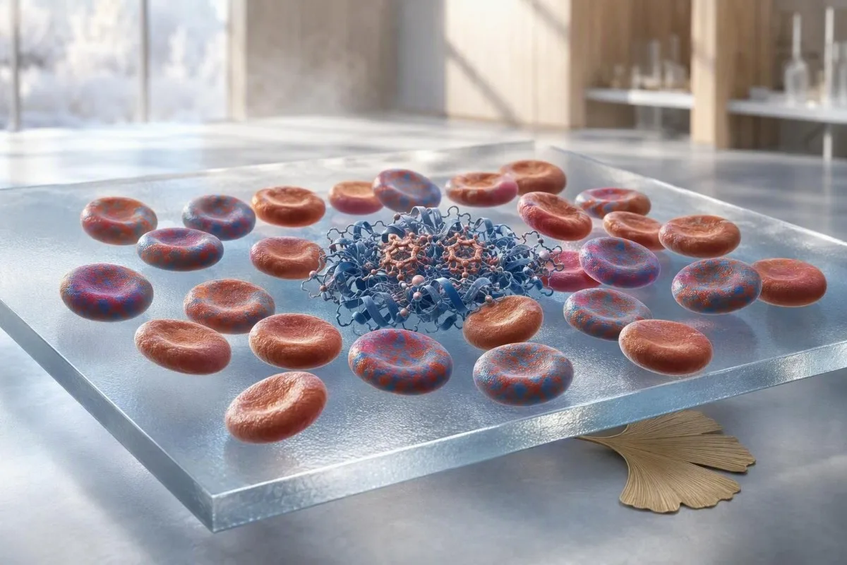
Donor red cells can temporarily replace the glucose history that an A1c is meant to measure. The safest next test depends on whether you need to diagnose diabetes, adjust treatment, or investigate symptoms today.


- Hemoglobin A1c is generally unreliable after a red-cell transfusion because the result mixes your cells with donor cells.
- False low A1c is common when donor cells came from a person without diabetes, but the direction is not guaranteed.
- Three months is a practical minimum before relying on A1c again after a substantial transfusion; 4 months gives greater confidence after multiple units.
- Fasting plasma glucose of 126 mg/dL (7.0 mmol/L) or higher on two occasions can diagnose diabetes when A1c is unsuitable.
- Random plasma glucose of 200 mg/dL (11.1 mmol/L) or higher with classic symptoms can diagnose diabetes without waiting for A1c.
- Fructosamine reflects roughly the prior 2-3 weeks and is not altered by donor red cells, although low albumin can distort it.
- Continuous glucose monitoring is especially useful after transfusion for people already using insulin because it shows current glucose rather than red-cell history.
- Tell the laboratory and clinician the transfusion date and number of units; this context changes how an A1c should be interpreted.
Can hemoglobin A1c be trusted after a transfusion?
Hemoglobin A1c should not be used as a reliable measure of your usual glucose control immediately after a red-cell transfusion. Donor red cells carry the donor's pre-existing glucose exposure, so the reported percentage becomes a mixture rather than your personal 8-12 week average. In practice, I use glucose-based testing now and reconsider A1c after about 3 months, sometimes 4 months after several units. As of August 29, 2026, that remains the clinically cautious approach.
A single unit of packed red cells contains about 250-300 mL of cellular material, enough to materially change an A1c in a smaller adult or anyone with significant anaemia. The result may look reassuring even while finger-stick or laboratory glucose remains high. This is why blood-test timing after illness matters as much as the printed percentage.
In my clinical work, the concerning scenario is the patient discharged after gastrointestinal fluid loss whose A1c has fallen from 8.7% to 6.2% within days. That is not an overnight metabolic recovery; it is usually a laboratory result diluted by donor cells and accelerated replacement of the patient's older cells.
Kantesti is an AI blood test analyzer that reads an A1c alongside haemoglobin, reticulocytes, transfusion history, and current glucose rather than treating one percentage as a verdict. Dr. Thomas Klein, our Chief Medical Officer, advises recording the transfusion date beside every post-hospital laboratory result because it prevents misleading trend graphs.
Why donor red cells change the A1c calculation
A1c measures glucose attached to haemoglobin inside circulating red cells, not glucose floating in the blood on test day. Since red cells normally survive about 120 days, their glycation records prior glucose exposure; transfused cells bring a different record into the same sample.
The laboratory reports the proportion of haemoglobin that is glycated, commonly by high-performance liquid chromatography or enzymatic methods. If half of circulating red-cell mass comes from donor cells with an A1c near 5.2%, a recipient whose own A1c was 9.0% can receive a deceptively intermediate result. The exact shift depends on transfused volume, recipient blood volume, and red-cell survival.
Spencer and colleagues documented clinically meaningful A1c decreases after transfusion in people with diabetes, particularly when the pre-transfusion A1c was high (Spencer et al., 2011). The lesson is not that every result becomes low; it is that the number loses its usual relationship to average glucose.
A reticulocyte count can provide useful context because reticulocytes are newly released red cells with little time for glycation. Our reticulocyte and blood-type guide explains why a high reticulocyte percentage after recovery can lower A1c even without transfusion.
Is a false low A1c the only possible result?
A false low A1c is the usual pattern after transfusion from a donor without diabetes, but transfusion does not guarantee a low result. The direction depends on the relative glycation and age of donor and recipient cells, making mathematical correction unsafe in routine care.
Most volunteer blood donors do not have marked chronic hyperglycaemia, so their cells often lower an elevated recipient A1c. Yet a donor can have prediabetes or diabetes, and stored red cells may undergo biochemical changes that complicate assumptions. No laboratory can reliably reconstruct your original A1c from the number of units alone.
Red-cell age matters too. Older recipient cells tend to have accumulated more glycation, while newly made cells after acute loss have accumulated less. A raised RDW, which signals a broader spread of cell sizes and often cell ages, should make clinicians cautious about interpreting A1c; see our RDW and red-cell index guide.
This is one of those areas where context beats a clever calculation. A reported A1c of 5.8% after 2 units may coexist with fasting glucose values of 154 mg/dL (8.6 mmol/L), and the glucose values should drive the immediate decision.
Does one unit of blood make A1c unreliable?
Even one unit can make hemoglobin A1c less trustworthy, especially in a person with low body weight, marked anaemia, or low pre-transfusion blood volume. Two or more units, exchange transfusion, and ongoing red-cell loss create even greater uncertainty.
An average 70-kg adult has roughly 5 litres of blood, but packed cells are concentrated and anaemia reduces the recipient's own red-cell mass before transfusion. In a 50-kg adult with haemoglobin of 6.5 g/dL, 1 unit can represent a substantial fraction of the circulating red cells measured days later.
A post-transfusion complete blood count cannot tell exactly how many donor cells remain, but it flags the setting in which A1c is vulnerable. Haemoglobin, haematocrit, MCV, RDW, bilirubin, and reticulocytes often tell a more useful story together than A1c alone; review the components in our full blood count explanation.
Kantesti AI flags a recent transfusion as a context warning when interpreting A1c, because a laboratory reference interval does not itself certify clinical validity. A value below 5.7% is normally considered below the prediabetes threshold, but that threshold should not be applied mechanically in this situation.
When does an A1c become meaningful again?
For most people, wait at least 3 months after a substantial red-cell transfusion before using A1c to judge usual glucose exposure. After multiple units, persistent anaemia, ongoing blood loss, or haemolysis, 4 months or a clinician-confirmed stable CBC is more defensible.
The 3-month interval is a practical approximation, not a magic switch. Red cells circulate for up to 120 days, but donor-cell survival after storage and transfusion varies, while the recipient is simultaneously making new cells. A normalising haemoglobin and a stable reticulocyte count add confidence that the next A1c represents the patient's own erythrocyte population.
If the A1c is being repeated to assess a lifestyle or medicine change, use the same laboratory method where possible and document the transfusion date. Our 90-day A1c retest plan describes why a full red-cell interval is usually more informative than weekly retesting.
Dr. Thomas Klein has seen patients become discouraged by an apparent A1c rise 3 months after transfusion. Often the glucose did not worsen; the laboratory result simply stopped being diluted by donor cells. The trend must be interpreted alongside home readings or CGM data.
Which blood tests can replace A1c after transfusion?
Fructosamine and glycated albumin can assess recent glucose exposure after transfusion because they measure glycated serum proteins rather than haemoglobin. Fructosamine reflects roughly 2-3 weeks, while glycated albumin reflects about 2-4 weeks.
Fructosamine is particularly practical when a clinician needs a near-term picture of glucose control after transfusion. Typical laboratory reference intervals vary, often around 200-285 micromol/L, so results must be interpreted using that laboratory's range rather than a universal diabetes cut-off.
Low albumin can make fructosamine appear lower than expected because there is less serum protein available to glycate. Nephrotic-range protein loss, severe liver disease, thyroid disease, and albumin below about 3.0 g/dL can therefore limit its value. Our serum protein reference guide helps place this test in context.
Glycated albumin is less available in some health systems but is attractive when albumin turnover is stable. Neither alternative is a perfect substitute for A1c, and neither should be used alone to diagnose diabetes in most guidelines.
When should a glucose test be used instead?
Use plasma glucose testing rather than A1c when diabetes must be diagnosed or excluded soon after transfusion. A fasting plasma glucose of 126 mg/dL (7.0 mmol/L) or higher on two separate days meets the diagnostic threshold for diabetes if no unequivocal symptoms are present.
The American Diabetes Association lists fasting plasma glucose, a 75-g oral glucose tolerance test, and A1c as diagnostic options, but acknowledges that altered red-cell turnover can invalidate A1c (American Diabetes Association, 2024). A 2-hour glucose of 200 mg/dL (11.1 mmol/L) or higher during a 75-g oral glucose tolerance test also meets the diagnostic threshold.
A random plasma glucose of 200 mg/dL (11.1 mmol/L) or higher with classic symptoms such as thirst, frequent urination, unexplained weight loss, or blurred vision can establish diabetes without a confirmatory second test. For interpretation of an unexpected result, see our random glucose thresholds guide.
Kantesti is an AI blood test interpretation platform that distinguishes a current glucose result from an A1c history marker and highlights when the two cannot reasonably be compared. That distinction is particularly useful in the first 12 weeks after hospital care.
Can home glucose checks or CGM guide care after transfusion?
Home glucose monitoring and continuous glucose monitoring can guide day-to-day treatment after transfusion because they measure current interstitial or capillary glucose, not donor-cell glycation. They are most valuable when readings are collected at consistent times and reviewed as patterns.
For many non-pregnant adults with diabetes, common targets are 80-130 mg/dL (4.4-7.2 mmol/L) before meals and below 180 mg/dL (10.0 mmol/L) 1-2 hours after starting a meal, though individual targets can differ. A 14-day CGM record with at least 70% of data captured is often enough to identify whether glucose is persistently high or variable.
CGM-derived glucose management indicator, or GMI, should not be mistaken for a laboratory A1c. It estimates an A1c-like number from average sensor glucose, but it is still useful precisely because it bypasses the transfusion problem. Readers can compare normal fasting and post-meal values in our glucose range guide.
Kantesti's AI-powered blood test analysis tool can organise laboratory glucose values with patient-entered CGM summaries and medication dates. It cannot replace a prescribing clinician, but it can make a post-discharge pattern visible before the follow-up appointment.
What if diabetes screening was planned after surgery or childbirth?
Delay A1c-based screening after a transfusion and use fasting glucose or an oral glucose tolerance test if the result is needed now. This is especially relevant after obstetric haemorrhage, major surgery, trauma, or treatment for severe anaemia.
Pregnancy and postpartum care deserve extra caution because blood loss and iron deficiency can each alter A1c independently of transfusion. For people with previous gestational diabetes, a 75-g oral glucose tolerance test at 4-12 weeks postpartum is generally preferred over A1c, and transfusion gives another reason not to rely on the percentage.
The diagnostic thresholds remain fasting glucose 126 mg/dL (7.0 mmol/L) or 2-hour glucose 200 mg/dL (11.1 mmol/L) for diabetes. Prediabetes includes fasting glucose of 100-125 mg/dL (5.6-6.9 mmol/L) and 2-hour glucose of 140-199 mg/dL (7.8-11.0 mmol/L).
Our post-gestational-diabetes testing guide explains the timing in more detail. A transfusion should be documented on the request form, not merely mentioned in passing at the visit.
Should diabetes medication be changed based on post-transfusion A1c?
Do not increase, stop, or reduce diabetes medication solely because of an A1c measured soon after transfusion. Treatment changes should be based on current plasma glucose, CGM trends, symptoms, food intake, kidney function, and hypoglycaemia risk.
A falsely low A1c can lead to inappropriate de-escalation of insulin or other glucose-lowering treatment. Conversely, a later rebound in A1c as donor cells disappear can provoke unnecessary intensification if the clinician does not know the transfusion history. The safer question is: what are glucose readings doing this week?
A fasting glucose repeatedly above 180 mg/dL (10.0 mmol/L), frequent readings above 250 mg/dL (13.9 mmol/L), or recurrent glucose below 70 mg/dL (3.9 mmol/L) deserves prompt clinical review regardless of A1c. Kidney function also influences choices for medicines such as metformin and SGLT2 inhibitors; our blood-work-after-metformin guide covers useful monitoring.
Kantesti AI helps users place the A1c beside creatinine, eGFR, glucose, and medication timing, but a prescriber must make dose decisions. The same post-transfusion result can mean very different things in a person on basal insulin versus someone being screened for diabetes.
Which other red-cell conditions make A1c unreliable?
Any condition that shortens red-cell survival can lower A1c relative to true average glucose, while iron deficiency may raise it in some people. Transfusion is therefore one part of a larger red-cell interpretation problem.
Haemolysis, sickle-cell disease, thalassaemia, recent acute blood loss, erythropoietin treatment, and recovery from anaemia can all produce unexpectedly low A1c because cells have less time to accumulate glucose attachment. Cohen and colleagues showed that differences in red-cell lifespan can shift A1c even among people without obvious blood disorders (Cohen et al., 2008).
Iron deficiency is less straightforward. A1c may rise modestly in iron deficiency and fall after iron replacement, but the size and direction of change vary by assay and severity. Ferritin, transferrin saturation, MCV, and reticulocytes provide more clinical context than assuming every low haemoglobin has the same effect.
If bilirubin is elevated, LDH is high, or haptoglobin is low, discuss possible haemolysis with a clinician before relying on A1c. Our haptoglobin results guide outlines why these markers are read together.
How should you track glucose results during the recovery period?
A useful post-transfusion record includes the transfusion date, units given, current glucose values, medication changes, and CBC recovery markers. This turns an apparently contradictory A1c into an understandable timeline rather than a source of anxiety.
Write down the date and number of units before leaving hospital, then keep fasting glucose and selected post-meal readings for 7-14 days if your clinician recommends monitoring. Add major events: steroids, intravenous nutrition, infection, surgery, reduced appetite, and iron treatment. Each can change glucose independent of transfusion.
A CBC repeated in 2-6 weeks may show whether haemoglobin and reticulocytes are stabilising, but it does not by itself clear A1c for use. A later A1c is most interpretable when compared with glucose data from the preceding month, not with the immediate post-transfusion value.
Kantesti supports this kind of longitudinal review by placing dated laboratory reports side by side; our personal lab baseline guide explains why a single outlier should not rewrite your history.
When does high or low glucose need urgent care?
Seek urgent medical advice for glucose above 300 mg/dL (16.7 mmol/L) with vomiting, dehydration, deep breathing, confusion, or ketones, regardless of your post-transfusion A1c. A falsely reassuring A1c must never delay assessment of an acute glucose emergency.
High glucose becomes especially concerning when paired with abdominal pain, rapid breathing, marked weakness, or positive urine or blood ketones. People taking SGLT2 inhibitors can develop ketoacidosis with glucose below 250 mg/dL (13.9 mmol/L), so symptoms and ketones matter as much as the number.
Glucose below 70 mg/dL (3.9 mmol/L) should be treated promptly with 15 g of fast-acting carbohydrate if the person is awake and able to swallow; recheck in 15 minutes. Loss of consciousness, seizure, or inability to swallow is an emergency and requires local emergency services rather than oral food or drink.
For a symptom-based triage framework, see our high glucose urgent-care guide. The transfusion explains an A1c discrepancy; it does not explain away dangerous symptoms.
What should you ask at your next appointment?
Ask whether your A1c was drawn before or after transfusion, whether it should be excluded from your trend, and which glucose-based test answers your immediate clinical question. These three questions often prevent both under-treatment and unnecessary repeat testing.
Bring the discharge summary if you have it, including the transfusion date, indication, and number of units. Ask whether fructosamine, glycated albumin, fasting glucose, an oral glucose tolerance test, or CGM is best for the next 2-12 weeks. The answer depends on whether the goal is diagnosis, medication safety, or monitoring.
Ask whether anaemia, iron deficiency, kidney disease, haemolysis, or a haemoglobin variant could continue to affect A1c after the transfusion window has passed. A useful laboratory review examines the full pattern, not merely whether an A1c is flagged high or low.
Kantesti is an AI biomarker interpretation platform built to surface these context questions from uploaded laboratory reports, while our medical validation standards and the Medical Advisory Board guide clinical oversight. Dr. Thomas Klein's practical rule is simple: after transfusion, trust present-tense glucose before trusting a red-cell history marker.
Frequently Asked Questions
How long after a blood transfusion should I wait for an A1c test?
Wait at least 3 months after a substantial red-cell transfusion before relying on hemoglobin A1c for usual glucose control. A 4-month interval may be more reliable after multiple units, ongoing blood loss, haemolysis, or continued anaemia because donor and newly produced red cells can still affect the result. If diabetes assessment is needed earlier, use fasting plasma glucose, a 75-g oral glucose tolerance test, fructosamine, or CGM data. The transfusion date and number of units should be documented with the result.
Can a blood transfusion cause a false low A1c?
Yes, a blood transfusion can cause a false low A1c when donor red cells have lower prior glucose exposure than the recipient's cells. This is common when a person with diabetes receives donor cells from someone without chronic hyperglycaemia, but the amount of change cannot be predicted safely from the number of units. One unit contains roughly 250-300 mL of packed cellular material and can matter greatly in severe anaemia. A low A1c after transfusion should be checked against current glucose values.
Can I use fructosamine after a blood transfusion?
Fructosamine can be used after a blood transfusion because it measures glucose attached to circulating serum proteins rather than haemoglobin inside red cells. It generally reflects the preceding 2-3 weeks and is not diluted by donor red cells. Results are less reliable when albumin is low, often below about 3.0 g/dL, or when protein turnover is abnormal from kidney, liver, or thyroid disease. Laboratories commonly use reference intervals near 200-285 micromol/L, although the local range should be used.
What glucose level diagnoses diabetes if A1c is inaccurate?
When A1c is inaccurate after transfusion, diabetes can be diagnosed with a fasting plasma glucose of 126 mg/dL (7.0 mmol/L) or higher on two separate tests, or a 2-hour value of 200 mg/dL (11.1 mmol/L) or higher during a 75-g oral glucose tolerance test. A random plasma glucose of 200 mg/dL (11.1 mmol/L) or higher with classic symptoms such as thirst, frequent urination, or unexplained weight loss can diagnose diabetes immediately. These thresholds come from the American Diabetes Association diagnostic criteria. A clinician should select the test based on the reason testing is needed.
Does a transfusion affect a continuous glucose monitor?
A standard red-cell transfusion does not directly make continuous glucose monitoring unreliable because CGM estimates current glucose in interstitial fluid rather than glycation of haemoglobin. CGM can therefore be useful during the 3-4 months when A1c is temporarily hard to interpret. A 14-day CGM review with at least 70% data capture can show meaningful glucose patterns, although dehydration, severe illness, and sensor limitations still require clinical judgment. CGM-derived GMI is not the same as a laboratory A1c.
Can anaemia affect hemoglobin A1c even without a transfusion?
Yes, anaemia can affect hemoglobin A1c even without transfusion because A1c depends on how long red cells circulate. Haemolysis, recent blood loss, or rapid red-cell production often lowers A1c relative to average glucose, while iron deficiency can raise A1c modestly in some people. A reticulocyte count, ferritin, bilirubin, LDH, and haptoglobin can help identify the reason an A1c and glucose values disagree. A clinician should interpret the whole blood-count pattern before making diabetes treatment changes.
Get AI-Powered Blood Test Analysis Today
Join over 2 million users worldwide who trust Kantesti for instant, accurate lab test analysis. Upload your blood test results and receive comprehensive interpretation of 15,000+ biomarkers in seconds.
📚 Referenced Research Publications
Klein, T., Mitchell, S., & Weber, H. (2026). Urobilinogen in Urine Test: Complete Urinalysis Guide 2026. Kantesti AI Medical Research.
Klein, T., Mitchell, S., & Weber, H. (2026). Iron Studies Guide: TIBC, Iron Saturation & Binding Capacity. Kantesti AI Medical Research.
📖 External Medical References
⚕️ Medical Disclaimer
This article is for educational purposes only and does not constitute medical advice. Always consult a qualified healthcare provider for diagnosis and treatment decisions.
E-E-A-T Trust Signals
Experience
Physician-led clinical review of lab interpretation workflows.
Expertise
Laboratory medicine focus on how biomarkers behave in clinical context.
Authoritativeness
Written by Dr. Thomas Klein with review by Dr. Sarah Mitchell and Prof. Dr. Hans Weber.
Trustworthiness
Evidence-based interpretation with clear follow-up pathways to reduce alarm.