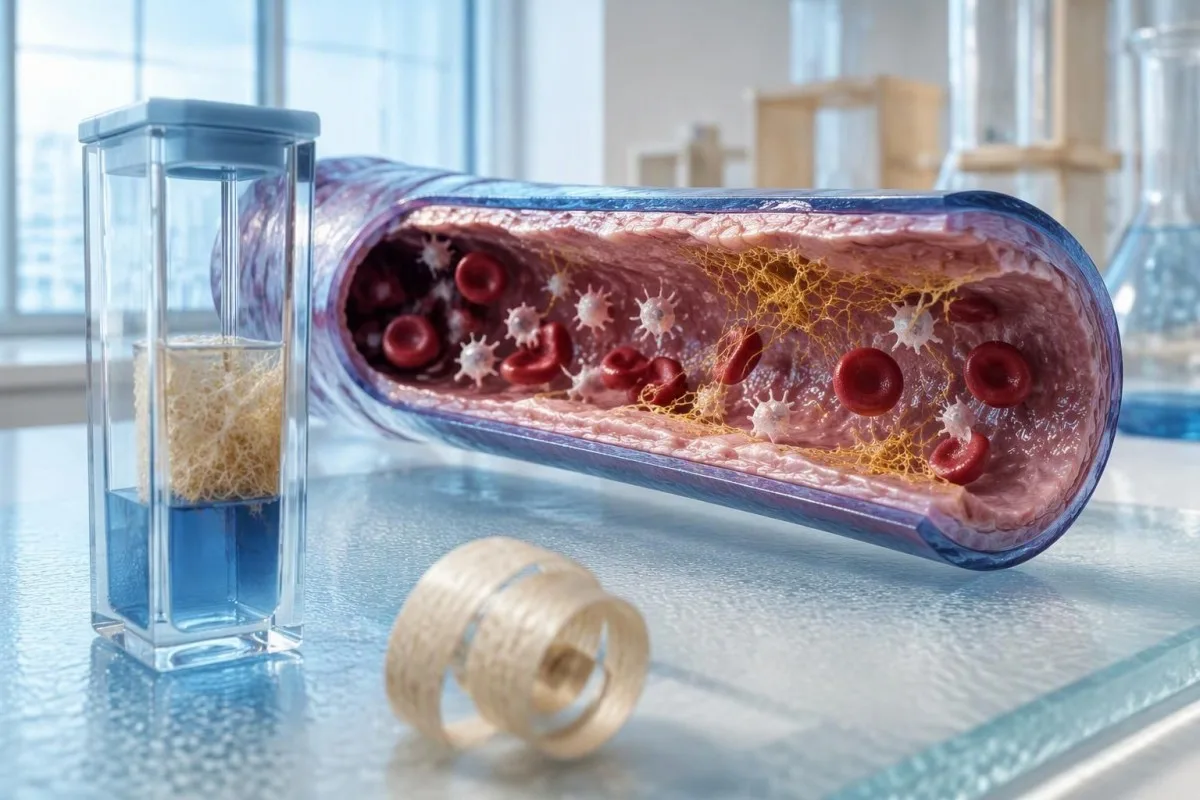
DIC is recognized by a changing combination of results—not one red flag. A normal fibrinogen result can coexist with a dangerous coagulation disorder.


- Combined trends matter most: falling platelets, increasing PT, elevated D-dimer, and falling fibrinogen support DIC in an appropriate clinical setting.
- Platelet count below 100 × 10^9/L contributes to the classic ISTH overt-DIC score, but a rapid decline above that threshold can still matter.
- Fibrinogen commonly measures about 2–4 g/L in adults; normal or elevated values do not exclude early DIC because inflammation increases production.
- Unit conversion matters: fibrinogen 1 g/L equals 100 mg/dL, and 2 g/L equals 200 mg/dL.
- Classic ISTH scoring uses platelets, fibrin-related markers, PT prolongation, and fibrinogen; a total of at least 5 supports overt DIC when an associated disorder is present.
- D-dimer has no universal DIC diagnostic cutoff; assay units and the laboratory's definition of a moderate or strong increase must be checked.
- Normal aPTT does not rule out DIC; increased factor VIII can partly conceal clotting-factor consumption.
- Emergency symptoms include uncontrolled bleeding, sudden breathlessness, stroke-like symptoms, collapse, or a suddenly cold, painful limb—do not wait for an app interpretation.
What combination of laboratory findings suggests DIC?
Disseminated intravascular coagulation labs typically show falling platelets, prolonged prothrombin time, elevated D-dimer, and falling fibrinogen in someone with a triggering illness. No single result diagnoses DIC, and initially normal fibrinogen does not exclude it; uncontrolled bleeding, sudden breathlessness, confusion, or a cold, painful limb requires emergency assessment.
DIC combines widespread coagulation activation with consumption of platelets and clotting proteins, so thrombosis and bleeding can occur together. The initial assessment usually brings together 4 core measurements—platelet count, PT, a fibrin-related marker, and fibrinogen—rather than treating each abnormal flag as a separate diagnosis.
A fall from 240 to 125 × 10^9/L represents an approximately 48% platelet decline, even though the later value is not profoundly low. That change carries more weight if PT is lengthening and D-dimer is rising; an isolated count of 125 has a much broader differential diagnosis.
Kantesti is an AI blood test analyzer that can help explain why these 4 measurements belong together, but it cannot establish DIC or assess circulation at the bedside. Our organization and clinical scope explain the distinction between educational interpretation and direct medical care.
As of October 13, 2026, this article distinguishes the classic 2001 ISTH scoring system from sepsis-focused assessment rather than presenting a single universal rule. Our coagulation panel limitations guide explains why apparently reassuring screening results cannot replace examination, treatment history, and repeat testing.
Why does the platelet trend matter more than one count?
Platelet consumption can produce a rapid count decline before severe thrombocytopenia develops. Adult platelet reference intervals commonly approximate 150–400 × 10^9/L, but the starting count, rate of change, and accompanying coagulation results determine whether a falling value suggests DIC.
A count of 85 × 10^9/L is abnormal, but it does not identify the cause: sepsis, medications, immune destruction, marrow disease, and dilution can all lower platelets. When I review a panel, I would ask whether yesterday's count was 95 or 250—those are very different trajectories.
A declining platelet count with rising PT suggests consumption more strongly than platelet decline alone, although liver dysfunction can generate a similar combination. Plateletcrit and MPV are not substitutes for platelet count in the classic 4-component DIC score; our plateletcrit interpretation guide separates these CBC measurements.
The percentage change can expose a misleadingly ordinary-looking number: a fall from 360 to 180 × 10^9/L is a 50% reduction despite remaining within many reference intervals. That decline deserves explanation in an acutely ill patient, but no percentage threshold by itself establishes DIC.
Recent transfusion can temporarily raise the count without resolving the underlying consumption, and large-volume fluids can lower it through dilution. Record the time of the 2 blood collections and any intervening platelet transfusion; otherwise, a superficially improved result may be mistaken for recovery from the coagulation disorder.
What do PT, INR, and aPTT reveal in suspected DIC?
PT prolongation supports DIC when it accompanies platelet consumption and increased fibrin-related markers. A normal aPTT does not exclude DIC, because increased factor VIII during an acute-phase response can offset the effect of consuming other clotting factors.
PT is often approximately 11–14 seconds and aPTT approximately 25–35 seconds, but reagent-specific reference intervals take priority. For classic ISTH scoring, the relevant measurement is PT prolongation above the laboratory's control or reference, not simply whether the printed result exceeds 14 seconds.
A PT of 17 seconds may represent 4 seconds of prolongation if the laboratory's comparison value is 13 seconds. That would contribute 1 point in the classic score; the same raw PT cannot be interpreted identically across laboratories using different reagents and comparison values.
INR standardizes PT chiefly for vitamin K antagonist monitoring; it is not a universal measure of DIC severity or bleeding risk. Our INR result explanation helps distinguish an INR of 1.5 caused by anticoagulation from a similar result during acute illness.
Heparin contamination, direct oral anticoagulants, vitamin K deficiency, and liver dysfunction can alter these tests without DIC. The 2009 British guideline emphasizes integrating coagulation results with clinical findings and repeating assessment as the condition evolves (Levi et al., 2009); our aPTT and clotting guide explains the additional assay limitations.
How should elevated D-dimer and fibrin degradation products be read?
D-dimer elevation indicates breakdown of cross-linked fibrin, but it does not identify DIC specifically. Clinicians interpret its magnitude and trajectory alongside at least 3 other core measurements: platelets, PT, and fibrinogen, plus the illness capable of triggering systemic coagulation activation.
Many venous-thromboembolism pathways use 500 ng/mL FEU as a conventional D-dimer threshold, but that is not a DIC diagnostic cutoff. Pregnancy, recent procedures, malignancy, inflammation, and an isolated deep vein thrombosis can all produce values above that threshold without disseminated coagulation.
FEU and DDU are different reporting conventions; FEU values are approximately twice DDU values for comparable material, although exact assay relationships vary. A result of 2,000 ng/mL FEU cannot be compared directly with 2,000 ng/mL DDU, and 1 mg/L FEU equals 1,000 ng/mL FEU.
The classic ISTH score assigns 2 points for a moderate increase in fibrin-related markers and 3 for a strong increase. Those categories require the laboratory's assay-specific interpretation; importing a cutoff from a different hospital can change the score without any biological change in the patient.
A decrease from 12 to 8 mg/L FEU may accompany improvement, yet both values can remain markedly elevated after coagulation activation has slowed. D-dimer alone should not determine discharge or treatment cessation; our D-dimer accuracy and units guide explains why positive results are easier to obtain than disease-specific answers.
Why can DIC fibrinogen levels initially remain normal?
DIC fibrinogen levels can remain normal or elevated early because the liver increases fibrinogen production during an acute-phase response while coagulation consumes it. A typical adult interval is approximately 2–4 g/L, so movement within that interval can conceal a clinically meaningful decline.
An illustrative patient with sepsis might have fibrinogen fall from 6.0 to 3.1 g/L over 24 hours. The later value looks reassuring in isolation, but the nearly 48% reduction becomes concerning if platelets also fall and PT lengthens; this example describes clinical reasoning, not a reported patient history.
The classic ISTH score gives only 1 point for fibrinogen below 1 g/L, equivalent to 100 mg/dL. That threshold identifies substantial depletion; it does not mean values of 1.1–2.0 g/L are safe during active bleeding, nor does a result above 1 g/L rule out DIC.
Functional fibrinogen is commonly measured using a Clauss assay, whereas some instruments estimate fibrinogen from PT-based methods. A discrepancy of even 1 g/L can influence interpretation, especially with anticoagulants or unusual fibrinogen function; request the measurement method when the result conflicts with the clinical picture.
Kantesti is an AI blood test interpretation platform that can place fibrinogen 3.1 g/L beside an earlier 6.0 g/L result rather than treating the later value as automatically reassuring. Acute illness also changes other circulating proteins, as our inflammation and transferrin explanation illustrates; protein trends reflect production as well as consumption.
Which additional tests reveal hemolysis and organ injury?
A peripheral smear, hemolysis tests, and organ-function measurements help assess consequences of DIC and identify alternative diagnoses. These tests sit outside the classic 4-component overt-DIC score, but they can reveal organ injury that makes a modest-looking coagulation panel more urgent.
Schistocytes are fragmented red cells produced by mechanical injury and may appear in DIC, thrombotic microangiopathy, or other settings. A schistocyte proportion above 1% raises concern for thrombotic microangiopathy when fragments are the main abnormality; our urgent schistocyte interpretation explains why morphology and context still matter.
LDH elevation, reduced haptoglobin, increasing indirect bilirubin, and falling hemoglobin can support hemolysis, but none distinguishes DIC from TTP alone. A hemoglobin decline from 110 to 90 g/L could also reflect bleeding or dilution, so clinicians examine the entire pattern before assigning a mechanism.
Creatinine changes can detect kidney injury before an estimated filtration category tells the full story. An increase of at least 0.3 mg/dL, approximately 26.5 µmol/L, within 48 hours meets one standard acute-kidney-injury criterion; new oliguria or shock increases concern even before that laboratory threshold is reached.
Lactate above 2 mmol/L can accompany impaired perfusion or sepsis, but it is not specific for DIC and may be influenced by other physiology. Serial lactate, urine output, oxygenation, and liver tests help track organ consequences; a falling DIC score does not cancel worsening confusion or circulation.
How do classic ISTH DIC diagnostic criteria support assessment?
The classic ISTH overt-DIC score supports assessment through 4 laboratory domains, but it is intended for patients with an underlying disorder associated with DIC. A total of at least 5 is compatible with overt DIC; a lower score does not exclude an evolving process.
Taylor and colleagues described the classic framework in 2001 to standardize recognition of overt DIC (Taylor et al., 2001). The score deliberately combines complementary evidence—consumption, delayed clot formation, and fibrin turnover—because an isolated abnormality has too many possible explanations to carry the diagnosis.
Common classic tables assign platelets >100 × 10^9/L 0 points, 50–100 1 point, and <50 2 points. Fibrin-related markers add 0, 2, or 3 points for no, moderate, or strong increase; institutions should follow their own boundary conventions and assay definitions.
PT prolongation below 3 seconds adds 0 points, 3–6 seconds adds 1, and above 6 seconds adds 2; fibrinogen below 1 g/L adds 1 point. In an illustrative panel, platelets 78, strongly increased D-dimer, PT prolonged 4 seconds, and fibrinogen 2.4 g/L total 5 points despite normal fibrinogen.
A score is a structured checkpoint, not permission to ignore new symptoms or a rapidly changing panel. Classic recommendations include daily reassessment for overt DIC and repeat assessment within 1–2 days when suspicion persists below the threshold; our sudden laboratory change guide explains why both biological change and measurement error deserve attention.
Why the score version must be named
The term ISTH DIC score should be accompanied by the version and local implementation; this article describes the classic 2001 overt-DIC framework. Sepsis-specific systems and later proposals may use different inputs, so a score of 5 cannot be interpreted accurately unless the scoring method is identified.
How does sepsis-induced coagulopathy scoring differ from overt DIC?
Sepsis-induced coagulopathy, or SIC, scoring combines platelet count, PT-INR, and selected SOFA organ-dysfunction components. Unlike the classic overt-DIC score, SIC does not require D-dimer or fibrinogen, and it is designed specifically for patients with sepsis rather than every possible DIC trigger.
The SIC framework was proposed in a retrospective nationwide analysis by Iba and colleagues in 2017, not as a universal replacement for all DIC criteria (Iba et al., 2017). A total of at least 4 is required, with platelet-plus-PT-INR points greater than 2.
In SIC scoring, platelets of 100–149 × 10^9/L receive 1 point and values below 100 receive 2 points. PT-INR above 1.2 through 1.4 receives 1 point, while PT-INR above 1.4 receives 2; the scoring context matters because anticoagulant exposure can distort these measurements.
The SIC organ component uses respiratory, cardiovascular, hepatic, and renal SOFA contributions, grouped into 0, 1, or 2 points. SIC positivity identifies a sepsis-associated coagulation problem; it does not prove overt DIC, establish the source of sepsis, or dictate anticoagulant treatment for every patient.
Procalcitonin cannot substitute for clinical sepsis assessment or either coagulation score, even when it rises above 0.5 ng/mL. Our procalcitonin and sepsis trends guide explains why biomarker results need timing and clinical context; urgent treatment should not wait for a second biomarker to become abnormal.
Which illnesses make DIC lab findings more concerning?
Sepsis, major trauma, obstetric emergencies, and certain cancers are important DIC triggers. The same 4 laboratory abnormalities have different diagnostic weight in a critically ill patient with a plausible trigger than in a well person undergoing routine outpatient testing.
Acute promyelocytic leukemia can cause a particularly dangerous coagulation disorder, sometimes with severe fibrinolysis and early bleeding. Abnormal promyelocytes on a smear plus platelets of 35 × 10^9/L and falling fibrinogen demand urgent specialist assessment; neither molecular confirmation nor a completed score should delay escalation.
Pregnancy changes the baseline: fibrinogen is often approximately 4–6 g/L late in pregnancy, although intervals vary. A fall to 2 g/L may therefore represent substantial depletion despite meeting a nonpregnant laboratory's lower limit; obstetric hemorrhage protocols commonly treat that concentration as clinically concerning.
HELLP syndrome and obstetric DIC overlap but are not interchangeable diagnoses; hemolysis, elevated liver enzymes, and low platelets can occur before overt coagulation-factor depletion. Our HELLP emergency laboratory patterns guide explains why pregnancy or recent delivery changes the response to even 1 concerning result.
Chronic or compensated DIC can occur with some malignancies or vascular abnormalities and may produce less dramatic PT or fibrinogen changes than acute DIC. Persistent D-dimer elevation with platelets fluctuating around 100–150 × 10^9/L still requires an explanation, but cancer-related elevation alone should not be labeled disseminated coagulation.
How do clinicians distinguish DIC from TTP, HIT, and liver disease?
DIC, TTP, HIT, and advanced liver disease can share low platelets, but their supporting patterns and urgent treatments differ. Comparing PT, aPTT, fibrinogen, hemolysis, medication timing, and the underlying illness is safer than applying a DIC label to 1 low platelet result.
TTP typically causes thrombocytopenia and microangiopathic hemolysis while routine PT and aPTT are often near normal. ADAMTS13 activity below 10% strongly supports TTP in the appropriate setting; our ADAMTS13 and urgent TTP assessment explains why clinicians may begin treatment before that result returns.
HIT often involves a platelet decline greater than 50% and thrombosis after heparin exposure, commonly within 5–10 days in a previously unexposed patient. A positive PF4 immunoassay does not independently establish HIT; our PF4 confirmation pathway separates antibody detection from clinical probability and functional confirmation.
Advanced liver disease may produce prolonged PT, reduced platelets, elevated D-dimer, and reduced fibrinogen without acute DIC. A longstanding platelet count near 80 × 10^9/L differs from a new fall to 80 during shock; our cirrhosis laboratory clues explains that baseline comparison.
Factor VIII is not a dependable single-test discriminator between liver disease and DIC because it can be elevated in both settings. When I review an ambiguous panel, I would prioritize the preceding 24–48 hours, medication exposure, smear findings, and organ deterioration rather than promise that 1 additional factor assay will settle it.
Which sample problems can mimic or conceal a DIC pattern?
Collection errors, platelet clumping, anticoagulant contamination, and assay interference can generate misleading DIC lab findings. A suspicious result should be checked against the specimen's quality and previous measurements, while clinical instability still receives immediate assessment rather than waiting for a redraw.
Standard citrate coagulation collection aims for approximately 9 parts specimen to 1 part citrate; underfilling leaves excess citrate relative to plasma and can falsely prolong clotting times. With hematocrit above 55%, laboratories generally adjust citrate volume because the reduced plasma fraction creates a similar imbalance.
EDTA-associated platelet clumping can produce a falsely low automated count, sometimes as low as 30 × 10^9/L despite adequate circulating platelets. Our EDTA platelet clumping explanation describes how smear review and a laboratory-directed alternative specimen help confirm pseudothrombocytopenia rather than genuine consumption.
Sampling through a heparin-exposed line can prolong aPTT disproportionately, creating an apparent coagulation abnormality that does not match the patient. A new aPTT of 90 seconds with unchanged platelets and fibrinogen should prompt a contamination check, although anticoagulant effect and real illness can coexist.
Optical clot detection can be affected by marked lipemia, hemolysis, or bilirubin, depending on the instrument and assay. Before interpreting a change from fibrinogen 3.5 to 1.2 g/L, ask whether either report carries an interference warning; repeat or alternative testing may clarify the result without delaying necessary clinical treatment.
Which bleeding or clotting symptoms require emergency care?
Uncontrolled bleeding, sudden breathlessness, stroke-like symptoms, collapse, or a suddenly cold and painful limb require emergency care. These symptoms can indicate severe bleeding, thrombosis, shock, or another emergency even when the latest fibrinogen is above 2 g/L or the DIC score is below 5.
Call your local emergency number for vomiting blood, coughing up a substantial amount of blood, heavy bleeding with faintness, or black tarry stool accompanied by weakness or collapse. Our stool bleeding warning signs explains the distinction between minor color changes and potentially serious bleeding; do not wait 24 hours for repeat laboratories.
Sudden chest pain with breathlessness, new weakness on 1 side, difficulty speaking, or sudden loss of vision warrants immediate emergency assessment. A suddenly cold, pale, painful hand or foot is also time-sensitive; these presentations require evaluation for clotting emergencies, but symptoms alone cannot identify DIC.
Rapidly spreading purpura, persistent oozing from several sites, confusion, markedly reduced urine output, or worsening mottled extremities during severe illness needs urgent in-person assessment. Visible bleeding is not required: microvascular thrombosis can injure organs while the platelet count remains above 100 × 10^9/L.
If a laboratory or clinician contacts you about critical coagulation results, follow their instructions immediately even if you feel relatively well. Bring your medication list and the last 2 available laboratory reports if doing so causes no delay; do not drive yourself when faint, confused, breathless, or experiencing stroke-like symptoms.
How do laboratory trends guide treatment and repeat testing?
DIC treatment addresses the underlying illness while supporting circulation and managing bleeding or thrombosis. Serial platelets, PT, fibrinogen, and clinical examination guide reassessment; a laboratory threshold such as platelets below 50 × 10^9/L informs decisions but does not automatically prescribe transfusion.
The British guideline advises considering platelet transfusion in DIC with bleeding or high bleeding risk when platelets are below 50 × 10^9/L (Levi et al., 2009). In a stable, nonbleeding patient, correcting an abnormal number alone is generally not the goal; the procedural plan and overall risk change the decision.
Severe fibrinogen depletion below 1 g/L that persists despite plasma may warrant cryoprecipitate or fibrinogen concentrate under specialist protocols. Many active major-bleeding protocols use higher targets, commonly at least 1.5 g/L and often 2 g/L in obstetric hemorrhage; these are treatment-context targets, not interchangeable DIC diagnostic criteria.
Repeat testing may occur every 4–6 hours during unstable active bleeding, whereas stable patients may be assessed less frequently according to the treating team. Anticoagulation decisions depend on whether thrombosis predominates, bleeding risk, and the trigger; neither a positive SIC score nor D-dimer elevation automatically mandates therapeutic anticoagulation.
Our AI interpretation methodology explains the intended role of computerized report interpretation, not continuous hospital monitoring. Kantesti's clinical standards and limitations should be read with that boundary in mind: no claimed accuracy percentage can replace examination or establish safety during the next 4 hours.
After discharge: monitoring and related research publications
After DIC, follow-up tracks recovery of platelets, coagulation tests, and affected organs rather than demanding immediate normalization of every marker. A D-dimer still above 500 ng/mL FEU can persist during recovery; new symptoms or reversal of an improving trend deserves clinical reassessment.
A useful handover lists the trigger, lowest platelet count, lowest fibrinogen, peak PT, transfusions, anticoagulants, and the next planned review. Recording that platelets improved from 42 to 118 × 10^9/L communicates more than writing 'still low'; the responsible team should also specify which symptoms require immediate return.
Kantesti is an AI-powered blood test analysis tool that can support explanation of dated follow-up reports after the urgent clinical phase has passed. I write as Thomas Klein, Chief Medical Officer, to emphasize a practical boundary: comparing 2 reports cannot determine whether a new symptom is safe, and educational scenarios here are not verified patient histories.
Thomas Klein's editorial approach in this article separates diagnostic evidence from related background reading, and our medical advisory board describes the clinical expertise behind the service. The classic score's 4 laboratory domains are supported by the external DIC publications listed below; the 2 supplied Zenodo guides are related educational resources, not DIC-validation studies.
The companion serum protein interpretation guide addresses protein context beyond fibrinogen. The complement and ANA guide covers a separate immune-testing framework; its presence here does not make C3, C4, or ANA part of the classic 4-component DIC score.
Formal citations for the 2 supplied Zenodo resources appear in the publication records below; author and publication-year metadata were not supplied, so the entries use title-first APA formatting and 'n.d.' rather than inventing details. ResearchGate and Academia.edu links are discovery searches, not claims that an authenticated copy is hosted on either service.
Frequently Asked Questions
What laboratory findings are most typical of DIC?
Typical DIC lab findings are falling platelets, prolonged PT, elevated D-dimer or other fibrin-related markers, and falling fibrinogen. These 4 measurements must be interpreted together in someone with an illness capable of triggering DIC. Platelets below 100 × 10^9/L and progressively prolonged PT increase concern, but neither result establishes the diagnosis alone. Initially normal fibrinogen does not exclude DIC.
Can you have DIC with normal fibrinogen?
Yes, DIC can occur with normal or elevated fibrinogen because inflammation increases fibrinogen production while coagulation consumes it. A decline from 6.0 to 3.0 g/L may therefore be clinically concerning even though 3.0 g/L falls within a common adult reference interval. Clinicians compare the trend with platelets, PT, D-dimer, and symptoms. Fibrinogen below 1 g/L contributes to the classic overt-DIC score, but that threshold is not a rule-out boundary.
What ISTH score is considered positive for overt DIC?
A classic ISTH overt-DIC score of at least 5 is compatible with overt DIC when an underlying disorder associated with DIC is present. The score combines platelet count, fibrin-related markers, PT prolongation, and fibrinogen. A result below 5 does not exclude evolving DIC and may require repeat assessment. The score version and laboratory definitions must be identified because different frameworks are not interchangeable.
What D-dimer level diagnoses DIC?
No single D-dimer concentration diagnoses DIC. The commonly used 500 ng/mL FEU threshold belongs to many venous-thromboembolism testing pathways, not a universal DIC diagnostic rule. Classic DIC scoring uses laboratory-defined moderate or strong increases alongside other coagulation findings. FEU and DDU values are not directly interchangeable, and FEU values are approximately twice DDU values for comparable material.
Can PT or aPTT be normal in early DIC?
PT or aPTT can be normal early in DIC, so normal screening times do not independently exclude the condition. Increased factor VIII can partly conceal consumption-related aPTT prolongation. A platelet decline from 240 to 125 × 10^9/L with rising D-dimer may warrant reassessment even before clotting times become clearly abnormal. Clinical deterioration requires urgent assessment rather than waiting for every laboratory measurement to change.
How often should DIC blood tests be repeated?
Repeat-testing frequency depends on clinical stability, active bleeding, treatment, and the underlying illness. During unstable major bleeding, clinicians may repeat key measurements every 4–6 hours or more frequently according to the local protocol. The classic ISTH framework recommends daily reassessment for overt DIC and repeat assessment within 1–2 days when the score is below the overt-DIC threshold but suspicion persists. Patients should follow the treating team's schedule rather than arrange routine outpatient testing for suspected acute DIC.
Which symptoms of DIC mean I should seek emergency help?
Uncontrolled bleeding, sudden breathlessness, collapse, stroke-like symptoms, or a suddenly cold and painful limb require emergency assessment. Rapidly spreading purpura, confusion, and markedly reduced urine output during severe illness are also urgent warning signs. These symptoms can be dangerous even with fibrinogen above 2 g/L or a DIC score below 5. Call your local emergency number and do not delay care to upload laboratory results.
Get AI-Powered Blood Test Analysis Today
Join over 2 million users worldwide who trust Kantesti for instant, accurate lab test analysis. Upload your blood test results and receive comprehensive interpretation of 15,000+ biomarkers in seconds.
📚 Referenced Research Publications
Klein, T., Mitchell, S., & Weber, H. (2026). Serum Proteins Guide: Globulins, Albumin & A/G Ratio Blood Test. Kantesti AI Medical Research.
Klein, T., Mitchell, S., & Weber, H. (2026). C3 C4 Complement Blood Test & ANA Titer Guide. Kantesti AI Medical Research.
📖 External Medical References
Taylor FB Jr et al. (2001). Towards definition, clinical and laboratory criteria, and a scoring system for disseminated intravascular coagulation. Thrombosis and Haemostasis.
Iba T et al. (2017). New criteria for sepsis-induced coagulopathy (SIC) following the revised sepsis definition: a retrospective analysis of a nationwide survey. BMJ Open.
⚕️ Medical Disclaimer
This article is for educational purposes only and does not constitute medical advice. Always consult a qualified healthcare provider for diagnosis and treatment decisions.
E-E-A-T Trust Signals
Experience
Physician-led clinical review of lab interpretation workflows.
Expertise
Laboratory medicine focus on how biomarkers behave in clinical context.
Authoritativeness
Written by Dr. Thomas Klein with review by Dr. Sarah Mitchell and Prof. Dr. Hans Weber.
Trustworthiness
Evidence-based interpretation with clear follow-up pathways to reduce alarm.