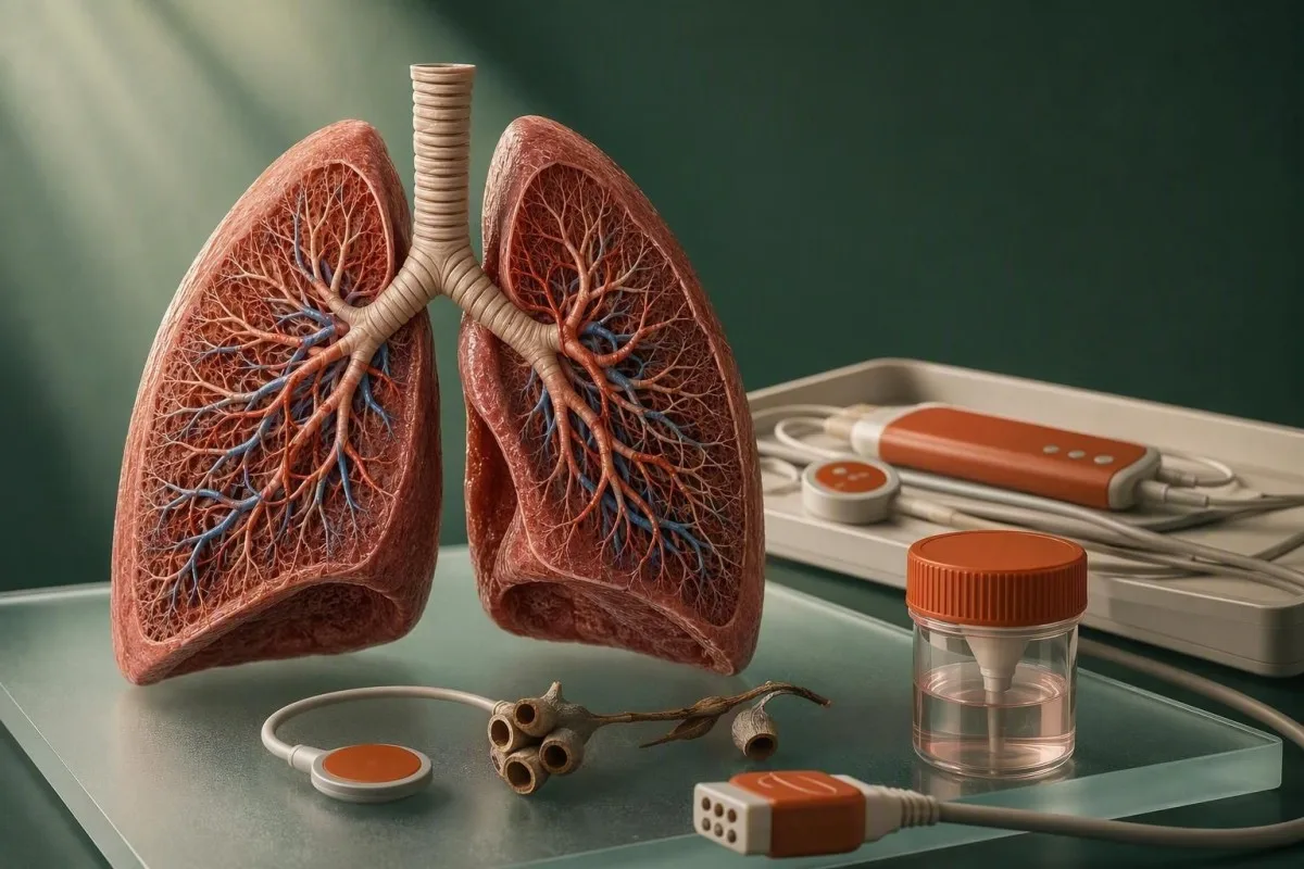
A blood test for shortness of breath can identify anemia, heart strain, infection, clot risk, abnormal glucose, kidney dysfunction, or acid-base disturbance—but normal results cannot exclude pulmonary embolism, asthma, pneumothorax, or a heart emergency. Sudden breathlessness at rest, chest pressure, fainting, blue-grey lips, confusion, coughing blood, or oxygen saturation below 90% needs emergency assessment rather than waiting on laboratory results.


- Emergency symptoms include breathlessness at rest, fainting, chest pressure, coughing blood, confusion, or an oxygen reading below 90%; blood work should not delay emergency care.
- Hemoglobin below 12.0 g/dL in most non-pregnant adult women or below 13.0 g/dL in most adult men meets WHO anemia criteria and can reduce oxygen delivery.
- Ferritin below 15 ng/mL strongly supports depleted iron stores; ferritin below 30 ng/mL is often clinically useful when symptoms and CBC findings fit.
- BNP below 100 pg/mL or NT-proBNP below 300 pg/mL makes acute heart failure less likely in many emergency presentations, not impossible.
- High-sensitivity troponin above the assay's 99th percentile signals myocardial injury but does not alone diagnose a heart attack.
- D-dimer below an age-adjusted threshold can help exclude pulmonary embolism only when pre-test probability is low or intermediate.
- Lactate of 2.0 mmol/L or higher during suspected infection or shock warrants prompt clinical reassessment and repeat measurement.
- Bicarbonate below 18 mmol/L with high glucose or ketones may indicate diabetic ketoacidosis, which can cause deep, rapid breathing.
- Normal blood work does not rule out asthma, pulmonary embolism, collapsed lung, airway obstruction, or early pneumonia.
When shortness of breath needs urgent care before blood work
Breathlessness with chest pain, fainting, confusion, blue-grey lips, coughing blood, or inability to speak full sentences needs emergency assessment now, not a routine blood test. In my clinical practice, the patient who says, “I feel fine sitting still but cannot walk across the room,” often needs a pulse oximeter, ECG, examination, and sometimes imaging before any lab result is back.
For most adults, an oxygen saturation below 90% is an emergency threshold; 90% to 92% also deserves same-day evaluation unless a clinician has given you a different target for chronic lung disease. A normal oxygen reading does not completely reassure me when there is sudden pleuritic chest pain, rapid breathing, or a new one-sided swollen calf, because pulmonary embolism can initially preserve saturation at rest.
The time course changes the decision. Breathlessness developing over minutes to hours after surgery, a long flight, childbirth, an allergic exposure, or stimulant use carries a different risk profile from gradually worsening exertional breathlessness over 3 months. Dr. Thomas Klein’s practical rule is simple: a dangerous symptom pattern outweighs a reassuring-looking CBC.
A laboratory panel is most helpful once the immediate danger has been assessed. If you are deciding whether a potassium, sodium, or bicarbonate result needs urgent care, our guide to urgent electrolyte patterns explains why a number must always be read alongside symptoms and an ECG.
Which initial blood tests help explain breathlessness?
A useful shortness of breath blood work panel usually includes a CBC, electrolytes and kidney function, glucose, and targeted tests such as BNP, troponin, D-dimer, CRP, or blood gas according to the clinical story. No single “breathlessness panel” fits everyone; the safest test selection follows risk, onset, examination findings, and oxygen level.
The complete blood count (CBC) measures hemoglobin, hematocrit, red-cell indices, white cells, and platelets; it is the fastest laboratory screen for anemia and some infectious patterns. A typical adult white-cell reference interval is about 4.0 to 11.0 × 10⁹/L, though the printed range from your own laboratory takes priority. See exactly what a CBC includes before interpreting an isolated flag.
Kantesti AI reads CBC, chemistry, and cardiac-marker patterns together, then highlights result combinations that merit clinical review rather than assigning a diagnosis from one number. Our biomarker reference guide is useful for checking units, which vary notably between UK, European, and US reports.
For a 68-year-old with ankle swelling and breathlessness when lying flat, BNP or NT-proBNP is usually more informative than D-dimer. For a 28-year-old with sudden sharp chest pain after a long journey, clot-risk assessment and imaging decisions may take precedence. The order form should reflect the question being asked.
Anemia and shortness of breath: CBC and iron results that matter
Anemia can cause exertional shortness of breath when hemoglobin falls enough to limit oxygen delivery, even if the lungs and heart are structurally normal. WHO cutoffs define anemia as hemoglobin below 12.0 g/dL in most non-pregnant adult women and below 13.0 g/dL in most adult men; symptoms depend greatly on how quickly the value fell.
A hemoglobin of 8.5 g/dL that has developed slowly may cause breathlessness climbing stairs, palpitations, and fatigue, whereas an abrupt fall from 14.0 to 10.5 g/dL can feel much more severe. MCV below 80 fL suggests microcytosis in adults, but early iron deficiency can still have a normal MCV. Low hemoglobin plus a rising RDW often appears before the red cells become clearly small.
Ferritin below 15 ng/mL is highly specific for iron deficiency in otherwise well adults, while ferritin below 30 ng/mL is a practical threshold many clinicians use in symptomatic patients. Ferritin rises with infection and chronic inflammation, so a ferritin below 100 ng/mL with transferrin saturation below 20% can still support iron-restricted erythropoiesis in inflammatory disease. Our detailed iron studies guide explains this often-missed distinction.
Kantesti is an AI blood test analyzer that flags the hemoglobin-ferritin-transferrin saturation pattern rather than treating a “normal” ferritin as a full stop. Before starting iron, ask why it is low—heavy menstrual loss, blood donation, dietary restriction, malabsorption, or gastrointestinal loss each change the follow-up plan. Food can help over time; our explanation of heme and non-heme iron covers absorption rather than promising a quick fix.
A low reticulocyte count in anemia suggests the marrow is not responding adequately, while a high count can point toward recovery, blood loss, or red-cell breakdown. Black stools, heavy bleeding, pregnancy, new dizziness, or hemoglobin near 7 g/dL require prompt clinician-led assessment; transfusion decisions depend on stability and cause, not a web threshold alone.
BNP test for breathlessness and signs of heart strain
BNP and NT-proBNP rise when the heart muscle is stretched by pressure or fluid volume, making them useful tests when breathlessness could be heart failure. In acute care, BNP below 100 pg/mL or NT-proBNP below 300 pg/mL makes acute heart failure less likely in many patients, although neither result replaces examination or echocardiography.
For non-acute outpatient assessment, the 2021 ESC heart-failure guideline uses BNP below 35 pg/mL or NT-proBNP below 125 pg/mL as thresholds that make chronic heart failure unlikely (McDonagh et al., 2021). Values increase with age, atrial fibrillation, reduced kidney function, pulmonary embolism, and sepsis; obesity can produce a misleadingly low BNP or NT-proBNP.
A BNP of 450 pg/mL in a person with new leg swelling, crackles on examination, and worsening orthopnea is far more meaningful than the same value after rapid atrial fibrillation or advanced kidney disease. I have also seen a patient with obesity and a BNP of 70 pg/mL who still had fluid overload—context wins. For a deeper explanation, read our guide to high NT-proBNP results.
Kantesti AI interprets BNP test for breathlessness results alongside creatinine, hemoglobin, age, and prior values so that a change from 180 to 1,400 pg/mL is not buried in a report. A raised peptide is a follow-up trigger, not proof that breathlessness is caused by heart failure; an ECG, chest imaging, and echocardiogram often answer the next question.
Troponin results: when breathlessness may reflect cardiac injury
High-sensitivity troponin above the laboratory’s 99th-percentile upper reference limit indicates myocardial injury, not automatically a heart attack. Shortness of breath can be the main symptom of acute coronary syndrome, particularly in older adults, people with diabetes, and some women who do not report classic crushing chest pain.
The number alone is less useful than the rise or fall over 1 to 3 hours, the ECG, symptoms, and the assay used. A stable high-sensitivity troponin T of 35 ng/L in chronic kidney disease has a different meaning from a rise from 8 to 62 ng/L during new breathlessness. Laboratories use assay-specific cutoffs, so never compare an hs-cTnI value directly with an hs-cTnT value.
Myocardial injury also occurs with pulmonary embolism, severe anemia, fast arrhythmia, myocarditis, sepsis, hypertensive crisis, and heart failure. The practical concern is that these are not benign alternatives. Our comparison of troponin I and troponin T explains why reference limits and serial testing differ.
New breathlessness with a raised or rising troponin needs same-day clinical assessment, particularly when paired with chest discomfort, sweating, nausea, or abnormal ECG findings. A normal first troponin drawn very early after symptoms begin may need repeat testing; this is why emergency departments use structured serial protocols rather than one isolated result.
D-dimer and pulmonary embolism: what the blood test can and cannot tell you
D-dimer is best used to rule out pulmonary embolism in people with low or intermediate clinical probability; a positive result does not diagnose a clot. For adults older than 50, an age-adjusted D-dimer threshold of age × 10 micrograms/L FEU can reduce unnecessary scans when the assay reports in FEU units.
A 72-year-old may have an age-adjusted rule-out threshold of 720 micrograms/L FEU, rather than the conventional 500 micrograms/L FEU, if pre-test probability is not high. The 2019 ESC pulmonary embolism guideline supports age-adjusted cutoffs in suitable patients (Konstantinides et al., 2020). Units matter: 500 ng/mL FEU equals 500 micrograms/L FEU, but DDU values are commonly about half the FEU value.
D-dimer rises after surgery, trauma, pregnancy, cancer, hospital admission, infection, liver disease, and simply with age. A value of 2,000 micrograms/L FEU may increase concern, but it cannot locate a clot; CT pulmonary angiography or another imaging pathway makes the diagnosis. Read our careful review of D-dimer accuracy before assuming an elevated result means thrombosis.
Do not wait for D-dimer if clinical probability is high—such as sudden unexplained breathlessness plus fainting, low oxygen, fast pulse, or signs of deep-vein thrombosis. In that setting, clinicians generally arrange urgent imaging and use blood work as supporting evidence. A negative D-dimer is not a safety net for a high-risk presentation.
table
subsections
extra
Why pre-test probability comes first
A patient with a clearly provoked, low-risk symptom pattern may safely avoid imaging after a negative validated D-dimer. A patient with high-risk features may need imaging despite a normal result because no blood test has zero false negatives.
Infection, pneumonia, and sepsis clues in blood work
White-cell count, CRP, procalcitonin, and lactate can support the assessment of infection-related breathlessness, but none can confirm or exclude pneumonia on its own. Chest imaging, oxygen saturation, respiratory rate, and examination remain central because pneumonia can occur with a normal white-cell count.
A white-cell count above 11.0 × 10⁹/L with neutrophilia may support bacterial infection, yet steroids, stress, smoking, and dehydration can also raise it. Conversely, a low white-cell count during acute illness can be concerning when paired with fever or low blood pressure. CRP is nonspecific; a CRP above 100 mg/L increases the likelihood of substantial inflammation but cannot tell us whether the source is lung, urinary tract, autoimmune disease, or something else.
Lactate of 2.0 mmol/L or higher during suspected infection identifies patients who need urgent reassessment of perfusion and repeat testing. The Surviving Sepsis Campaign recommends measuring lactate in suspected sepsis and using it within a broader resuscitation assessment, not as a stand-alone diagnosis (Evans et al., 2021). Our sepsis marker guide covers why normal blood pressure can still coexist with serious illness.
Procalcitonin below 0.25 ng/mL can make significant bacterial infection less likely in selected settings, but it should not be used to withhold initial treatment from a clinically unstable person. Viral infections, early bacterial infection, localized pneumonia, and kidney dysfunction muddy the picture. If breathlessness follows fever and worsening cough, a same-day review is sensible even with “only mildly raised” markers.
Glucose, electrolytes, and acid-base causes of rapid breathing
High glucose with ketones, low bicarbonate, kidney dysfunction, or major electrolyte disturbance can cause rapid or labored breathing even when the lungs are not the primary problem. A serum bicarbonate below 18 mmol/L with elevated glucose and ketones raises concern for diabetic ketoacidosis and needs urgent assessment.
The body increases ventilation to lower carbon dioxide when acid accumulates. In diabetic ketoacidosis, glucose is often above 250 mg/dL (13.9 mmol/L), bicarbonate is below 18 mmol/L, and the anion gap is raised—yet glucose can be lower with SGLT2 medicines. Deep, regular breathing is a compensatory response, not anxiety by default.
Potassium below 3.0 mmol/L or above 6.0 mmol/L can impair heart rhythm and muscle function; symptoms may include weakness, palpitations, or breathlessness. Low phosphate below 0.8 mmol/L (2.5 mg/dL) can weaken respiratory muscles in severe cases, particularly after malnutrition, alcoholism, or refeeding. The basic metabolic panel is often more informative when sodium, chloride, bicarbonate, glucose, urea, and creatinine are read as one pattern.
Kantesti is an AI blood test interpretation platform that maps bicarbonate, anion-gap components, glucose, and renal markers together, then directs urgent-pattern results to clinical care. A venous blood gas usually gives a useful pH and bicarbonate estimate; an arterial gas is more useful when oxygenation or carbon-dioxide retention is the immediate question.
Kidney function, albumin, and fluid balance markers
Reduced kidney function and low albumin can contribute to fluid retention and breathlessness, but neither marker can diagnose fluid in the lungs by itself. Creatinine must be interpreted with eGFR, body size, muscle mass, medication use, urine findings, blood pressure, and the direction of change.
An eGFR below 60 mL/min/1.73 m² for at least 3 months supports chronic kidney disease, while an abrupt creatinine rise can signal acute kidney injury. Creatinine may look deceptively normal in frail or low-muscle adults; cystatin C can occasionally clarify kidney function when the clinical picture and creatinine disagree. Our chronic kidney disease guide explains why one eGFR result does not establish a stage by itself.
Albumin below 30 g/L (3.0 g/dL) can reduce oncotic pressure and contribute to swelling, but liver disease, nephrotic-range protein loss, malnutrition, and inflammation require different investigations. I worry most when low albumin appears with new edema, reduced urine output, rising creatinine, and increasing breathlessness—those clues together point toward a fluid problem.
Urea can rise with dehydration, gastrointestinal bleeding, steroid treatment, and kidney impairment, so the urea-to-creatinine relationship is only a clue. A person taking high-dose diuretics may have breathlessness from heart failure while also appearing biochemically dehydrated. That seeming contradiction is common enough to fool a simplistic interpretation.
Thyroid and less obvious hormonal causes of breathlessness
Thyroid dysfunction can worsen breathlessness through heart-rate changes, anemia, muscle weakness, or fluid retention, but TSH is rarely the first emergency test for sudden symptoms. Overt hyperthyroidism usually shows suppressed TSH with elevated free T4 and/or free T3, while primary hypothyroidism generally shows raised TSH with low free T4.
A TSH below 0.1 mIU/L with high free T4 may contribute to atrial fibrillation, tremor, weight loss, and exertional breathlessness. A TSH above 10 mIU/L with low free T4 supports overt hypothyroidism, which can be associated with fatigue, anemia, pleural fluid, or reduced exercise capacity. These are associations, not shortcuts around examining the heart and lungs.
Biotin supplements can interfere with some thyroid immunoassays and falsely lower TSH or raise free T4 results. Stopping high-dose biotin for at least 48 hours before testing is often advised, although the required interval varies by dose and assay. Our explanation of thyroid test abbreviations helps patients check which tests were actually ordered.
Other selective tests include cortisol for suspected adrenal crisis, creatine kinase for inflammatory muscle disease, and pregnancy testing where clinically appropriate. These should answer a specific question. Ordering every hormone available can generate false alarms without explaining why someone is breathless.
How medicines, exercise, pregnancy, and timing alter results
Recent exercise, pregnancy, altitude exposure, medicines, and the timing of symptoms can shift breathlessness-related blood tests enough to change interpretation. The most useful laboratory report includes what happened in the previous 48 hours, not merely the flagged values.
Strenuous endurance exercise can transiently raise troponin, BNP, creatinine, white cells, and creatine kinase; a marathon finisher may have a troponin result above the reference limit without coronary occlusion. That does not make chest symptoms safe to ignore. The speed of biomarker normalization, ECG, symptoms, and cardiovascular risk determine what happens next.
Beta-blockers can blunt the expected fast heart rate in anemia, sepsis, or pulmonary embolism. Diuretics can lower sodium and potassium while improving congestion; inhaled beta-agonists may lower potassium and raise lactate slightly. Keep a precise medication and supplement list, especially if your result changed after a new blood-test timeline.
Pregnancy lowers hemoglobin through plasma-volume expansion and raises D-dimer progressively, making non-pregnant reference assumptions unsafe. Breathlessness is common in pregnancy, but sudden onset, chest pain, fainting, calf swelling, or oxygen saturation decline still requires urgent obstetric or emergency evaluation.
Why normal blood tests cannot rule out a lung emergency
Normal blood work cannot rule out asthma, early pneumonia, pulmonary embolism in a high-risk patient, pneumothorax, airway obstruction, or many rhythm disorders. Blood tests measure indirect consequences; they do not show lung expansion, airway narrowing, a clot’s location, or an abnormal electrical rhythm.
A pulmonary embolism can produce normal CBC, CRP, troponin, BNP, and even D-dimer under the wrong circumstances, especially if symptoms are prolonged, probability is high, or anticoagulation has already begun. A chest X-ray can miss some emboli; CT pulmonary angiography or ventilation-perfusion imaging answers a different question. This is why clinicians begin with probability, not a blanket lab panel.
Asthma may have completely normal blood results between attacks. Spirometry, peak-flow variation, response to bronchodilator, wheeze, triggers, and nocturnal symptoms carry more diagnostic value; eosinophils can support a phenotype but do not establish asthma. In my experience, patients are often surprised that a “normal blood test” says almost nothing about airway caliber.
Kantesti’s clinical validation approach is built around this limitation: an AI interpretation should identify laboratory patterns and safety prompts, never substitute for emergency examination, imaging, or a clinician’s judgment. If your symptom is new, escalating, or severe, the correct next test may not be another blood draw.
How to read breathlessness blood work as a pattern
The most informative shortness of breath blood work patterns combine values, symptoms, and change over time rather than treating one red flag as a diagnosis. Hemoglobin 9.2 g/dL with ferritin 7 ng/mL points in a different direction from hemoglobin 13.5 g/dL with NT-proBNP 1,800 pg/mL and rising creatinine.
Look for concordance. Low hemoglobin, low MCV, high RDW, ferritin below 15 ng/mL, and low transferrin saturation form a coherent iron-deficiency pattern; any one result alone is weaker evidence. Similarly, high BNP, worsening kidney function, low sodium, weight gain, and edema make congestion more plausible than BNP alone.
A delta can matter even inside the reference interval. Hemoglobin falling from 15.0 to 12.2 g/dL in 6 weeks deserves more attention than a stable 12.1 g/dL when symptoms are new, particularly if there is bleeding risk. Use our guide to meaningful lab changes to separate normal biological variation from a clinically relevant shift.
Kantesti is an AI-powered blood test analysis tool that compares prior laboratory values, units, and related markers in a structured report. The way our interpretation technology works matters: it is designed to surface patterns and questions for your clinician, not to declare that a particular pattern is a confirmed disease.
What to check before repeating a breathlessness blood test
Before repeating blood work for breathlessness, verify the exact test, units, collection time, current medicines, recent exertion, and whether the sample was hemolyzed or delayed. A poor-quality specimen can falsely raise potassium, LDH, and sometimes other analytes, creating an alarming but avoidable result.
Potassium above 6.0 mmol/L requires urgent attention unless the laboratory quickly confirms hemolysis or a repeat sample proves it was spurious. Do not dismiss it yourself: true hyperkalemia can cause dangerous rhythm disturbances without dramatic symptoms. Our article on hemolyzed potassium results explains why the redraw process is time-sensitive.
Fasting is not usually required for CBC, BNP, troponin, D-dimer, or CRP, but hydration and acute illness still influence results. Iron studies are more variable after supplementation and across the day; if your clinician needs comparability, use the same laboratory and similar collection conditions. This small detail reduces noise in serial ferritin and transferrin saturation measurements.
As of August 15, 2026, I advise patients to save the PDF, not just a screenshot of a highlighted number. Dr. Thomas Klein recommends taking prior values, symptoms, home oxygen readings if available, medications, and a timeline to the appointment—five pieces of context that frequently change the interpretation.
What to do after a blood test for shortness of breath
The next step after a blood test for shortness of breath depends on symptom severity and the pattern: emergency assessment for red flags, same-day review for new significant abnormalities, and planned follow-up for stable chronic findings. Do not self-treat a potentially serious result with iron, diuretics, or supplements before identifying the cause.
Seek emergency care now for severe or rapidly worsening breathlessness, oxygen saturation below 90% for most adults, fainting, chest pressure, confusion, coughing blood, or a new blue-grey colour around the lips. Call emergency services rather than driving yourself if symptoms are significant. For people with established COPD, a clinician may provide an individualized oxygen target that differs from the general threshold.
Arrange same-day medical advice for hemoglobin below 8 g/dL, a new positive troponin, potassium above 6.0 mmol/L, bicarbonate below 18 mmol/L with diabetes symptoms, lactate 2.0 mmol/L or higher in suspected infection, or a markedly elevated BNP with new swelling or orthopnea. These numbers are prompts for assessment, not home-treatment instructions.
Kantesti’s Medical Advisory Board supports a clinician-first approach to AI-assisted laboratory interpretation. Most patients find it helpful to bring a concise question: “Could this pattern explain my breathlessness, and what test would rule out the dangerous alternatives?” That question gets to the point.
Frequently Asked Questions
Can a blood test show why I am short of breath?
A blood test can identify several important causes of shortness of breath, including anemia, heart strain, cardiac injury, infection, metabolic acidosis, kidney dysfunction, and clot-related risk. Hemoglobin below 12.0 g/dL in most non-pregnant women or below 13.0 g/dL in most men supports anemia, while BNP below 100 pg/mL can make acute heart failure less likely in many emergency presentations. Blood work cannot directly diagnose asthma, a collapsed lung, or every pulmonary embolism. Sudden or severe breathlessness still needs examination, oxygen measurement, ECG, and sometimes imaging.
What blood test checks for shortness of breath from anemia?
A complete blood count is the first blood test for anemia-related breathlessness because it measures hemoglobin, hematocrit, MCV, RDW, and red-cell count. Hemoglobin below 12.0 g/dL in most non-pregnant women or below 13.0 g/dL in most men meets standard anemia criteria, and ferritin below 15 ng/mL strongly supports iron deficiency. Transferrin saturation below 20% adds useful evidence when ferritin is affected by inflammation. The rate of hemoglobin decline and symptoms determine urgency more reliably than one cutoff.
What BNP level suggests heart failure with breathlessness?
In acute assessment, BNP below 100 pg/mL or NT-proBNP below 300 pg/mL makes acute heart failure less likely for many patients, but these tests do not exclude every case. In non-acute settings, BNP of 35 pg/mL or higher and NT-proBNP of 125 pg/mL or higher usually justify further evaluation when symptoms fit. Kidney dysfunction, age, atrial fibrillation, pulmonary embolism, and sepsis can raise BNP or NT-proBNP without primary heart failure. Obesity can produce lower-than-expected natriuretic peptide values.
Can a normal D-dimer rule out a blood clot in the lung?
A negative D-dimer can help rule out pulmonary embolism only when clinical probability is low or intermediate and a validated diagnostic pathway is used. For adults older than 50, many pathways use an age-adjusted cutoff of age × 10 micrograms/L FEU; a 70-year-old may therefore use 700 micrograms/L FEU. A normal D-dimer does not safely exclude pulmonary embolism in a high-risk presentation, after anticoagulant treatment has started, or when symptoms and examination strongly suggest a clot. Sudden breathlessness, fainting, chest pain, or one-sided leg swelling needs urgent clinical assessment.
Can infection blood tests be normal with pneumonia?
Yes, pneumonia can occur with a normal white-cell count and a modest or even normal CRP, especially early in illness, in older adults, or in people with a reduced immune response. A white-cell count above about 11.0 × 10⁹/L and CRP above 100 mg/L support significant inflammation but do not identify the source. Oxygen saturation, respiratory rate, examination, and chest imaging often provide more direct evidence of pneumonia. Fever with worsening breathlessness should be assessed promptly regardless of one normal laboratory result.
What blood results can cause fast deep breathing?
Low bicarbonate below 18 mmol/L can cause fast, deep breathing because the lungs compensate for metabolic acid accumulation by lowering carbon dioxide. Diabetic ketoacidosis often includes glucose above 250 mg/dL or 13.9 mmol/L, raised ketones, and an increased anion gap, although SGLT2 medicines can cause ketoacidosis at lower glucose levels. Lactate of 2.0 mmol/L or higher during suspected infection or poor circulation also warrants urgent reassessment. These patterns require clinician-led evaluation rather than an attempt to correct them at home.
Get AI-Powered Blood Test Analysis Today
Join over 2 million users worldwide who trust Kantesti for instant, accurate lab test analysis. Upload your blood test results and receive comprehensive interpretation of 15,000+ biomarkers in seconds.
📚 Referenced Research Publications
Klein, T., Mitchell, S., & Weber, H. (2026). AI Blood Test Analyzer: 2.5M Tests Analyzed | Global Health Report 2026. Kantesti AI Medical Research.
Klein, T., Mitchell, S., & Weber, H. (2026). RDW Blood Test: Complete Guide to RDW-CV, MCV & MCHC. Kantesti AI Medical Research.
📖 External Medical References
⚕️ Medical Disclaimer
This article is for educational purposes only and does not constitute medical advice. Always consult a qualified healthcare provider for diagnosis and treatment decisions.
E-E-A-T Trust Signals
Experience
Physician-led clinical review of lab interpretation workflows.
Expertise
Laboratory medicine focus on how biomarkers behave in clinical context.
Authoritativeness
Written by Dr. Thomas Klein with review by Dr. Sarah Mitchell and Prof. Dr. Hans Weber.
Trustworthiness
Evidence-based interpretation with clear follow-up pathways to reduce alarm.