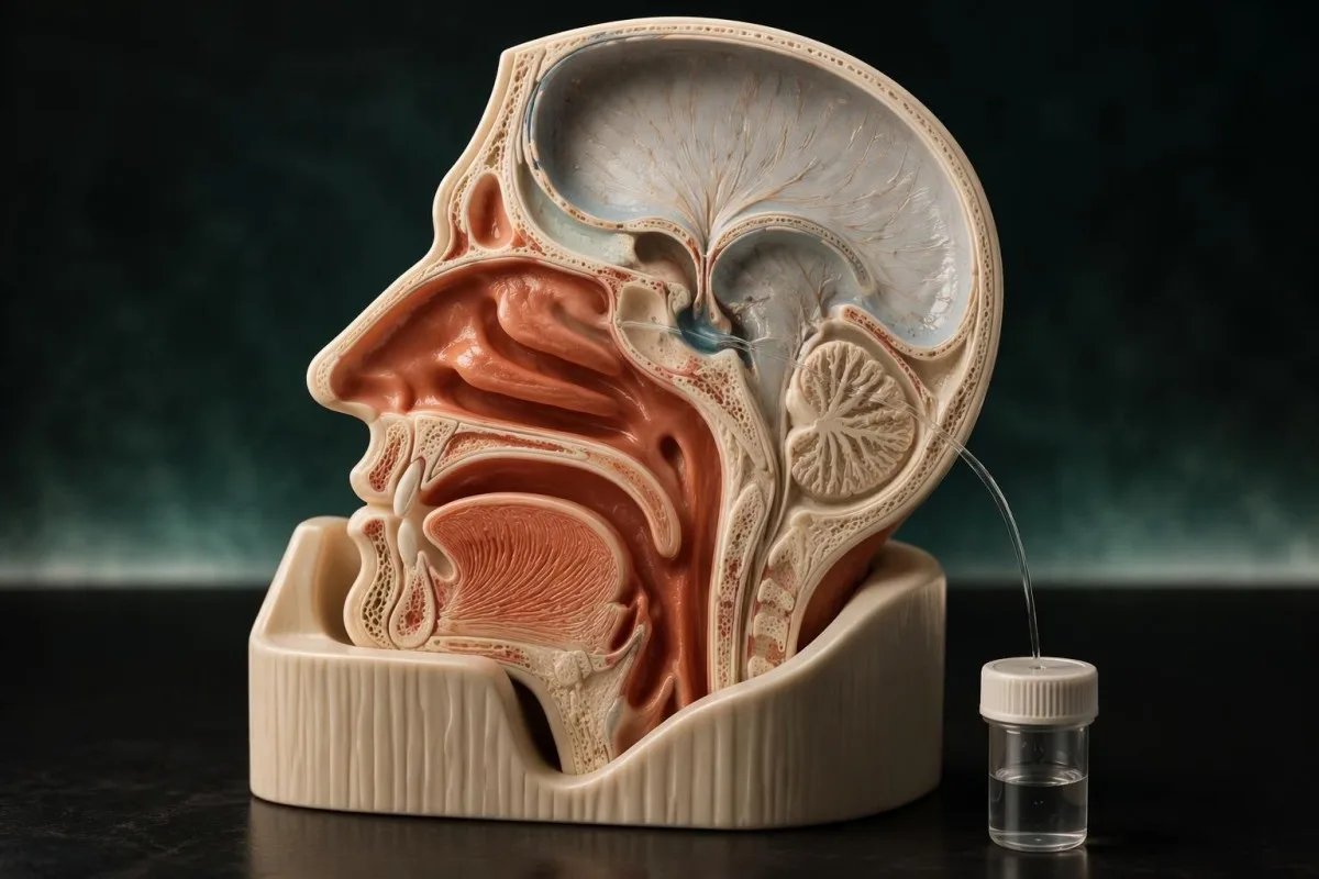
The beta-2 transferrin test checks nasal or ear fluid for a marker associated with cerebrospinal fluid; clear drainage alone does not confirm a leak. A positive finding usually supports a cranial CSF leak, but intermittent leakage or an unsuitable specimen can produce a negative result.

Key takeaways
- Specimen type matters: this test examines nasal or ear drainage, not a routine blood sample or standard iron panel.
- Clear drainage from 1 nostril can raise suspicion, especially after injury or surgery, but allergies and other nasal conditions can look similar.
- Positive findings usually support CSF in the submitted fluid; the test does not locate the opening or measure its size.
- Negative findings do not completely exclude an intermittent leak, particularly when the collected fluid was mostly mucus or insufficient for analysis.
- Collection volume is laboratory-specific; some services request approximately 0.5–1 mL, while others need a different amount or collection method.
- Storage instructions must come from the receiving laboratory; refrigeration at 2–8°C is appropriate only when its protocol specifies refrigerated transport.
- Emergency symptoms include severe headache with fever, neck stiffness, confusion, or new neurological changes; a temperature of 38°C or higher adds concern but is not required.
- Next steps may include repeat collection and targeted skull-base imaging; neither home glucose strips nor a halo on tissue reliably diagnoses a CSF leak.
Can the beta-2 transferrin test identify a CSF leak?
The beta-2 transferrin test can help identify cerebrospinal fluid in nasal or ear drainage, but clear fluid alone cannot confirm a leak. A positive finding usually supports a cranial CSF leak; a negative finding must be interpreted alongside symptoms, collection quality, and imaging.
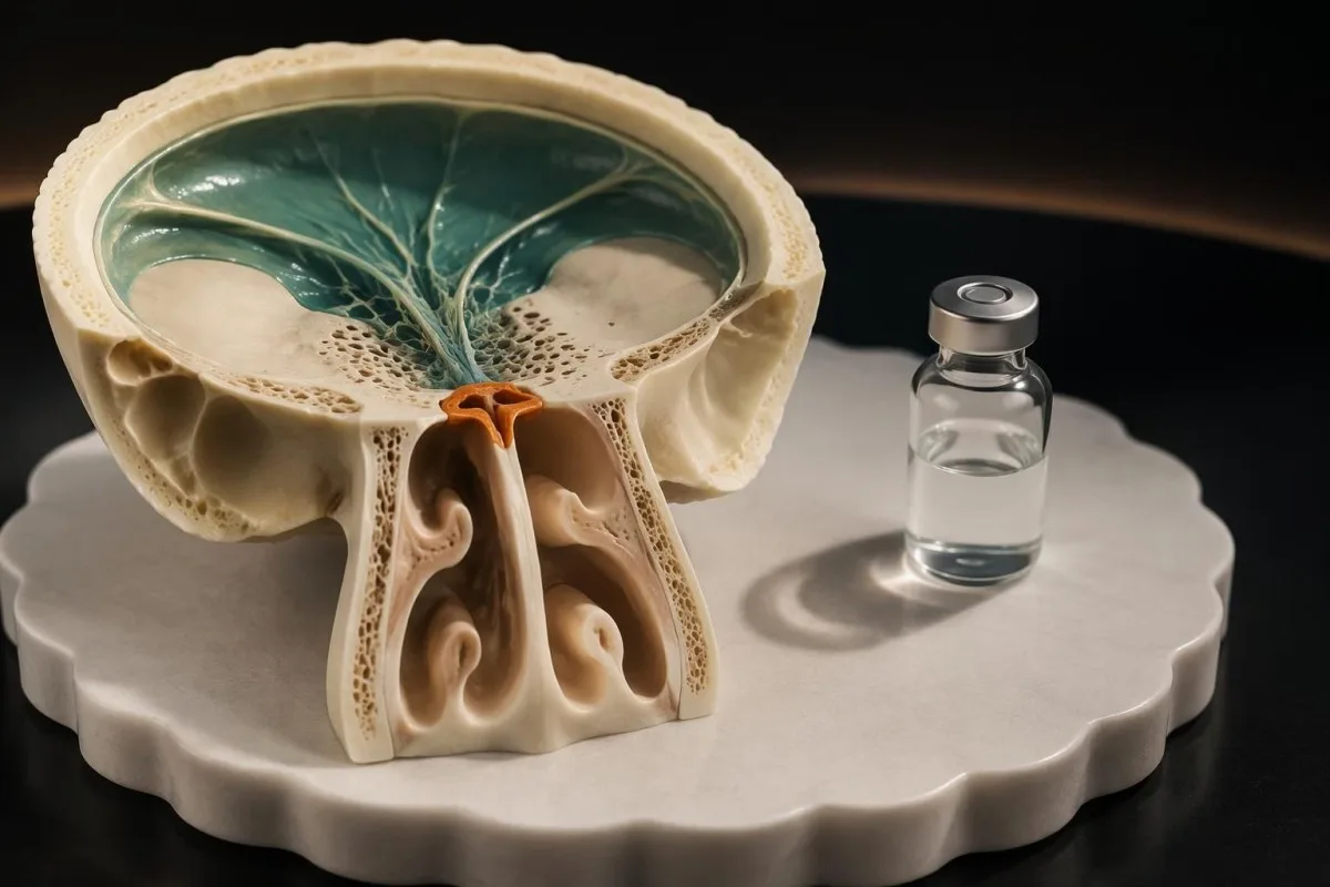
Beta-2 transferrin is a particular form of the transferrin protein, not an infection test or a measure of iron stores. The investigation answers 1 biochemical question: does this submitted drainage contain a protein pattern compatible with CSF? It cannot independently explain why the fluid is escaping.
A suspected cranial leak means fluid may be passing through an abnormal connection between the CSF-containing space and the nose or middle ear. Adults have approximately 150 mL of CSF at a given time, while producing roughly 500 mL daily; the amount visible on a tissue does not measure the size or danger of that connection.
Oakley et al. recommended beta-2 transferrin as the most reliable confirmatory laboratory test in their 2016 evidence-based review of CSF rhinorrhea. Their diagnostic framework separates 2 tasks—confirming the fluid's identity and locating the defect—because a laboratory result and a scan answer different questions.
Kantesti is an AI blood test analyzer that helps explain laboratory reports, but a suspected CSF leak requires examination and specimen-specific testing rather than routine bloodwork interpretation. Our organization and clinical scope explain that distinction: no blood panel can replace the nasal or ear fluid investigation described here.
Which drainage patterns raise suspicion without proving a leak?
Persistent or recurrent watery drainage from 1 nostril, particularly after head injury or skull-base surgery, warrants assessment for a possible CSF leak. Sneezing, itching, and bilateral congestion often favor rhinitis, but neither the appearance nor the side of drainage establishes its source.
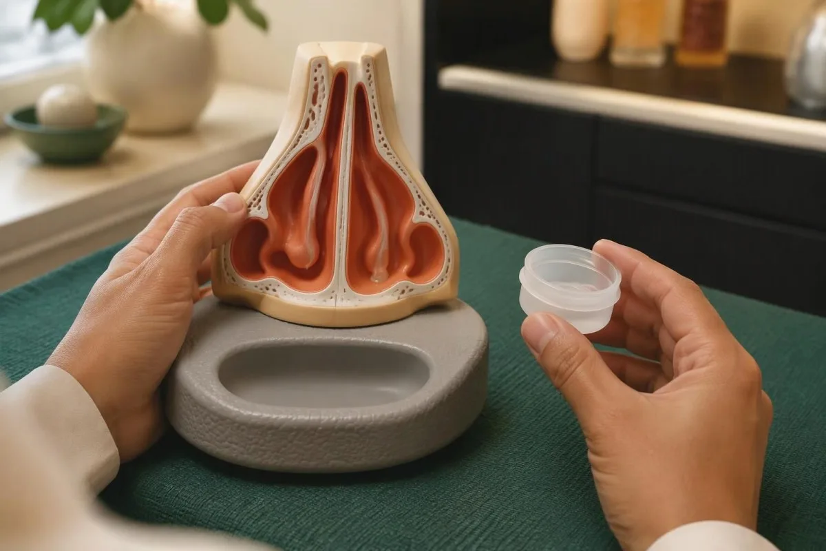
A salty taste, drainage noticed when bending forward, or fluid that seems unusually thin may contribute to the history. These clues have limited specificity: ordinary nasal secretions can produce similar experiences, and CSF may mix with mucus rather than look completely clear. Do not repeatedly bend or strain to test the pattern yourself.
An ear leak may present differently from a nasal leak. With an intact eardrum, fluid can accumulate behind it and travel toward the nose through the Eustachian tube; visible ear drainage may occur when there is a perforation or ventilation tube. Hearing changes therefore deserve attention even when there is no obvious nasal drip.
Allergic symptoms and suspected leakage can coexist, so an allergy diagnosis does not automatically settle a new, distinctly unilateral pattern. Our explanation of component-based allergy testing describes what allergy investigations can establish; those tests do not determine whether nasal fluid contains CSF.
For example, consider a hypothetical 46-year-old with seasonal itching who develops a new continuous drip from 1 nostril after sinus surgery. The change in timing and character matters more than the existing allergy label. Contacting the operating team promptly is safer than assuming that an antihistamine response will confirm or exclude a leak.
What does beta-2 transferrin detect—and what is it not?
Beta-2 transferrin is a low-sialic-acid form of transferrin found in CSF and certain eye and inner-ear fluids, but not normally in ordinary nasal secretions. It is different from serum transferrin, transferrin saturation, beta-2 microglobulin, and other similarly named laboratory measurements.

Most beta-2 transferrin reports describe a characteristic electrophoretic pattern rather than a concentration with a standard ng/mL reference interval. There is no universal patient-facing numerical cutoff that can be borrowed from an iron panel. A report headed 'transferrin' should therefore be checked for its full test name and specimen source.
Serum transferrin transports iron and is commonly interpreted with ferritin, iron, and transferrin saturation. Our guide to serum transferrin and inflammation concerns that blood measurement; a low serum result neither diagnoses a CSF leak nor means that beta-2 transferrin will be absent from leaked CSF.
Beta-2 transferrin is not completely exclusive to CSF because it is also present in perilymph and ocular fluids. That distinction is particularly relevant after eye or ear trauma, where the collection site and clinical findings help separate possible sources. A positive result from 1 specimen cannot resolve every anatomical possibility by itself.
Other CSF investigations have different purposes and collection routes. For instance, oligoclonal bands in CSF assess an immune-protein pattern, usually in lumbar-puncture fluid; finding or excluding those bands does not identify whether a nasal drip represents a cranial leak.
What should you arrange before beta-2 transferrin sample collection?
Beta-2 transferrin sample collection should follow the receiving laboratory's instructions, ideally obtained before fluid is collected. Confirm the accepted container, minimum volume, storage temperature, and delivery deadline; collection requirements are not identical across laboratories.
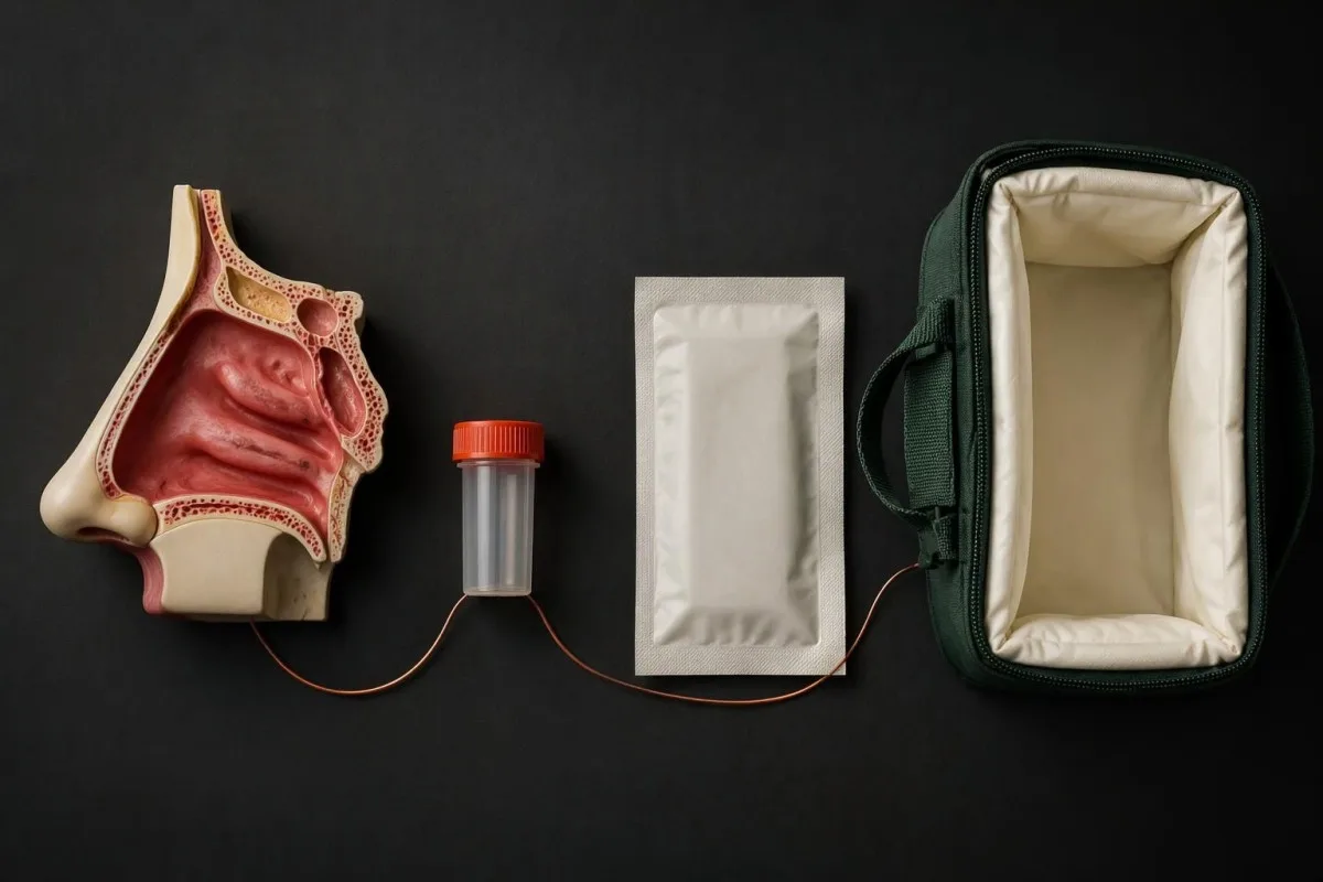
Ask the ordering clinician which laboratory will perform the assay, rather than relying only on a generic collection kit. Some services request approximately 0.5–1 mL, while others request more or accept specially collected material. The receiving service's minimum is decisive; these example volumes are not universal acceptance thresholds.
A clean, sterile, tightly closing container without additives is commonly requested for freely dripping fluid. Swabs, gauze, or other absorbent material may be accepted by some laboratories and rejected by others, particularly if extraction methods differ. Check before using them, and never add water or saline to reach a requested volume.
Identify at least 3 practical details before collection: the exact fluid source, the collection time, and whether the drainage followed trauma or a procedure. These details help the laboratory and clinician interpret an unusual pattern. If both sides drain, ask whether separate containers are required rather than combining them automatically.
Temperature-sensitive assays illustrate why specimen instructions cannot be generalized. Our guide to test-specific specimen handling discusses ACTH as a separate example; its blood-tube and cooling rules must not be transferred to nasal fluid. Ask for instructions specifically naming beta-2 transferrin and the intended specimen.
How should nasal or ear fluid be collected safely?
Collect naturally draining fluid only as instructed by the clinician and laboratory, preferably during an active episode. Avoid adding saline, mixing unrelated secretions, or inserting collection devices deep into the nose or ear; never provoke drainage by forceful straining.
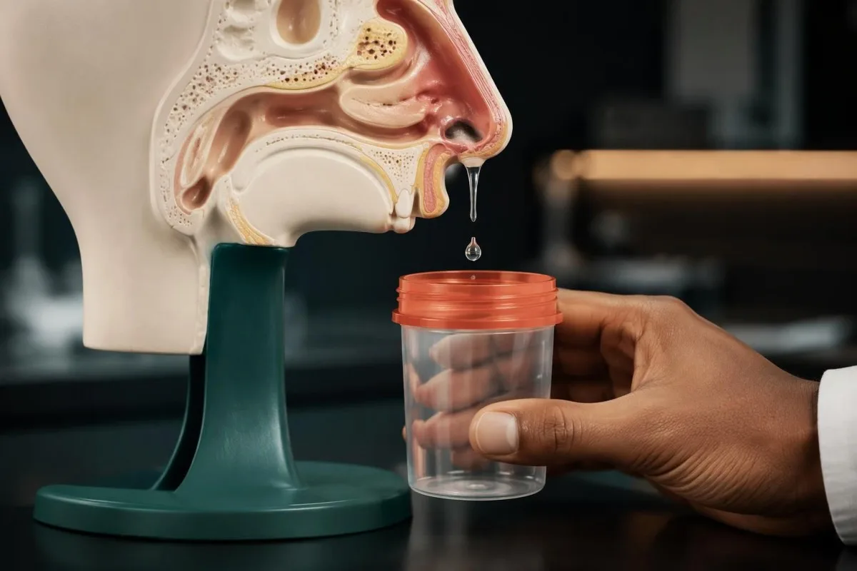
For freely dripping nasal fluid, a clinician may recommend allowing drops to enter the supplied container without touching its rim to nasal tissue. Nasal irrigation immediately before collection can dilute the specimen, while heavy mucus can make the sample less representative. Do not stop prescribed treatment without asking the ordering team.
Ear specimens require particular care because the anatomy and collection route vary. Do not insert a cotton bud, swab, or other object into the ear canal to obtain fluid; a clinician may need to collect from an existing drainage site. This is especially relevant after an ear procedure or when 1 ear has new hearing loss.
A hypothetical specimen containing 0.7 mL of drainage could be acceptable to a laboratory whose minimum is 0.5 mL, but inadequate to a service requiring 1 mL. Volume alone also does not prove quality: a larger sample composed mostly of mucus may be less useful than a smaller, correctly collected active drip.
The right biological material can matter more than the number of tests ordered. Our discussion of choosing the correct specimen explains that principle for a different condition. For suspected cranial leakage, substituting a routine blood sample for the actual nasal or ear drainage misses the diagnostic target.
How do storage and transport affect the specimen?
Storage and transport should follow the laboratory's beta-2 transferrin protocol, because leakage, contamination, evaporation, and unsuitable handling can compromise testing. A rejected specimen is not a negative result; it means the laboratory could not provide a valid answer from that material.
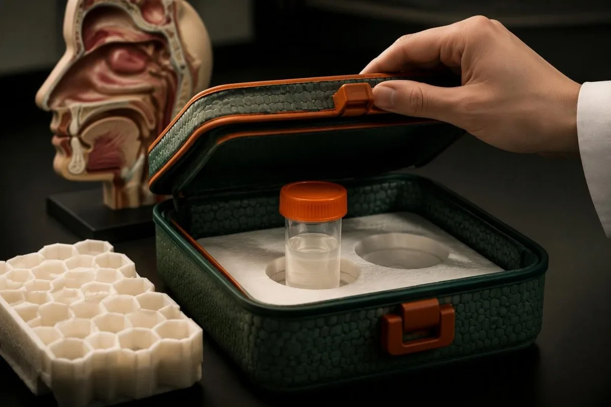
Some laboratories specify refrigerated storage at 2–8°C, while others provide different validated instructions or permit particular frozen specimens. Do not assume that colder is always better, and do not freeze a container unless the receiving service instructs you to do so. Its protocol should also specify packaging and allowable storage duration.
Close the container promptly and keep it upright in the supplied transport packaging. Do not repeatedly open it to inspect the fluid, and avoid pooling new episodes into an older specimen without permission. Collection across more than 1 episode can obscure which event was sampled and may violate the service's acceptance rules.
Turnaround commonly takes several days because testing may be performed by a specialist or referral laboratory, but there is no universal 24-hour reporting promise. Ask whether the estimate starts at collection, local receipt, or arrival at the performing laboratory. Urgent symptoms require care before any of these reporting milestones.
Sample integrity and clinical interpretation are separate questions. Our explanation of sample interference and reliability describes this distinction for blood specimens; nasal fluid has its own acceptance criteria. If a report says 'insufficient,' 'unsuitable,' or 'unable to interpret,' ask what specifically prevented testing before arranging another collection.
What does a beta-2 transferrin positive result mean?
A beta-2 transferrin positive result usually means the submitted nasal or ear fluid contains a protein pattern supporting CSF leakage. It does not identify the defect's location, measure the leak rate, establish meningitis, or determine whether a particular treatment is necessary.
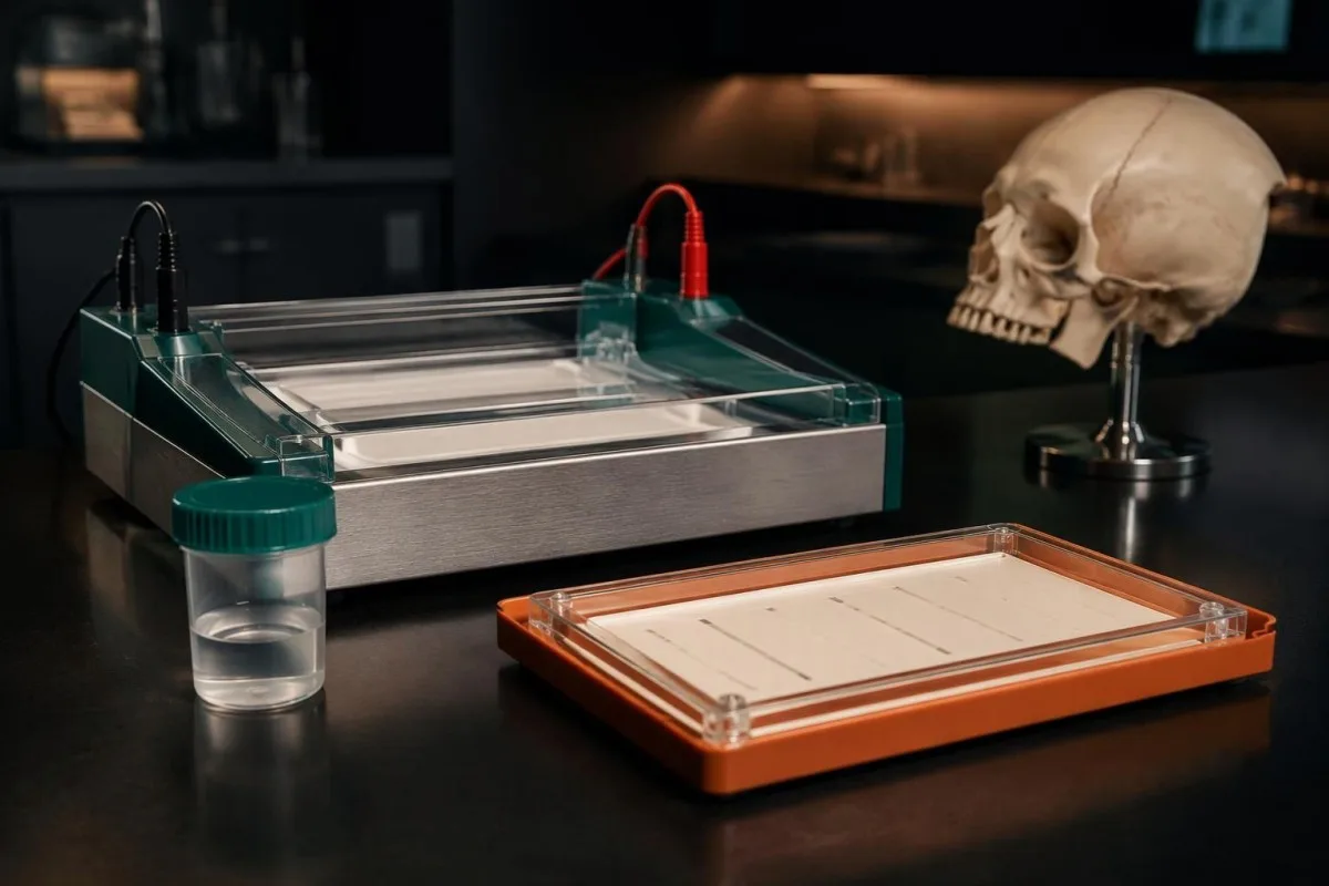
The clinician first checks whether the result fits the specimen source and circumstances. A characteristic finding in unilateral nasal drainage is different from an unusual pattern in fluid obtained after complex eye or ear trauma. Rare transferrin variants and analytical interference may require discussion with the laboratory, rather than assuming every positive has 1 explanation.
A positive result needs timely follow-up even if the dripping has stopped, because an intermittent connection may remain. However, stable patients do not all require the same immediate intervention. Recent injury, fever, neurological changes, imaging findings, and the suspected cause determine whether emergency assessment or an expedited specialist pathway is appropriate.
Consider a clearly hypothetical assay with 95% sensitivity and 95% specificity: among 100 people with a 10% pretest likelihood, about 9.5 true positives and 4.5 false positives would occur. The positive predictive value would be approximately 68%, illustrating why context matters; these are teaching assumptions, not measured performance figures for beta-2 transferrin.
Kantesti is an AI lab test interpretation service that can help explain the difference between a detected finding and a numerical measurement, but it cannot independently verify a CSF source. Our guide to qualitative versus quantitative results explains why 'detected' should not be translated into a leak-volume estimate.
Can a negative beta-2 transferrin result miss a leak?
A negative beta-2 transferrin result does not completely exclude a CSF leak, especially when leakage is intermittent or the specimen is diluted, contaminated, or collected between episodes. Persistent clinical suspicion can justify repeat collection and targeted imaging despite a negative report.

An intermittent leak creates a sampling problem before the assay even begins. A container collected during a dry period may contain ordinary mucus rather than the fluid responsible for the concerning episodes. For example, a hypothetical patient who drips for 10 minutes each morning might submit an afternoon specimen that does not capture the event.
Dilution can reduce the amount of the target protein reaching the assay. Recent rinsing, substantial mucus, or a small proportion of CSF mixed with other secretions may affect detectability; laboratories also differ in analytical methods and limits. There is no universal minimum percentage of CSF that patients can calculate reliably at home.
Oakley et al.'s 2016 review supports combining biochemical confirmation with anatomical investigation rather than treating a single laboratory finding as the whole diagnosis. Our article on negative neurological test results illustrates a separate condition in which the sampled target and disease form also influence what a negative test can exclude.
Repeat testing is useful when it corrects a specific problem, not merely when it duplicates the same poor collection. Ask whether the next specimen should be obtained during active drainage, collected by a clinician, or replaced by imaging. Similar reasoning applies to negative cultures and diagnosis, although the mechanisms and treatments are entirely different.
Why might the result be indeterminate or misleading?
Indeterminate beta-2 transferrin findings can arise from an atypical protein pattern, contamination, or insufficient specimen quality. Rare inherited transferrin variants and some conditions affecting transferrin glycosylation can complicate interpretation; laboratory review is more useful than treating an unclear band as a definite leak.
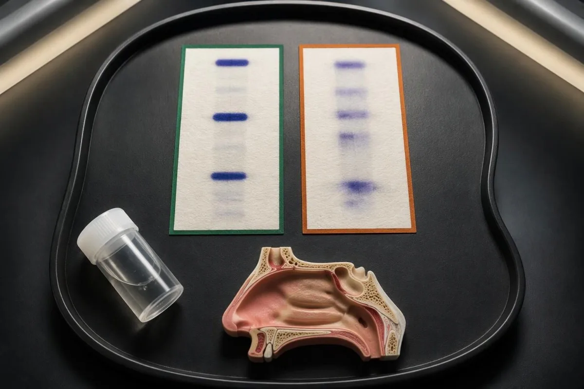
Heavy blood contamination can complicate electrophoretic interpretation, although the precise effect depends on the assay and specimen composition. Some laboratories may request a paired serum sample when a transferrin variant is suspected. That blood sample is a comparison tool—it does not replace the original drainage specimen or independently diagnose the leak.
Microbial enzymes can alter transferrin's sugar groups and produce atypical patterns in contaminated material. Conversely, some forms of contamination or degradation can make the expected finding harder to identify. These are laboratory-level issues: they do not mean that patients should infer an infection, a genetic disorder, or a false result from 1 unclear report.
Beta-trace protein is an alternative CSF-associated marker used by some services, but its interpretation is not interchangeable with beta-2 transferrin. Beta-trace can be measured quantitatively, and blood concentrations, kidney function, and local methods influence its interpretation. A cutoff from 1 laboratory should not be applied to another service's beta-2 transferrin report.
Protein-pattern testing often requires more interpretation than a simple high-or-low flag. Our serum protein interpretation guide explains the broader distinction between protein quantity and pattern, but its blood reference ranges do not apply here. Ask whether the report means 'not detected,' 'atypical,' or 'not technically assessable'—3 different outcomes.
What tests locate a suspected cranial CSF leak?
High-resolution skull-base CT and selected MRI techniques help locate a suspected cranial CSF leak. Beta-2 transferrin identifies a biochemical marker in the drainage; imaging evaluates bone, soft tissues, and the possible route of leakage, so the investigations complement rather than replace one another.
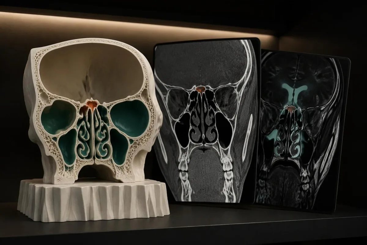
Thin-section CT can reveal small skull-base bone defects; protocols commonly use slices around 0.5–1 mm, although the imaging service selects the appropriate acquisition. An ordinary head CT may not provide the same detail. A visible bone gap also requires clinical correlation, because not every anatomical defect is actively leaking.
Heavily T2-weighted MRI can help show CSF signal and associated soft-tissue abnormalities, including a meningocele or encephalocele. CT and MRI answer different anatomical questions. Imaging may still be inconclusive when drainage is intermittent or more than 1 possible site exists, so a normal scan does not always finish the evaluation.
The 2021 International Consensus Statement on spontaneous CSF rhinorrhea emphasizes specialist assessment, imaging, and evaluation for associated intracranial-pressure problems (Georgalas et al., 2021). Selected difficult cases may need more invasive localization methods; intrathecal contrast or fluorescein is not a routine first step and requires an experienced team's risk assessment.
Lumbar puncture is not automatically required just to test nasal drainage for beta-2 transferrin. Its role depends on a separate clinical question, such as pressure assessment or suspected meningitis. Our explanation of CSF, MRI, and EEG describes another neurological workup; those tests should not be copied wholesale into a leak investigation.
Which symptoms need urgent evaluation before the result returns?
Seek emergency care for clear nasal or ear drainage after significant head injury, or for drainage accompanied by severe headache, neck stiffness, fever, confusion, or new neurological symptoms. Do not wait for beta-2 transferrin results or try to collect a complete specimen first.

A temperature of 38°C or higher with severe headache or neck stiffness raises concern for meningitis, particularly when a cranial leak is suspected. However, early meningitis may not produce every classic symptom, and fever can be absent. Light sensitivity, repeated vomiting, unusual drowsiness, or rapid deterioration also require urgent clinical assessment.
Sudden weakness, facial drooping, speech difficulty, a seizure, or a rapidly changing level of alertness is an emergency regardless of whether drainage is present. Our guide to stroke warning signs explains why these symptoms require immediate action. Do not let 1 pending laboratory result distract from a time-sensitive neurological problem.
New clear drainage after a head injury warrants emergency assessment even if there is little discomfort or the fluid stops. Blood-based injury markers address different questions; our explanation of GFAP after head injury discusses CT decision support, not confirmation or exclusion of CSF in nasal fluid.
After recent sinus, ear, or skull-base surgery, contact the operating team immediately about new watery drainage and follow its escalation instructions. Severe symptoms should go directly to emergency care. For recurrent unexplained drainage without emergency features, seek prompt medical assessment rather than waiting through multiple episodes or relying on a home test.
What should you do while waiting, and how are leaks treated?
Follow the treating team's precautions while awaiting investigation, and do not self-treat a suspected CSF leak with antibiotics or pressure-lowering medication. Management depends on whether the leak followed trauma, arose spontaneously, or occurred after a procedure; conservative care and repair are not interchangeable choices.
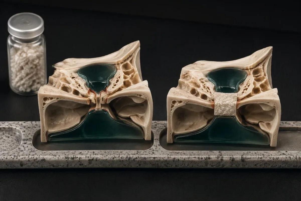
Clinicians commonly advise avoiding forceful nose blowing, heavy lifting, and straining when a cranial leak is suspected, but individual restrictions depend on the situation. Do not repeatedly reproduce the drainage by bending forward. Ask about activity, travel, nasal rinses, and prescribed positive-airway-pressure equipment rather than making several treatment changes independently.
Some traumatic leaks are initially managed conservatively under specialist supervision, whereas persistent or spontaneous leaks often need repair. Georgalas et al.'s 2021 consensus also addresses associated intracranial-pressure abnormalities. Stopping the drip is only 1 management objective; reducing recurrence and protecting against intracranial infection are separate concerns.
Acetazolamide may be used in selected pressure-related cases, but it is not appropriate for every suspected leak and should not be started from an online recommendation. Kidney function and electrolytes influence its safety, and dose decisions require clinical oversight. Our guide to electrolyte monitoring and safety explains why changes in bicarbonate, sodium, or potassium can matter during treatment.
Routine preventive antibiotics are not a substitute for evaluation or repair, and recommendations vary with the clinical context. A confirmed CSF leak can also affect pneumococcal-vaccination eligibility; the appropriate schedule depends on country, age, and previous doses. Ask the team about these 2 prevention questions rather than assuming either intervention is automatic.
Can routine bloodwork or AI interpretation rule out a CSF leak?
Routine bloodwork and AI blood test interpretation cannot rule out a cranial CSF leak. Blood counts and inflammatory markers may contribute to other clinical questions, but they do not establish the source of nasal drainage; beta-2 transferrin requires the relevant fluid specimen.

A normal white-cell count or CRP does not prove that a suspected leak is harmless, and abnormal values do not confirm that the drip contains CSF. Interpretation involves at least 2 separate questions: whether a connection exists and whether complications are present. Bloodwork may inform the second question without resolving the first.
Cranial and spinal CSF leaks also need different diagnostic pathways. A spinal leak usually does not produce nasal or ear drainage and may cause headache that worsens upright and improves lying down, although that pattern is not universal. A negative nasal-fluid test cannot exclude a spinal leak because it samples the wrong location.
Kantesti is an AI blood test interpretation platform that explains supported blood biomarkers in clinical context; it does not detect a skull-base defect or replace specialist examination. Our medical validation framework describes methodology boundaries, while our guide to AI report source checks explains why the specimen name and original laboratory wording should remain visible.
Kantesti's role here is educational, not anatomical confirmation: no interpretation speed changes the need to collect the right fluid and assess warning symptoms. Our responsible AI principles distinguish assistance from diagnosis. If an explanation conflicts with a clinician's urgent instructions, follow the clinical escalation plan rather than seeking another automated opinion.
Research publications and the limits of the evidence
Evidence supporting CSF-leak diagnosis comes from condition-specific laboratory and clinical research, not general AI blood-test benchmarks. The 2 Figshare publications listed below concern our blood-report interpretation engine and must not be presented as validation of beta-2 transferrin testing or CSF-leak detection.
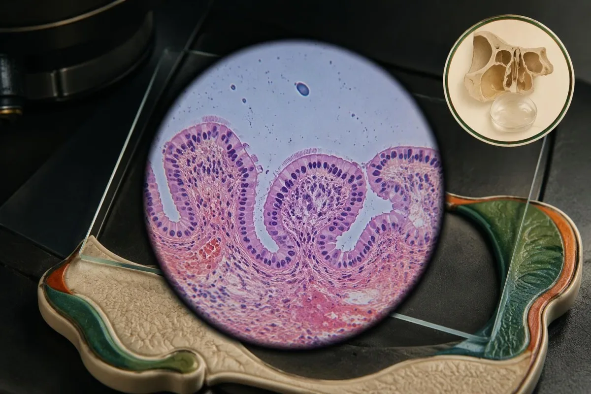
Oakley et al.'s 2016 evidence-based review addresses the diagnostic sequence for CSF rhinorrhea, including biochemical confirmation and imaging. Georgalas et al.'s 2021 consensus focuses specifically on spontaneous leakage and its management. Neither publication makes clear drainage alone diagnostic, and the spontaneous-leak recommendations should not automatically replace a trauma-specific pathway.
Reported assay performance depends on the analytical technique, patient selection, and how the final diagnosis was established. Studies containing obvious high-volume leaks may not reflect patients with tiny intermittent episodes or heavily mixed secretions. A single sensitivity percentage therefore cannot provide an individualized guarantee after 1 negative specimen.
The Kantesti deployment publication describes multilingual clinical decision support for hantavirus triage across 50,000 interpreted blood-test reports. The separate technical benchmark describes 100,000 synthetic test cases; synthetic cases are not patients, and technical scoring is not equivalent to prospective clinical outcome validation. Neither title should be read as evidence that our software confirms a CSF leak.
Kantesti separates these evidence categories through our medical content standards, and our blood-test benchmark methods describe the scope of engine testing. For a suspected leak, the practical evidence remains the drainage specimen, its collection quality, the examination, and targeted imaging—not the presence of an AI research citation.
Frequently asked questions
Does clear fluid from one nostril mean I have a CSF leak?
Clear fluid from 1 nostril does not by itself confirm a CSF leak; rhinitis and other nasal conditions can produce watery drainage. Persistent unilateral dripping, a new pattern after head injury or surgery, or associated neurological symptoms increases the need for assessment. The beta-2 transferrin test examines the drainage for a CSF-associated protein form. New drainage after significant head injury or drainage with severe headache, neck stiffness, confusion, or fever requires urgent care rather than home observation.
Is beta-2 transferrin tested in blood or nasal fluid?
The beta-2 transferrin test for a suspected cranial CSF leak is performed on nasal or ear fluid, not a routine blood sample. Serum transferrin and transferrin saturation are separate blood measurements related to iron handling. Some laboratories may request 1 paired serum specimen to investigate an unusual transferrin pattern, but that comparison does not replace the drainage sample. Check that the order names both beta-2 transferrin and the correct fluid source.
How much fluid is needed for beta-2 transferrin testing?
The required volume for beta-2 transferrin testing depends on the performing laboratory and its validated collection method. Some services request approximately 0.5–1 mL, while others request a different amount or accept particular absorbent specimens. Obtain the receiving laboratory's instructions before collecting, and do not add water or saline to reach the volume. An inadequate or rejected specimen is not the same as a negative result.
Can a beta-2 transferrin test be negative even with a CSF leak?
A beta-2 transferrin test can be negative despite a CSF leak if the submitted specimen does not capture active leakage or is unsuitable for the assay. Intermittent drainage, dilution, mucus admixture, and analytical limitations can contribute. A specimen collected between 2 drainage episodes may contain ordinary nasal secretion rather than the fluid of concern. Persistent suspicion may require repeat collection during active drainage and specialist-directed imaging.
Does a positive beta-2 transferrin result mean I need surgery?
A positive beta-2 transferrin result usually supports CSF in the submitted drainage, but it does not determine treatment by itself. The clinician considers the cause, symptoms, imaging, duration, and possible complications before choosing management. Some traumatic leaks receive supervised conservative care, while persistent or spontaneous leaks often require repair. The 2021 international consensus on spontaneous CSF rhinorrhea also addresses associated intracranial-pressure problems.
Can glucose strips or a halo on tissue diagnose a CSF leak?
Glucose strips and a halo pattern on tissue cannot reliably diagnose or exclude a CSF leak. Glucose can be present in other secretions, and several fluid mixtures can form a ring, so neither method is a substitute for laboratory fluid testing and clinical assessment. A temperature of 38°C or higher with severe headache or neck stiffness adds concern for meningitis, but serious illness can occur without that temperature. Do not delay emergency evaluation to perform either home test.
Get your blood test results explained
Upload your lab report and Kantesti's AI explains each biomarker in plain language within seconds. Used by more than 2 million people worldwide.
References
- Klein, T., Mitchell, S., & Weber, H. (2026). Multilingual AI Assisted Clinical Decision Support for Early Hantavirus Triage: Design, Engineering Validation, and Real-World Deployment Across 50,000 Interpreted Blood Test Reports. Kantesti AI Medical Research. DOI
- Klein, T., Mitchell, S., & Weber, H. (2026). A Pre-Registered, Rubric-Based Automated Technical Benchmark of the Kantesti Blood-Test Interpretation Engine on 100,000 Synthetic Test Cases. Kantesti AI Medical Research. DOI
- Oakley GM et al. (2016). Diagnosis of cerebrospinal fluid rhinorrhea: an evidence-based review with recommendations. International Forum of Allergy & Rhinology.
- Georgalas C et al. (2021). International Consensus Statement: Spontaneous Cerebrospinal Fluid Rhinorrhea. International Forum of Allergy & Rhinology.
- 2M+Tests analysed
- 127+Countries
- 100+Languages
About the medical team


E-E-A-T Trust Signals
Experience
Physician-led clinical review of lab interpretation workflows.
Expertise
Laboratory medicine focus on how biomarkers behave in clinical context.
Authoritativeness
Medically reviewed by Dr. Thomas Klein, with contributions from Dr. Sarah Mitchell and Prof. Dr. Hans Weber.
Trustworthiness
Evidence-based interpretation with clear follow-up pathways to reduce alarm.





Medical disclaimer
This article is for educational purposes only and does not constitute medical advice. Always consult a qualified healthcare provider for diagnosis and treatment decisions.
Published · Contact the editorial team
Published by Kantesti Ltd, registered in England & Wales, company no. 17090423, London, United Kingdom.
