
A GFAP blood test can help clinicians assess the likelihood of CT-visible brain injury in selected adults. A negative result does not exclude concussion or cancel the need for urgent assessment.

Key takeaways
- GFAP blood test measures a protein associated with astrocytes; its acute head-injury role is supporting assessment for CT-visible intracranial injury, not diagnosing every concussion.
- GFAP and UCH-L1 are interpreted together on some assays; either marker meeting its assay-specific cutoff can produce an elevated result.
- Sampling window depends on the test: established pathways include collection within 12 hours, while a particular whole-blood assay permits collection within 24 hours.
- Assay thresholds are not interchangeable: examples include GFAP cutoffs of 22, 30, and 65 pg/mL on different testing systems.
- Negative results substantially lower the likelihood of CT-visible injury in eligible patients, but false negatives occur and concussion may still be present.
- High GFAP levels do not identify the location of an injury, prove that bleeding is present, or determine whether an operation is necessary.
- Urgent symptoms such as worsening confusion, repeated vomiting, a seizure, or new weakness require emergency assessment regardless of the result.
- Units matter: 30 pg/mL equals 0.030 ng/mL; a cutoff copied from a different assay can change the interpretation incorrectly.
What does a GFAP blood test show after head injury?
A GFAP blood test measures glial fibrillary acidic protein released from brain-supporting cells and can help clinicians assess the likelihood of CT-visible intracranial injury after suspected mild traumatic brain injury. Used within its approved sampling window—often 12 or 24 hours—it can support a decision about head CT, but cannot exclude concussion.
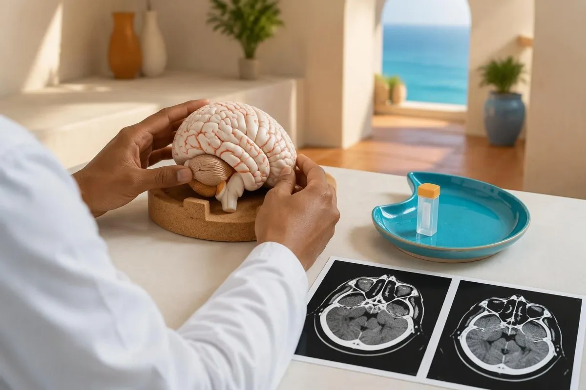
The clinical target is an imaging finding, not every consequence of a head impact. A person can have a Glasgow Coma Scale score of 15, no abnormality on CT, and substantial problems with memory, balance, or light sensitivity; those symptoms may still support a concussion diagnosis after clinical assessment.
A result is only useful inside a defined decision pathway. The clinician needs the injury time, examination findings, medications, and exact assay—not simply a GFAP concentration such as 25 pg/mL—because the same number can fall on different sides of different test cutoffs.
Kantesti is an AI blood test analyzer that can help readers organize laboratory terminology, but emergency head-injury decisions require an examining clinician. Our company and clinical purpose are separate from the assay manufacturer's intended use; an interpretation delivered in approximately 60 seconds is not clearance to avoid emergency care.
Why are GFAP and UCH-L1 sometimes tested together?
GFAP and UCH-L1 reflect different cellular sources: GFAP is associated mainly with astrocytes, while ubiquitin carboxyl-terminal hydrolase L1 is enriched in neurons. Paired assays combine these signals to help assess CT-visible injury, usually through a rule that classifies the test as elevated when either marker reaches its cutoff.

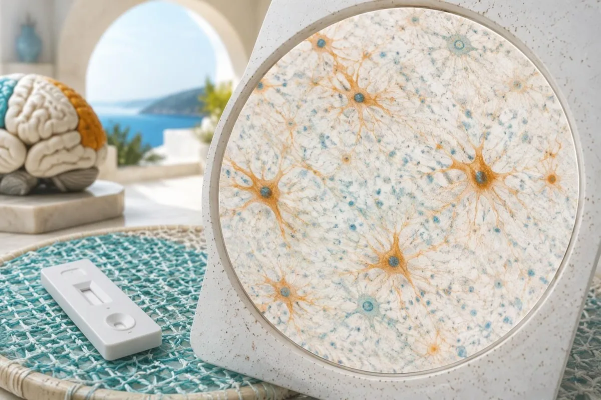
Astrocytes support neurons and help maintain the brain's local environment. GFAP forms part of their internal filament network; a circulating concentration of 30 pg/mL is therefore a protein measurement, not a count of damaged cells or a percentage of brain tissue affected.
UCH-L1 adds a neuronal signal, but two markers do not create a universal concussion test. On a paired assay, a GFAP result below its cutoff cannot make the overall result negative if UCH-L1 is elevated; the report's combined classification matters more than reading only one line.
Other neurological biomarkers answer different questions. Our explanation of neurofilament light chain describes an axonal marker whose interpretation often involves longer timescales; neither its reference interval nor a GFAP research cutoff can be substituted for a validated 12-hour acute-injury algorithm.
Who is eligible for acute GFAP testing?
Acute GFAP-based CT-triage assays are generally intended for adults aged 18 years or older with suspected traumatic brain injury who meet the specific test's clinical and timing requirements. Many rapid paired assays address mild injury with a Glasgow Coma Scale score of 13–15; eligibility is not established by age alone.
The original authorized paired serum test covered a broader GCS range of 9–15. That historical indication should not be copied onto another device: a rapid assay labeled for GCS 13–15 has a different intended population, even when both measure the same two proteins.
Children require pediatric assessment rather than borrowed adult thresholds. A 14-year-old athlete is outside an assay restricted to adults aged 18 or older; our guide to children's laboratory interpretation explains why an apparently familiar number does not establish pediatric validity.
Guideline age boundaries and assay age boundaries can differ. NICE separates several head-CT recommendations at age 16, whereas commonly used acute biomarker assays start at age 18; a 17-year-old may therefore follow the older-person imaging pathway without being eligible for an adult-labeled GFAP test.
When must the sample be collected after the injury?
The GFAP sampling window is assay-specific: established paired serum and rapid plasma pathways use collection within 12 hours, while a particular paired whole-blood assay allows collection within 24 hours. The clock starts at the injury, not at hospital registration, and eligibility depends on the current instructions for the actual test.
Protein detectability and validated clinical use are different concepts. Papa et al. reported different GFAP and UCH-L1 trajectories over 7 days in JAMA Neurology (2016); detecting GFAP several days later does not validate using an acute 12-hour rule to decide against CT.
An uncertain injury time makes a negative result harder to use safely. For example, a hypothetical fall discovered at 09:00 may have occurred 2 hours earlier or overnight; recording only discovery time could incorrectly place the sample inside a validated window.
Collection time is not the same as reporting time. A sample obtained 11 hours after injury and analyzed later may meet the collection requirement if handling instructions are satisfied, whereas a newly collected 13-hour sample may not; our blood test timing guide explains why timestamps belong beside the result.
Which GFAP thresholds apply to your laboratory report?
There is no universal normal range for acute GFAP CT triage. Representative decision cutoffs include 22 pg/mL for a particular serum assay, 30 pg/mL for a rapid plasma assay, and 65 pg/mL for a particular whole-blood assay; these values must never be transferred between testing systems.
These are decision thresholds, not general wellness reference intervals. They were selected to identify patients unlikely to have the target CT finding while maintaining high sensitivity, so a GFAP concentration of 35 pg/mL does not translate into a universally defined mild, moderate, or severe injury category.
Both markers usually must remain below their respective thresholds for a paired result to be negative. A hypothetical rapid-plasma report with GFAP 20 pg/mL and UCH-L1 400 pg/mL is elevated despite the low GFAP; our guide to qualitative and quantitative results explains why a concentration and a combined classification are different outputs.
Kantesti's explanatory approach treats units, sample type, and assay identity as necessary context. Because 1 ng/mL equals 1,000 pg/mL, a report showing GFAP 0.030 ng/mL represents 30 pg/mL—not a thousandfold lower biological signal—and borderline values should be read against the laboratory's stated rule.
| Level | Range | What it means |
|---|---|---|
| Representative paired serum assay | GFAP 22 pg/mL; UCH-L1 327 pg/mL | Original authorized pathway: adults, GCS 9–15, collection within 12 hours. Either marker at or above its cutoff produces an elevated classification. |
| Representative rapid paired plasma assay | GFAP 30 pg/mL; UCH-L1 360 pg/mL | Adult mild-injury pathway with collection within 12 hours. These thresholds apply only to the corresponding assay. |
| Representative paired whole-blood assay | GFAP 65 pg/mL; UCH-L1 360 pg/mL | A particular adult mild-injury pathway permits collection within 24 hours. Do not substitute its GFAP cutoff for a plasma or serum cutoff. |
How reassuring is a negative GFAP and UCH-L1 result?
A negative paired result can substantially reduce the likelihood of CT-visible intracranial injury in the intended population, but does not make that likelihood zero. In the 1,959-patient ALERT-TBI study, the evaluated serum test had 97.6% sensitivity and 99.6% negative predictive value for its target CT findings.
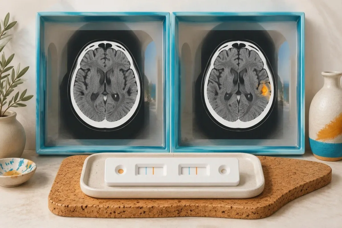
ALERT-TBI recorded 3 CT-positive cases among 671 negative biomarker results. Bazarian et al., in The Lancet Neurology (2018), also reported specificity of approximately 36.4%, illustrating the tradeoff: high sensitivity can coexist with many elevated results in people whose CT does not show the target injury.
Negative predictive value depends on the population's starting risk. In a clearly hypothetical group of 10,000 people with a 5% CT-positive prevalence, applying 97.6% sensitivity and 36.4% specificity would produce approximately 12 false negatives among 3,470 negative tests—a residual probability around 0.35%, not a guarantee.
A negative test cannot rescue an unsafe clinical assessment. Our article on normal tests missing appendicitis explains the broader diagnostic principle; Kantesti's validation and clinical oversight should not be mistaken for evidence that our AI can independently rule out intracranial injury.
What do high GFAP levels actually mean?
High GFAP levels indicate an increased circulating astroglial protein signal, but they do not identify a particular injury or establish that bleeding is present. After an acute impact, a result above the relevant cutoff means the biomarker pathway cannot provide its negative reassurance; CT decisions still depend on the examination and clinical rules.

The positive predictive value of a screening test can be modest. In ALERT-TBI, approximately 9.5% of elevated paired results corresponded to the target CT finding; that figure applies to its study population and serum assay, not automatically to every rapid test or every emergency department.
GFAP can also be elevated in non-traumatic neurological conditions. A concentration of 80 pg/mL without a recent impact needs a different clinical question, because stroke, neurodegenerative processes, and other central nervous system conditions can influence GFAP; it should not be interpreted through an acute 12-hour injury algorithm.
A GFAP number is not a brain-growth screening result. Our discussion of brain symptom evaluation explains why imaging and neurological assessment answer structural questions; neither a single value of 65 pg/mL nor a much higher result can locate an abnormality or predict the need for an operation.
Which symptoms override a negative biomarker result?
Worsening confusion, repeated vomiting, a seizure, new weakness, or difficulty staying awake require urgent assessment regardless of GFAP. NICE NG232 recommends head CT within 1 hour for several high-risk findings in people aged 16 or older, including focal neurological deficit, post-traumatic seizure, and more than 1 vomiting episode.
Reduced consciousness is itself a CT trigger. NICE's Head injury guideline (2023) includes an initial GCS of 12 or less, or a GCS below 15 at 2 hours after injury, among indications for CT within 1 hour; suspected open or depressed skull fracture and signs of basal skull fracture also qualify.
Anticoagulant use changes the assessment even when symptoms seem mild. NICE advises considering CT for people taking anticoagulants or antiplatelets other than aspirin alone, typically within 8 hours of injury or within 1 hour when presentation is more than 8 hours later; a warfarin INR result cannot replace imaging assessment.
A normal routine coagulation result does not prove that medication-related bleeding risk is absent. Our coagulation testing guide explains why individual tests have limited targets; if someone becomes difficult to wake or develops new weakness, call emergency services rather than waiting for a biomarker result.
Can a negative GFAP concussion test exclude concussion?
No: a negative GFAP concussion test does not exclude concussion. Concussion is a clinical diagnosis involving symptoms and functional changes after injury, and conventional head CT often appears normal; a negative acute biomarker result addresses the likelihood of its target CT findings, not recovery, cognition, or fitness to return to sport.

A GCS score of 15 does not establish normal attention or memory. A hypothetical adult with headache, dizziness, and slowed thinking after an impact may have both a negative biomarker result and concussion; comparing the new symptoms with the person's pre-injury baseline helps distinguish an acute change from a longstanding condition.
New post-injury brain fog is not equivalent to lifelong attention difficulty. Our guide to adult attention symptom patterns discusses that distinction; symptoms beginning within 24 hours of an impact deserve a head-injury assessment rather than being dismissed because GFAP is low.
Return-to-activity decisions cannot be read from a pg/mL value. After assessment, concussion care commonly starts with 24–48 hours of relative rest followed by symptom-limited activity; contact sport, hazardous work, and driving require separate clinical advice, especially when dizziness, slowed reactions, or visual symptoms persist.
How do age, kidney function, and medication affect interpretation?
Age, kidney function, and medication history can change the clinical meaning of a GFAP result, but there is no universal age-adjusted or kidney-adjusted acute CT-triage cutoff. An assay's validated 12-hour or 24-hour decision rule should not be rewritten using an improvised correction formula.

Older age can increase both background biomarker concentrations and clinical injury risk. NICE uses age 65 or older as an additional CT risk factor after loss of consciousness or amnesia; a low biomarker result does not erase that history, and a raised value should not automatically be blamed on aging.
Kidney function is a source of uncertainty, not a validated adjustment calculator. Associations between renal function and circulating neurological biomarkers vary by marker, population, and assay; a hypothetical eGFR of 35 mL/min/1.73 m² should prompt contextual review, not a mathematical reduction of a GFAP value such as 90 pg/mL.
Kantesti is an AI biomarker interpretation platform that can help organize accompanying laboratory findings, but no general report-analysis feature establishes an age-specific GFAP cutoff. Our guide to older-adult medication effects explains why a complete medication list—especially anticoagulants and sedating medicines—belongs in the assessment.
Can sample handling or assay interference mislead the result?
Sample type, processing, and assay interference can invalidate a GFAP interpretation. Serum, plasma, and whole blood are not interchangeable specimens, and an apparently precise concentration such as 29 pg/mL cannot be trusted as a negative rapid-plasma result if the laboratory reports an invalid run or unsuitable sample.
Hemolysis and other sample-quality problems have assay-dependent effects. Our explanation of a high hemolysis index describes why laboratory flags need attention; the direction and size of any effect on GFAP or UCH-L1 must come from that assay's interference data, not a generic rule.
Processing requirements may include specific tubes, separation times, storage temperatures, and limits on freeze-thaw cycles. A 12-hour injury window says nothing about whether a specimen was stored correctly; our sample handling explanation illustrates how pre-analytical requirements can be as consequential as the reported concentration.
Immunoassay interference should be investigated when the result conflicts with the clinical picture. Ask the laboratory about applicable heterophile-antibody or supplement-related interference rather than assuming every GFAP method is affected by biotin; repeating a sample 2 hours later also changes the injury-to-collection interval and may move it outside eligibility.
How should you read a borderline or discordant result?
Borderline GFAP results must be interpreted using the exact assay's combined rule, not a universal severity scale. On the representative rapid-plasma pathway, GFAP 20 pg/mL with UCH-L1 300 pg/mL is below both cutoffs, while GFAP 32 pg/mL with the same UCH-L1 is elevated.
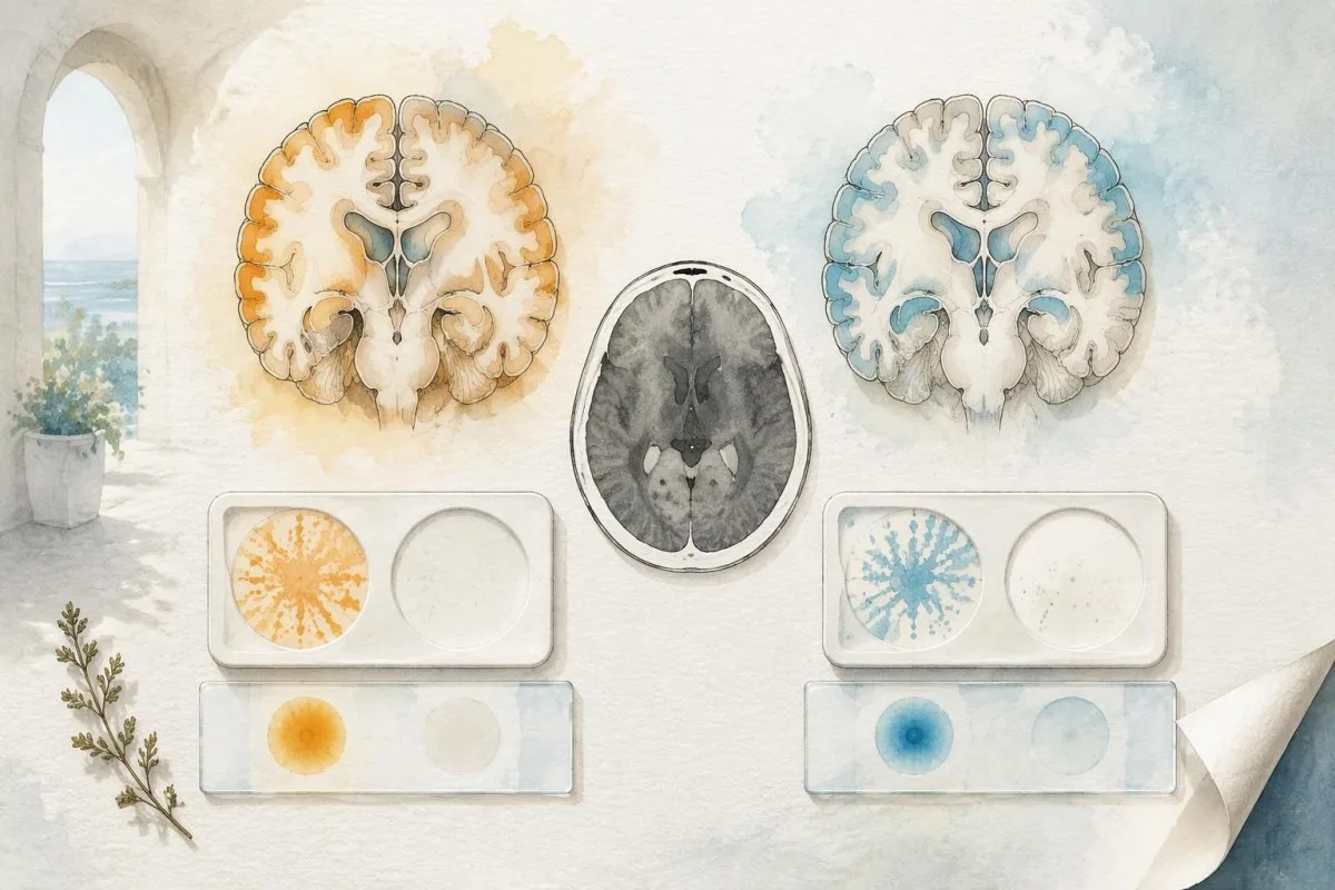
Example 1 is hypothetical: a 28-year-old is assessed 4 hours after a sports impact. GFAP 20 pg/mL and UCH-L1 300 pg/mL on the matching rapid-plasma assay may support avoiding CT if the clinician finds no overriding indication, but headache and dizziness can still require a concussion care plan.
Example 2 is hypothetical: a 52-year-old has GFAP 32 pg/mL and UCH-L1 300 pg/mL at 6 hours. The paired rapid-plasma classification is elevated because GFAP crosses 30 pg/mL; the result does not prove a CT abnormality, and describing it as only slightly elevated cannot safely convert it to negative.
Example 3 is hypothetical: an 80-year-old taking an anticoagulant reports worsening drowsiness despite low markers. Urgent clinical reassessment takes priority; our guide to differences between laboratory visits also explains why a shift from 29 to 31 pg/mL may reflect analytical variation without authorizing anyone to disregard the stated cutoff.
What should your clinician check before using the result?
A usable GFAP result requires four linked checks: clinical eligibility, injury-to-collection interval, assay identity, and laboratory validity. A concentration, a negative label, or a report explanation by itself cannot establish that an adult safely fits a 12-hour or 24-hour CT-triage pathway.
The original report should preserve both concentrations and the combined interpretation when available. Useful details include sample type, units, collection timestamp, decision limits, and quality flags; a screenshot containing only GFAP 20 pg/mL omits the UCH-L1 value that might change a paired result.
Kantesti is an AI blood test interpretation platform that explains laboratory reports, rather than an emergency imaging decision-maker. Our AI technology guide describes report-analysis methods, but an approximately 60-second explanation cannot verify the neurological examination or confirm eligibility for a particular GFAP assay.
Consent and source verification remain necessary when sharing results. Our privacy and data practices explain data-handling choices; our separate guide to checking AI report explanations recommends checking values and units against the original report before acting on an interpretation.
What does the research establish—and what remains uncertain?
Clinical research supports GFAP-based assessment for selected acute CT-triage populations, not universal concussion diagnosis or self-clearance after injury. ALERT-TBI's 1,959-patient serum study and Papa et al.'s 7-day biomarker time-course study answer different questions; neither supplies a transferable cutoff for every assay, age group, or collection time.

Sensitivity, specificity, and operational usefulness are separate outcomes. A test with 97.6% sensitivity can still miss target injuries, and whether a hospital reduces unnecessary CT depends on patient selection, turnaround time, established decision rules, and how clinicians respond to elevated results.
The two Figshare publications listed below are separate organizational research references, not evidence validating acute GFAP testing. The women's health guide does not address traumatic brain injury, and the multilingual hantavirus decision-support report concerns a different clinical condition; its 50,000 interpreted reports do not establish GFAP accuracy or justify a 12-hour imaging decision.
Kantesti distinguishes its research portfolio from evidence supporting a medical claim. Our medical content standards explain that distinction; for a personal report, the practical questions remain the assay name, the collection interval, both marker results, and whether any symptom requires urgent assessment regardless of the number.
Frequently asked questions
Does a GFAP blood test diagnose concussion?
A GFAP blood test does not independently diagnose or exclude concussion. Acute GFAP-based assays help assess the likelihood of CT-visible intracranial injury in selected patients, often within a 12-hour or assay-specific 24-hour collection window. Concussion can produce headache, dizziness, memory difficulty, and slowed thinking despite a negative biomarker result and normal CT. Diagnosis and return-to-activity advice require clinical assessment.
What GFAP level is considered high after a head injury?
A high GFAP result depends on the specific assay rather than a universal normal range. Representative acute decision cutoffs include 22 pg/mL on a particular serum test, 30 pg/mL on a rapid plasma test, and 65 pg/mL on a particular whole-blood test. Paired tests also consider UCH-L1, and either marker meeting its cutoff can make the combined result elevated. These values do not define mild, moderate, or severe brain injury.
How long after a head injury can GFAP be tested?
The validated sampling window depends on the GFAP assay: established paired serum and rapid plasma pathways use collection within 12 hours, while a particular whole-blood pathway permits collection within 24 hours. The interval runs from injury to sample collection, not from arrival at hospital to result reporting. GFAP may remain detectable beyond these periods, but detectability does not validate using an acute rule outside its intended window. Check the laboratory's current assay instructions.
Can a negative GFAP and UCH-L1 result mean I do not need a CT?
A negative GFAP and UCH-L1 result may support a clinician's decision not to obtain CT when the patient meets the assay's eligibility requirements and has no overriding imaging indication. In ALERT-TBI, the studied serum test had a negative predictive value of 99.6%, but 3 CT-positive cases occurred among 671 negative results. Those figures are population- and assay-specific, not a personal guarantee. New weakness, seizure, repeated vomiting, or worsening consciousness requires urgent assessment regardless of the biomarker result.
Can GFAP be elevated without traumatic brain injury?
GFAP can be elevated in non-traumatic neurological conditions, including stroke and some neurodegenerative processes. A concentration such as 80 pg/mL does not identify the cause, location, or severity of a brain abnormality. Acute head-injury cutoffs should not be applied to an unrelated neurological investigation or a sample collected outside the validated window. Interpretation requires the reason for testing, assay details, and clinical findings.
Is GFAP testing suitable for children or people taking anticoagulants?
Commonly used acute GFAP-based CT-triage assays are labeled for adults aged 18 years or older, so adult cutoffs should not be borrowed for children. Anticoagulant use requires separate head-injury risk assessment and cannot be dismissed by a negative biomarker result. NICE advises considering CT within 8 hours of injury for relevant anticoagulant or antiplatelet use, or within 1 hour when presentation is more than 8 hours after injury. Urgent symptoms require immediate assessment at any age.
Get your blood test results explained
Upload your lab report and Kantesti's AI explains each biomarker in plain language within seconds. Used by more than 2 million people worldwide.
References
- Klein, T., Mitchell, S., & Weber, H. (2026). Women's Health Guide: Ovulation, Menopause & Hormonal Symptoms. Kantesti AI Medical Research. DOI
- Klein, T., Mitchell, S., & Weber, H. (2026). Multilingual AI Assisted Clinical Decision Support for Early Hantavirus Triage: Design, Engineering Validation, and Real-World Deployment Across 50,000 Interpreted Blood Test Reports. Kantesti AI Medical Research. DOI
- Bazarian JJ et al. (2018). Serum GFAP and UCH-L1 for prediction of absence of intracranial injuries on head CT (ALERT-TBI): a multicentre observational study. The Lancet Neurology. DOI
- Papa L et al. (2016). Time Course and Diagnostic Accuracy of Glial and Neuronal Blood Biomarkers GFAP and UCH-L1 in a Large Cohort of Trauma Patients With and Without Mild Traumatic Brain Injury. JAMA Neurology. DOI
- National Institute for Health and Care Excellence (2023). Head injury: assessment and early management (NG232). NICE guideline.
- 2M+Tests analysed
- 127+Countries
- 100+Languages
About the medical team


E-E-A-T Trust Signals
Experience
Physician-led clinical review of lab interpretation workflows.
Expertise
Laboratory medicine focus on how biomarkers behave in clinical context.
Authoritativeness
Medically reviewed by Dr. Thomas Klein, with contributions from Dr. Sarah Mitchell and Prof. Dr. Hans Weber.
Trustworthiness
Evidence-based interpretation with clear follow-up pathways to reduce alarm.

Medical disclaimer
This article is for educational purposes only and does not constitute medical advice. Always consult a qualified healthcare provider for diagnosis and treatment decisions.
Published · Contact the editorial team
Published by Kantesti Ltd, registered in England & Wales, company no. 17090423, London, United Kingdom.
