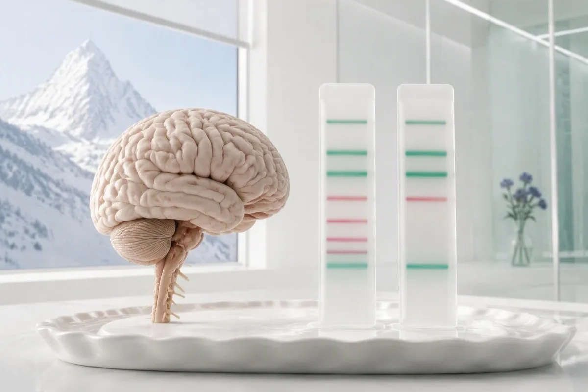
Positive CSF-restricted bands support antibody production within the central nervous system—not a diagnosis of multiple sclerosis by themselves. Matching serum bands usually point to a different explanation.


- CSF-restricted bands are IgG bands present in cerebrospinal fluid but absent from paired serum; many laboratories define positivity as at least 2 unique bands.
- Matched bands occur in both CSF and serum and do not, by themselves, establish antibody production within the central nervous system.
- Five laboratory patterns distinguish absent bands, CSF-restricted bands, mixed restricted and matched bands, mirror patterns, and monoclonal-looking patterns.
- Multiple sclerosis has CSF-restricted bands in approximately 90–95% of established cases in many predominantly European cohorts, but infections and other inflammatory disorders can also produce them.
- One isolated band is usually insufficient for a definite positive result; some laboratories also classify 2 bands as borderline.
- MRI interpretation depends on lesion location, appearance, and evolution—not simply the number of white-matter spots.
- CSF white cells are commonly 0–5 cells/µL in adults; counts above 50 cells/µL are unusual for typical MS and prompt a broader differential diagnosis.
- Treatment decisions require a clinical diagnosis or a defined high-risk state; positive bands alone do not justify steroids or MS medication.
What do positive oligoclonal bands actually mean?
Positive oligoclonal bands in CSF indicate a pattern of immunoglobulin G antibodies, but their meaning depends on comparison with serum. At least 2 CSF-only bands meet the positive threshold in many laboratories and support antibody production within the central nervous system; matching bands do not carry the same interpretation.
A positive result is evidence, not a diagnosis. Multiple sclerosis, certain nervous-system infections, and other inflammatory conditions can produce CSF-restricted bands, so the next 3 questions concern symptoms, MRI findings, and the rest of the spinal-fluid profile. Our explanation of positive antibody results addresses the same distinction between detecting an immune signal and identifying its cause.
The report's wording matters more than the word positive. A result stating “4 bands in CSF and 4 matching bands in serum” means something different from “4 CSF-restricted bands,” even though both may sound alarming when read quickly. I would first look for the laboratory's interpretive comment, whether a serum sample was tested, and whether additional unmatched bands were identified.
Kantesti is an AI blood test analyzer that helps explain accompanying laboratory results, but a 2-sample CSF band comparison belongs in specialist neurological interpretation. I’m Thomas Klein, MD, Chief Medical Officer at Kantesti LTD; this article is educational, not a substitute for examination or MRI review. Our organization and clinical focus explain the distinction between laboratory education and diagnosing neurological disease.
Why CSF and serum must be tested together
CSF oligoclonal bands interpretation requires paired cerebrospinal fluid and serum because antibodies circulating in blood can also appear in spinal fluid. Comparing the 2 specimens reveals whether bands are restricted to CSF, shared across both compartments, or a mixture of shared and CSF-only bands.
Serum is the liquid remaining after a blood sample clots; it is not another name for spinal fluid. The distinction matters because the same IgG antibody can be detected in 2 different specimen types without proving that it was made inside the nervous system. Our serum versus plasma explanation helps decode specimen terminology on the report.
Most laboratories prefer serum collected close to the lumbar puncture, often on the same day. A specimen collected months earlier introduces avoidable uncertainty because treatment, infection, or another immune event may change the circulating antibody pattern between the 2 collections. If the report lists only CSF, ask whether serum was processed separately, omitted from the patient portal, or never submitted.
Oligoclonal band testing is predominantly a pattern-based assay, not a concentration measurement. The laboratory usually separates IgG by isoelectric focusing and detects the resulting bands with an immunological technique; a count of 5 bands is therefore not “five times the normal antibody level.” The difference resembles qualitative versus quantitative testing, although a band count adds limited descriptive information.
The five oligoclonal band patterns neurologists distinguish
The standard 5-pattern classification separates CSF-restricted antibody production from systemic antibody patterns. Types 2 and 3 support intrathecal IgG synthesis, type 4 shows matching bands without that evidence, and type 5 suggests a monoclonal systemic pattern that needs a different laboratory assessment.
Type 1 means no oligoclonal bands are identified in either specimen, while type 2 means bands appear only in CSF. Type 3 combines shared serum-CSF bands with additional CSF-restricted bands, so it still supports local antibody production. Deisenhammer et al. described this classification in their 2019 review, emphasizing that paired testing—not merely seeing bands—is necessary for interpretation.
Type 4 is the mirror pattern: corresponding bands appear in both CSF and serum without additional restricted bands. Type 5 is a matching monoclonal-looking pattern, which can occur with a circulating monoclonal immunoglobulin and should not be treated as an MS signature. Ask the clinician to translate the report into one of these 5 patterns if the laboratory uses different wording.
The distinction between type 3 and type 4 is whether unmatched CSF bands remain after comparison. For example, 3 shared bands plus 2 additional CSF-only bands may support intrathecal synthesis, whereas 5 shared bands alone do not. Reading IgG, IgA, and IgM together can clarify the systemic immune background, but total serum IgG cannot determine the CSF pattern.
What CSF-restricted bands reveal about local immunity
CSF-restricted oligoclonal bands support IgG production within the central nervous system's immune compartment. This is called intrathecal synthesis; it does not identify the antibody's target, establish the cause, or measure how much myelin has been affected, even when 10 or more bands are reported.
Oligoclonal describes a limited set of antibody-producing cell populations, not a specific disease. IgG molecules from different populations separate into visible bands because their electrical properties differ; the assay is not reading an MS-specific antigen. A result with 8 restricted bands therefore says more about the diversity of the antibody response than about a patient's disability or future treatment needs.
Band counts are not a validated severity scale for multiple sclerosis. A patient with 3 restricted bands can have clinically significant disease, while someone with 12 bands may have a different inflammatory diagnosis or little current clinical activity. This is one of those situations where the reassuring or worrying part comes from the neurological pattern, not from ranking the laboratory count.
CSF oligoclonal testing and serum immunofixation answer different questions, despite both producing bands. The former commonly examines paired IgG patterns using isoelectric focusing; the latter helps identify monoclonal proteins and their immunoglobulin type. Our immunofixation band interpretation explains why a type 5 pattern may require a systemic protein assessment rather than an automatic MS workup.
Can you have oligoclonal bands without multiple sclerosis?
Yes—oligoclonal bands without multiple sclerosis occur in infections, autoimmune neurological disorders, and other inflammatory conditions. Approximately 90–95% of patients with established MS have CSF-restricted bands in many predominantly European cohorts, but that statistic cannot be reversed to mean that 90–95% of positive results represent MS.
The probability of MS after a positive result depends on why the test was ordered. Someone with typical optic neuritis and characteristic MRI changes starts with a different probability from someone with fever, neck stiffness, and marked CSF pleocytosis, even if both have 6 restricted bands. Deisenhammer et al. (2019) explain that specificity falls when MS is compared with other inflammatory neurological diseases.
Neuroborreliosis, neurosyphilis, HIV-associated neurological disease, and some viral nervous-system infections can produce intrathecal antibody responses. Neurosarcoidosis and selected autoimmune disorders belong in the differential too, but testing should follow the clinical clues rather than an indiscriminate panel of 20 antibodies. An ACE result in sarcoidosis is only supporting evidence; a normal serum ACE does not exclude neurosarcoidosis.
Systemic autoimmune clues can redirect the investigation before MS treatment is considered. Dry eyes, dry mouth, inflammatory joint symptoms, rash, or kidney abnormalities may justify selected tests such as ANA and SSA, while 1 positive autoantibody alone still cannot explain a neurological syndrome. Our SSA antibody interpretation separates clinically meaningful combinations from isolated laboratory positivity.
What matched serum and CSF bands mean
Matched serum and CSF bands usually reflect a circulating antibody pattern appearing in both compartments rather than demonstrable CSF-restricted synthesis. A type 4 mirror pattern does not fulfill the same role as CSF-restricted bands in MS diagnostic criteria, even when the laboratory detects 5 or more shared bands.
A mirror pattern can occur with systemic immune activation and may coexist with altered barrier function. It does not, by itself, prove that the blood-CSF barrier is abnormal; clinicians need measures such as the albumin quotient and the rest of the CSF profile to assess that possibility. Reviewing albumin and globulin patterns can explain why serum protein context matters across the 2 specimens.
Matched bands are not equivalent to a completely normal neurological evaluation. A patient with a type 4 result may still have an inflammatory or noninflammatory neurological disorder, and MS cannot be ruled out solely because restricted bands are absent. The practical interpretation is narrower: this particular result does not provide the usual CSF-restricted IgG evidence, so MRI and symptoms must carry their own weight.
A type 5 pattern deserves a different question: is there a circulating monoclonal protein? Depending on the report and clinical history, the next steps may include serum electrophoresis, immunofixation, and serum free light chains—not assuming cancer or MS from the band image. Our serum light-chain ratio guide concerns systemic protein assessment, which is distinct from a CSF kappa free-light-chain index.
How MRI and diagnostic criteria change the interpretation
Neurologists combine CSF findings with MRI features, examination findings, and the absence of a better explanation. Under the 2017 McDonald criteria, CSF-restricted bands could substitute for dissemination in time in a typical clinically isolated syndrome with MRI dissemination in space; the 2024 revisions provide additional diagnostic pathways.
MRI dissemination in space means characteristic abnormalities occur in distinct central nervous system regions—not simply that several white spots exist. The 2017 framework emphasized periventricular, cortical or juxtacortical, infratentorial, and spinal cord regions, while the 2024 revisions add the optic nerve as a fifth region. Migraine- or vascular-associated spots should not be counted as MS findings just because they are multiple.
CSF bands do not demonstrate that a new MRI abnormality appeared on a particular date. Their diagnostic role under the 2017 framework was an accepted substitute for temporal evidence in a carefully selected clinical setting, not permission to diagnose anyone with positive bands and nonspecific symptoms. Thompson et al. (2018) explicitly anchored that framework to a typical presentation and no better explanation.
The 2024 revisions broaden the use of imaging and CSF biomarkers, including intrathecal kappa free light chains in defined settings. Montalban et al. (2025) describe these updated criteria; as of October 11, 2026, the exact pathway should be identified rather than mixing old and new rules. Kantesti's clinical validation framework provides context for interpretation standards, not validation of an automated MS diagnosis.
Which symptoms make positive bands more concerning?
Positive bands carry more diagnostic weight when symptoms and examination suggest a central nervous system demyelinating event. Examples include typical optic neuritis, a partial spinal cord syndrome, or a brainstem syndrome; a classic MS relapse generally lasts at least 24 hours without fever or infection explaining the change.
An illustrative 29-year-old with painful one-eye visual loss over several days needs a different assessment from someone with brief, shifting tingling. The first scenario may fit optic neuritis, but alternative causes such as MOG antibody-associated disease or aquaporin-4 antibody-positive disease still need consideration when clinical features warrant it. The second scenario requires examination and history before positive bands are assigned a causal role.
Vitamin B12 deficiency can cause sensory changes and spinal cord dysfunction without being MS. A serum B12 result around 200–300 pg/mL is often considered borderline, although laboratory cutoffs differ, and methylmalonic acid may help resolve uncertainty in selected patients. Our borderline B12 follow-up guide explains why neurological symptoms can matter even when anemia is absent.
Copper deficiency is another treatable mimic, particularly after gastrointestinal surgery or prolonged excessive zinc intake. It can affect gait and sensation, but correcting copper deficiency would not automatically explain 4 CSF-restricted bands; clinicians must avoid forcing separate abnormalities into one diagnosis. Our low copper assessment guide outlines the history and laboratory combinations that make this possibility more plausible.
How white cells, protein, glucose, and IgG index help
The rest of the CSF profile can support an inflammatory interpretation or reveal findings atypical for MS. Adult CSF commonly contains 0–5 white cells/µL; counts above 50 cells/µL, markedly increased protein, or substantially reduced glucose should prompt consideration of infections and other diagnoses rather than being explained away by positive bands.
CSF protein is often reported against an adult interval near 15–45 mg/dL, but age and laboratory method change the appropriate upper limit. A value above 100 mg/dL is less typical of uncomplicated MS and deserves a broader explanation, although it is not a standalone diagnosis. Deisenhammer et al. (2019) describe how substantial pleocytosis and unusual chemistry should shift attention toward alternative neurological disorders.
CSF glucose must be interpreted with a paired serum glucose rather than against an isolated fixed number. CSF glucose is commonly around 60% of serum glucose, so 50 mg/dL may mean something different when serum glucose is 90 versus 200 mg/dL. A ratio below about 0.4 is concerning in the appropriate clinical setting, but sample timing, metabolic changes, and the full differential still matter.
The IgG index adjusts the CSF-to-serum IgG ratio using the CSF-to-serum albumin ratio; many laboratories use an upper limit near 0.7. A normal index does not negate genuine CSF-restricted bands, and an elevated index is not MS-specific. Similarly, WBC and CRP disagreement reminds readers that normal systemic inflammatory markers cannot exclude a process confined mainly to the nervous system.
What one band, borderline bands, or negative results mean
One CSF-restricted band is generally insufficient for definite oligoclonal band positivity, and laboratories differ over whether 2 bands are positive or borderline. A negative result lowers support for typical MS but does not exclude it; the correct next step depends on assay quality, clinical findings, and MRI evidence.
A report of 2 bands cannot be interpreted without the laboratory's cutoff. Some centers use at least 2 unique CSF bands, while others require at least 3 for a definite positive classification, particularly when faint bands create uncertainty. The threshold exists to balance detection of low-level intrathecal synthesis against overcalling weak, ambiguous, or technically inconsistent patterns; it is not a biological switch.
Treatment and sample quality can complicate interpretation, but negative bands should not casually be blamed on medication. Recent immunotherapy, technical limitations, or an incomplete paired comparison may warrant discussion, especially when MRI and examination strongly disagree with the result. Kantesti is an AI lab test interpretation service that explains these report-level limitations; 1 negative laboratory result cannot replace a neurological examination.
Unexpected discordance deserves reassessment before a lifelong diagnostic label is attached. In a suspected autoimmune neurological syndrome, antigen-specific testing may be appropriate, and its specimen choice can differ from oligoclonal band testing; a high serum antibody result is not automatically proof of central nervous system involvement. Our GAD65 neurological interpretation illustrates why titer, phenotype, and sometimes CSF findings must be considered together.
Do you need another lumbar puncture or repeat band test?
A repeat lumbar puncture is not routinely needed simply because oligoclonal bands are positive. Bands can persist for years, and repeating them every 6 or 12 months is not an established method of monitoring MS activity; repetition is usually considered only when it answers a specific unresolved diagnostic question.
A lumbar puncture samples the lumbar CSF space, usually at L3–L4 or L4–L5, below where the adult spinal cord commonly ends. Before another procedure is proposed, ask whether the original serum comparison can be completed using stored material and whether a missing report can be recovered. Sometimes the apparent need for repetition is a documentation problem rather than a biological uncertainty.
A traumatic sample can complicate cell counts and quantitative protein interpretation because circulating material enters the specimen. Paired qualitative band analysis may still be useful, but the laboratory needs to assess the specimen and interpret any limitations; a raised red-cell count alone does not automatically invalidate all CSF findings. Review the original document using our PDF report checking guide so specimen names and comments are not lost.
Post-lumbar-puncture headache often begins within the first few days and typically worsens upright. Severe or persistent headache, fever, new weakness, confusion, or difficulty walking should be assessed promptly rather than attributed to the band result, especially if symptoms change over 24–48 hours. Follow the procedure team's discharge instructions; a neurological change after lumbar puncture and an abnormal antibody pattern are separate clinical questions.
What happens next after a positive CSF result?
The next step is a neurological review that reconciles the band pattern with symptoms, examination, MRI, and other CSF results. Positive bands alone do not require steroids or disease-modifying MS therapy; if diagnostic criteria are not met, the plan may involve observation and an individualized MRI interval, sometimes 6–12 months.
A positive result with a normal MRI does not automatically establish MS. Depending on the presentation, the neurologist may review imaging quality, obtain spinal or optic-nerve assessment, investigate another inflammatory cause, or arrange follow-up rather than immediately starting treatment. Ask which uncertainty the next test addresses; 3 targeted investigations can be more useful than a broad collection of poorly chosen screening tests.
Incidental MRI findings require their own diagnostic framework rather than assuming symptoms are unnecessary or always required. The 2024 revisions permit diagnosis in selected circumstances beyond the classic symptomatic presentation, but this depends on specific combinations of evidence—not CSF bands in isolation. A patient should be told whether the working assessment is MS, a clinically isolated syndrome, a radiologically isolated syndrome, or a different condition.
Medication risk testing comes after the treatment question is defined. For example, the JCV antibody index helps assess progressive multifocal leukoencephalopathy risk in particular treatment settings, especially natalizumab, but it neither confirms MS nor explains why 7 restricted bands appeared. Our JCV index risk explanation clarifies this separate decision; do not start or stop prescribed neurological medication based on an isolated report.
Questions to ask your neurologist—and where AI stops
The most useful appointment questions identify the exact band pattern, the diagnostic framework, and the unresolved alternatives. Bring both specimen reports and the MRI interpretation, then ask 3 questions: are the bands CSF-restricted, which findings support the proposed diagnosis, and what would change the current follow-up plan?
Kantesti is an AI blood test interpretation platform that can help organize accompanying blood results, but it cannot supply the neurological examination or original MRI review. A 60-second explanation should not be mistaken for a 60-second MS diagnosis, and serum free light chains should not be relabeled as a CSF kappa index. Our AI technology guide describes how laboratory explanation differs from specialist diagnostic judgment.
Protecting a report means checking more than the patient's name. Remove unnecessary identifiers, confirm whose results are being discussed, and retain specimen dates and types because the timing of the 2 paired samples is clinically useful. Kantesti's report privacy checklist offers practical preparation steps; avoid sharing another family member's neurological records without their permission.
I’m Thomas Klein, MD, and my priority here is to separate 3 statements that often get collapsed: antibodies were detected, local synthesis is supported, and MS is diagnosed. Those statements are not interchangeable, and no universal conversion percentage can tell an individual their prognosis from a positive result alone. Sudden weakness, speech difficulty, rapidly worsening vision, fever with neck stiffness, or new confusion warrants urgent assessment rather than waiting for a routine interpretation.
Research sources, related publications, and evidence limits
The directly relevant evidence comes from MS diagnostic criteria and neurological CSF research, not general blood-test guides. The 3 external references below support the diagnostic and laboratory discussion; the 2 related Kantesti publications concern accompanying CBC and kidney results and do not validate an oligoclonal-band diagnosis.
The 2017 and 2024 McDonald frameworks should be identified by year when discussing a diagnostic decision. Thompson et al. (2018) explain the earlier CSF role, Montalban et al. (2025) describe the revisions, and Deisenhammer et al. (2019) provide detailed laboratory context. Our medical advisory board page describes clinical governance; this educational article does not replace individualized specialist review.
A DOI makes a publication identifiable; it does not establish peer review or diagnostic accuracy. Related publication 1 is Kantesti LTD. (n.d.). RDW Blood Test: Complete Guide to RDW-CV, MCV & MCHC. Zenodo. https://doi.org/10.5281/zenodo.18202598. The accompanying RDW and CBC guide can help explain anemia-related findings during neurological assessment, but RDW neither confirms nor excludes MS.
Kidney-function results may inform medication planning, but they do not classify CSF bands. Related publication 2 is Kantesti LTD. (n.d.). BUN/Creatinine Ratio Explained: Kidney Function Test Guide. Zenodo. https://doi.org/10.5281/zenodo.18207872. Our BUN and creatinine guide addresses that separate question; the publication records below include DOI links and repository-search links, not claims of independent neurological validation.
Frequently Asked Questions
Do positive oligoclonal bands in CSF mean I have multiple sclerosis?
Positive CSF-restricted oligoclonal bands support antibody production within the central nervous system, but they do not diagnose multiple sclerosis by themselves. Many laboratories require at least 2 unique CSF bands, while others use a threshold of 3. Nervous-system infections and other inflammatory conditions can also produce restricted bands. A neurologist must interpret the result alongside symptoms, examination, MRI, and the rest of the CSF profile.
What is the difference between CSF-restricted and matched oligoclonal bands?
CSF-restricted bands appear in cerebrospinal fluid but not in the paired serum specimen and support intrathecal IgG synthesis. Matched bands appear in both specimens and do not, by themselves, establish local antibody production. A type 3 pattern contains both matched bands and additional CSF-restricted bands, whereas a type 4 pattern contains matching bands without additional restricted bands. The 2 specimens should ideally be collected close together and compared by the laboratory.
Can you have oligoclonal bands with a normal MRI?
CSF-restricted oligoclonal bands can occur when a brain MRI is normal, but that combination does not automatically establish multiple sclerosis. The neurologist may consider spinal imaging, optic-nerve assessment, alternative inflammatory causes, or follow-up depending on the presentation. Some patients need an individualized repeat MRI interval, such as 6–12 months, rather than immediate treatment. A normal brain MRI also cannot substitute for examining the patient or reviewing whether the imaging addressed the symptomatic region.
What does one oligoclonal band mean?
One CSF-restricted band is generally insufficient for a definite positive oligoclonal-band classification. Laboratories differ over whether 2 bands are positive or borderline, while some require at least 3 unique CSF bands. A faint or isolated band should be interpreted using the laboratory's method, paired serum comparison, and explanatory comment. It is not a measure of neurological disease severity.
Can multiple sclerosis occur without oligoclonal bands?
Multiple sclerosis can occur without detectable CSF-restricted oligoclonal bands. Approximately 90–95% of established MS cases have restricted bands in many predominantly European cohorts, so a minority do not. However, a negative result should prompt careful reconsideration when the clinical presentation or MRI is atypical. Diagnosis depends on the applicable criteria and exclusion of a better explanation, not one laboratory finding.
Do oligoclonal bands disappear after treatment?
Oligoclonal bands can persist for years despite changes in symptoms, MRI activity, or treatment. Band counts are not a validated measure of MS activity, and repeating the test every 6 or 12 months is not routine monitoring. Another lumbar puncture is usually considered only when a specific diagnostic uncertainty remains. Treatment response is assessed using clinical findings and other appropriate measures rather than expecting bands to disappear.
Is a positive oligoclonal band result an emergency?
An isolated positive oligoclonal-band result is not usually an emergency and does not by itself require steroids or MS medication. Urgency depends on the accompanying symptoms, especially sudden weakness, speech difficulty, rapidly worsening vision, fever with neck stiffness, or confusion. A typical MS relapse definition often includes symptoms lasting at least 24 hours, but that is not a reason to delay assessment of sudden neurological symptoms. Contact the requesting clinician for the interpretation plan, and seek urgent care for acute warning signs.
Get AI-Powered Blood Test Analysis Today
Join over 2 million users worldwide who trust Kantesti for instant, accurate lab test analysis. Upload your blood test results and receive comprehensive interpretation of 15,000+ biomarkers in seconds.
📚 Referenced Research Publications
Klein, T., Mitchell, S., & Weber, H. (2026). RDW Blood Test: Complete Guide to RDW-CV, MCV & MCHC. Kantesti AI Medical Research.
Klein, T., Mitchell, S., & Weber, H. (2026). BUN/Creatinine Ratio Explained: Kidney Function Test Guide. Kantesti AI Medical Research.
📖 External Medical References
Montalban X et al. (2025). Diagnosis of multiple sclerosis: 2024 revisions of the McDonald criteria. The Lancet Neurology.
Deisenhammer F et al. (2019). The Cerebrospinal Fluid in Multiple Sclerosis. Frontiers in Immunology.
⚕️ Medical Disclaimer
This article is for educational purposes only and does not constitute medical advice. Always consult a qualified healthcare provider for diagnosis and treatment decisions.
E-E-A-T Trust Signals
Experience
Physician-led clinical review of lab interpretation workflows.
Expertise
Laboratory medicine focus on how biomarkers behave in clinical context.
Authoritativeness
Written by Dr. Thomas Klein with review by Dr. Sarah Mitchell and Prof. Dr. Hans Weber.
Trustworthiness
Evidence-based interpretation with clear follow-up pathways to reduce alarm.