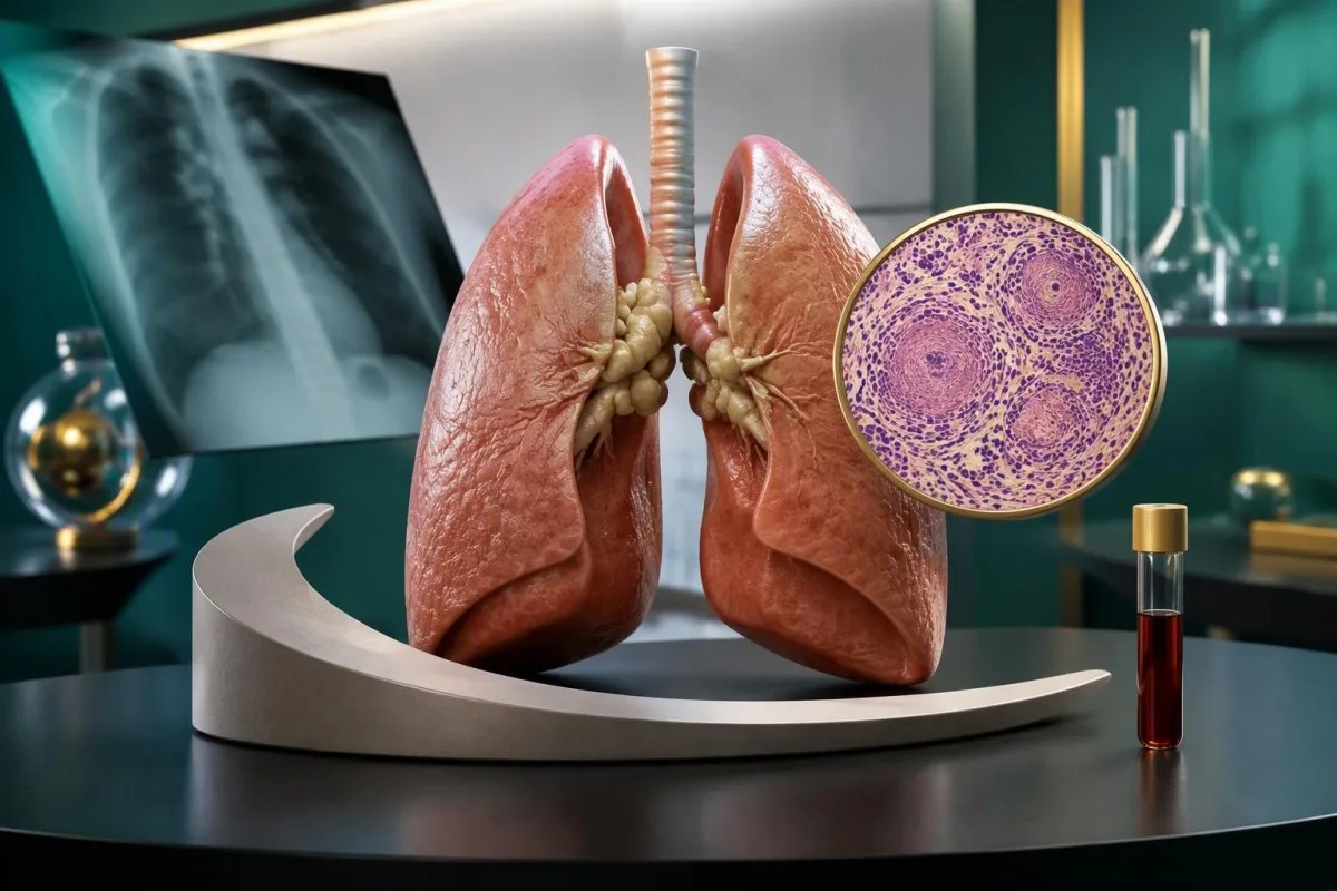
Elevated ACE does not confirm sarcoidosis, and a normal result cannot exclude it. Your medication list, organ symptoms, chest imaging, and sometimes tissue examination provide stronger evidence.


- Diagnostic limits: A meta-analysis estimated serum ACE sensitivity at 60% and specificity at 93%; neither a high nor a normal result settles the diagnosis.
- Reference interval: Some laboratories use an adult ACE range of 16–85 U/L, but your own report's interval and assay method take priority.
- ACE inhibitors: Lisinopril, ramipril, enalapril, and related medicines can suppress measured ACE activity; do not stop prescribed treatment to improve a test result.
- Probability matters: With an assumed 10% chance of sarcoidosis before testing, 60% sensitivity and 93% specificity produce a positive predictive value of approximately 49%.
- Imaging: Chest X-ray and selected CT examinations can identify enlarged chest lymph nodes and lung patterns that ACE cannot locate.
- Biopsy: Sarcoidosis diagnosis generally combines a compatible clinical picture, nonnecrotizing granulomas when tissue is required, and exclusion of alternative causes.
- Calcium: A calcium result of 12 mg/dL, equivalent to 3.0 mmol/L, needs prompt clinical assessment, particularly with dehydration, vomiting, confusion, or kidney problems.
- Monitoring: An ACE change from 110 to 70 U/L does not prove recovery if breathing, lung function, or another affected organ is worsening.
What does a high ACE result actually mean?
The ACE blood test for sarcoidosis measures an enzyme that can increase when granulomas are present, but high ACE levels do not confirm sarcoidosis. A normal result cannot exclude it either: pooled sensitivity is approximately 60%, and ACE-inhibitor medicines can suppress the measurement even when clinically significant disease is present.
A flagged result is a clue, not a diagnosis. An ACE of 92 U/L against a laboratory upper limit of 85 U/L deserves a different conversation from persistent elevation accompanied by enlarged chest lymph nodes, eye symptoms, or abnormal calcium; our explanation of out-of-range results separates laboratory flags from clinical decisions.
Consider an illustrative 46-year-old with a persistent cough and an ACE of 105 U/L: the next useful question is whether chest imaging and examination suggest granulomatous disease, not whether the number crosses an internet cutoff. By contrast, the same value in someone without symptoms or imaging abnormalities may justify reviewing medications, the assay, and alternative explanations before pursuing invasive testing.
Kantesti is an AI blood test analyzer that can help explain an ACE result alongside the other values on a laboratory report, without turning that result into a diagnosis. I am Thomas Klein, MD, the Chief Medical Officer named for this article; my clinical emphasis here is restraint rather than premature labeling, and readers can find our organizational details on About Us.
What does the angiotensin converting enzyme test measure?
The angiotensin converting enzyme test usually measures serum ACE activity, rather than counting granulomas or directly measuring lung damage. ACE converts angiotensin I into angiotensin II and also breaks down bradykinin; these functions explain why medicines acting on ACE can alter the laboratory result as well as blood pressure.
Granulomas contain organized collections of immune cells, including macrophages and epithelioid cells that can produce ACE. More granulomatous tissue may increase circulating enzyme activity, but production varies between people, and a small collection in a clinically sensitive organ can matter more than a larger collection elsewhere; no U/L value identifies the affected location.
A serum enzyme activity result is not interchangeable with a protein concentration reported in mg/L. Differences in substrate, reaction conditions, calibration, and laboratory technique mean that two ACE measurements cannot always be compared directly, much as other serum protein measurements need their own units and biological context.
Blood pressure and serum ACE activity answer different questions. Someone can have hypertension with an ACE result inside the laboratory interval, or elevated ACE with normal blood pressure; the test is not a substitute for repeated pressure measurements, kidney assessment, or evaluation of cardiovascular risk, and it does not reveal the body's entire renin–angiotensin system.
What is the normal ACE range in adults?
There is no universal normal ACE range: some laboratories use approximately 16–85 U/L for adults, while others use substantially different limits. Interpret sarcoidosis ACE levels against the interval printed on the actual report, because the assay method, age group, and local reference population affect what counts as elevated.
An ACE of 70 U/L could be normal in one laboratory and high in another. Dividing a value by that laboratory's upper limit can make the degree of elevation easier to describe—for example, 102 U/L divided by 85 U/L is 1.2 times the upper limit—but it does not create a validated diagnostic or treatment threshold.
Children often have higher ACE activity than adults, so an adult cutoff should not be pasted onto a pediatric report. Puberty, the laboratory's pediatric interval, symptoms, and the reason for testing all matter; our discussion of children's laboratory interpretation explains why age-specific review is more than a technical detail.
A reference interval describes results in a selected reference population; it is not a line separating sarcoidosis from everyone without it. There is also no accepted ACE concentration that automatically means an emergency, so a report marked high should lead to clinical review rather than assumptions about disease stage, permanent damage, or an immediate need for steroids.
How accurate is ACE for diagnosing sarcoidosis?
Serum ACE has insufficient sensitivity to exclude sarcoidosis and insufficient specificity to confirm it alone. Hu et al. (2022), in a meta-analysis published in Biomolecules, estimated pooled diagnostic sensitivity of approximately 60% and specificity of approximately 93%, with substantial variation between studies, populations, and testing methods.
Sensitivity of 60% means approximately 40 of every 100 people with sarcoidosis could have a non-elevated result under those study conditions. Specificity of 93% means approximately 7 of every 100 people without sarcoidosis could test positive; neither percentage is a personalized prediction, particularly when medicines or differences in patient selection change test performance.
Imagine 1,000 people whose estimated chance of sarcoidosis before testing is 10%, using those pooled figures purely for illustration. There would be about 60 true-positive and 63 false-positive results, making the probability of sarcoidosis after a positive test approximately 49%; among negative results, the remaining probability would be about 4.6%, not zero.
At a pretest probability of just 1%, the same assumptions yield a positive predictive value of approximately 8%. This is why broad testing for unexplained fatigue can generate misleading alarms, while a result obtained after suggestive imaging has a different meaning; the same probability principle appears in our explanation of D-dimer false positives, although the tests serve different clinical purposes.
How do ACE-inhibitor medicines affect the result?
ACE inhibitors can markedly lower measured serum ACE activity, making the result unreliable for assessing sarcoidosis in someone taking these medicines. Lisinopril, ramipril, enalapril, perindopril, and captopril are examples; the laboratory and ordering clinician need the actual medication names, not simply the description 'a blood-pressure tablet.'
A hypothetical patient taking lisinopril 10 mg daily could have low measured ACE despite active granulomatous disease; the dose is an example, not a threshold below which interference disappears. ACE inhibitors can also cause a dry cough, so both the symptom and the laboratory result need a medication review rather than an automatic sarcoidosis label.
Do not stop an ACE inhibitor to obtain a more reassuring or more interpretable number. These medicines may protect the heart or kidneys, and any interruption requires the prescriber's decision; there is no single patient-directed washout interval that works for every drug and assay, a recurring concern in medication-affected laboratory results.
Angiotensin receptor blockers such as losartan do not directly inhibit ACE in the way ACE inhibitors do, so the two drug classes should not be treated as identical assay confounders. Corticosteroids can lower ACE by reducing granulomatous activity, and recording treatment start dates alongside results makes medication safety trends more useful than comparing two isolated numbers.
What else can cause high ACE levels?
High ACE levels are not specific to sarcoidosis and may occur in other granulomatous disorders, Gaucher disease, and some endocrine or metabolic conditions. Genetic variation also influences baseline ACE activity; the sensible differential depends on exposure history, symptoms, examination, and accompanying abnormalities rather than a long list of rare possibilities.
Tuberculosis and certain fungal diseases can resemble sarcoidosis clinically or on tissue examination, so geography, travel, immune suppression, and exposure history matter. A positive tuberculosis IGRA indicates immune sensitization, not proof that chest findings represent active tuberculosis; a negative result also cannot reliably exclude active disease, as our IGRA interpretation guide explains.
Chronic beryllium disease can closely resemble pulmonary sarcoidosis, particularly after work involving aerospace components, electronics, or metal processing. A clinician may consider a beryllium lymphocyte proliferation test when the history fits; ACE activity cannot distinguish these conditions, and even a remote occupational exposure can be more informative than a second elevated enzyme result.
Hyperthyroidism and diabetes have been associated with increased ACE in some settings, but an elevated result does not establish either diagnosis. Thyroid evaluation starts with the appropriate hormone pattern rather than ACE, and our explanation of T4 and thyroid testing helps distinguish a relevant endocrine clue from a coincidental laboratory finding; routine ACE genotyping is not a standard sarcoidosis diagnostic step.
Can you have sarcoidosis with a normal ACE result?
Yes—sarcoidosis can be present with normal serum ACE, including disease affecting the lungs, eyes, heart, or nervous system. With pooled sensitivity near 60%, normal ACE leaves a meaningful false-negative problem; localized disease, individual biology, ACE-inhibitor treatment, and previous anti-inflammatory treatment can all weaken the usefulness of a negative result.
Consider an illustrative 39-year-old with breathlessness, bilateral hilar lymph-node enlargement, and ACE of 42 U/L within the local reference interval. The imaging pattern still needs assessment, because the enzyme result neither explains the breathlessness nor rules out sarcoidosis; pulmonary function, oxygenation, exposure history, and the need for tissue sampling are separate questions.
Shortness of breath deserves its own work-up, not repeated ACE measurements until one becomes abnormal. Anemia, asthma, heart disease, pulmonary embolism, and other lung conditions can coexist with or mimic sarcoidosis, and our guide to breathlessness blood tests explains why laboratory testing is only one part of that assessment.
Normal ACE is especially unhelpful as reassurance when the concern is an organ where small amounts of disease can cause substantial harm. New visual disturbance, unexplained fainting, or a focal neurological symptom should be evaluated on its clinical merits; waiting for an ACE elevation may delay the examination or imaging that actually answers the urgent question.
When are chest X-ray, CT, or cardiac imaging more useful?
Chest imaging is more useful than ACE for locating thoracic abnormalities, including enlarged hilar lymph nodes and changes in lung tissue. Chest X-ray is often the initial study, while CT can clarify an atypical or uncertain pattern; ACE cannot identify the distribution of disease or distinguish active tissue changes from established scarring.
The BTS Clinical Statement on pulmonary sarcoidosis emphasizes interpreting imaging within a compatible clinical presentation (Thillai et al., 2021). Symmetric hilar or mediastinal lymph-node enlargement and a perilymphatic distribution of small lung nodules can support suspicion, but these patterns are not unique to sarcoidosis; CT should answer a clinical question rather than follow automatically from a mildly elevated ACE.
Chest X-ray stages I through IV describe radiographic patterns, not a simple ladder of current symptoms or treatment need. Stage I refers to hilar lymphadenopathy without visible lung infiltrates, whereas stage IV indicates fibrosis; a person with little on a plain film can still have important extrapulmonary disease, and radiographic stage does not replace lung-function assessment.
For suspected cardiac involvement, ECG findings, rhythm monitoring, cardiac MRI, and selected FDG-PET examinations may provide evidence that ACE cannot. FDG uptake is not specific to sarcoidosis and requires preparation and specialist interpretation; our discussion of myocarditis test limitations similarly explains why a normal or abnormal blood marker cannot settle a cardiac imaging question.
When does biopsy provide stronger diagnostic evidence?
Biopsy can provide stronger evidence by demonstrating granulomas, but granulomas alone do not prove sarcoidosis. The ATS diagnostic guideline describes three central requirements: a compatible clinical presentation, nonnecrotizing granulomatous inflammation when tissue is needed, and exclusion of alternative causes; even with these elements, diagnostic certainty is not absolute (Crouser et al., 2020).
The best sampling site is often the safest accessible site likely to answer the question, such as an appropriate skin lesion or peripheral lymph node. When chest lymph-node sampling is required, the ATS guideline favors endobronchial ultrasound-guided sampling over mediastinoscopy as the initial approach in suitable patients; that recommendation does not mean every elevated ACE warrants bronchoscopy.
A pathology report saying nonnecrotizing granulomas still requires a conversation about infections, occupational exposures, drug reactions, and other granulomatous conditions. Cultures, special stains, and additional tests depend on the specimen and history; lymph-node disorders also have distinct confirmation pathways, illustrated in our guide to Bartonella antibody results, rather than a single universal blood test.
Some highly characteristic presentations can be assessed without immediate tissue sampling after specialist review. Löfgren syndrome—classically erythema nodosum, bilateral hilar lymphadenopathy, and ankle arthritis—may support that approach, while asymptomatic bilateral hilar lymphadenopathy requires an individualized decision; choosing observation instead of biopsy should include follow-up, not an assumption that normal ACE makes the issue disappear.
Which other blood and urine tests matter more?
Calcium, kidney function, liver tests, and a complete blood count can identify organ effects that are more actionable than ACE alone. The ATS guideline recommends baseline serum calcium assessment in sarcoidosis and suggests several other baseline checks, including creatinine, alkaline phosphatase, ECG, and an eye examination, tailored to the clinical setting (Crouser et al., 2020).
Granuloma-associated macrophages can convert vitamin D into its active form outside normal kidney regulation, increasing calcium absorption. A low 25-hydroxyvitamin D result therefore does not automatically justify high-dose replacement in suspected sarcoidosis; when supplementation is being considered, calcium and sometimes both vitamin D metabolites need review, particularly with slightly elevated calcium.
Serum calcium can be normal while urinary calcium is elevated, especially when stones or kidney symptoms raise concern. A properly collected 24-hour urine calcium test may answer a specific question, but collection errors and dietary factors affect interpretation; our guide to high urinary calcium explains why this result is not simply a second version of serum calcium.
Kantesti is an AI blood test interpretation platform that helps readers organize laboratory abnormalities, including ACE, calcium, creatinine, and liver markers, for discussion with a clinician. Our blood biomarker guide supports that report-reading task; no blood-panel interpretation can replace the ECG, eye examination, imaging, or tissue evidence needed when an organ-specific complication is suspected.
When should you see a specialist or seek urgent care?
Specialist assessment is appropriate when suspected sarcoidosis involves abnormal imaging, persistent respiratory symptoms, or possible eye, heart, kidney, or nervous-system disease. Urgency is determined by symptoms and organ function, not by the height of ACE; new vision loss, fainting with palpitations, severe breathlessness, or focal neurological symptoms need prompt evaluation.
A painful red eye, light sensitivity, or new blurred vision can indicate uveitis and warrants urgent ophthalmic assessment. Sarcoidosis is one possible cause, not the only one; our discussion of HLA-B27 and uveitis shows why different systemic conditions can lead to similar eye symptoms, and a normal ACE should not delay an eye examination.
Palpitations accompanied by fainting, chest discomfort, or marked dizziness need urgent cardiac evaluation because rhythm or conduction problems can be consequential. An ECG and appropriate monitoring are more informative than repeating ACE, while our guide to palpitations laboratory assessment explains the supporting role of electrolytes, thyroid tests, and other selected investigations.
Calcium of 12 mg/dL, equivalent to 3.0 mmol/L, needs prompt assessment, especially with vomiting, dehydration, confusion, or worsening kidney function; values around 14 mg/dL, or 3.5 mmol/L, generally represent severe hypercalcemia. Emergency symptoms should trigger local emergency care, whereas isolated ACE elevation without concerning symptoms usually allows an organized outpatient assessment rather than an emergency visit.
Can ACE levels track sarcoidosis treatment or relapse?
ACE may help follow selected patients whose levels were elevated before treatment, but it is an unreliable stand-alone measure of remission, relapse, or treatment need. Symptoms, pulmonary function, imaging when indicated, and organ-specific findings take priority; an improving enzyme result does not prove that lung scarring, cardiac involvement, or eye disease has improved.
An illustrative ACE fall from 110 to 70 U/L after treatment may support reduced granulomatous activity if the same assay was used and medicines were unchanged. If breathlessness or diffusion capacity worsens at the same time, the discordance matters more than the reassuring number; ACE can fall while another process, established fibrosis, or a complication still limits function.
The BTS statement includes baseline ACE testing with non-unanimous consensus, which reflects genuine disagreement about its usefulness rather than a requirement to track it in everyone (Thillai et al., 2021). There is no universal ACE change percentage that defines a flare, and follow-up intervals should reflect the affected organ and treatment plan rather than a fixed internet retesting schedule.
Treatment is aimed at protecting organs and improving meaningful symptoms, not normalizing ACE on its own. Before significant immunosuppression, clinicians may assess infection risks according to exposure history; for patients with relevant residence or travel, our guide to Strongyloides before immunosuppression explains a safety issue that can be more consequential than whether ACE has fallen.
How should you prepare for an ACE result review?
Bring the original report, its reference interval, your complete medication list, and any imaging or previous results to an ACE review. Fasting is not generally required for an isolated ACE test unless your laboratory gives different instructions or other ordered tests require it; do not alter medicines, supplements, or treatment simply to influence the number.
Four details often change interpretation: the actual value and units, the laboratory's interval, whether an ACE inhibitor is being taken, and whether corticosteroids or other treatment preceded the sample. Also note the timing of cough, breathlessness, eye symptoms, stones, or palpitations; a result obtained before a medication change is not necessarily comparable with one obtained afterward.
Kantesti is an AI-powered blood test analysis tool that can help explain an uploaded report, but the original document should remain the source of truth for values and units. Our AI technology guide describes the interpretation workflow; users should still verify transcription, supply relevant context, and avoid treating an AI explanation as confirmation of sarcoidosis.
As Thomas Klein, MD, I would prioritize a clear question for the treating clinician: 'What evidence would change the plan—imaging, a specialist examination, tissue sampling, or observation?' Kantesti's technical validation information should be read within its stated scope, because a blood-report interpretation benchmark does not establish diagnostic accuracy for ACE, sarcoidosis, or every clinical decision that follows.
Research publications and the limits of AI evidence
The medical evidence for ACE interpretation comes from sarcoidosis guidelines and diagnostic studies, not general AI engineering benchmarks. As of October 11, 2026, the publications listed below should be read according to their own methods and scope; neither a hantavirus deployment report nor a synthetic-case benchmark validates ACE-based diagnosis of sarcoidosis.
The ATS guideline by Crouser et al. (2020), the BTS statement by Thillai et al. (2021), and the meta-analysis by Hu et al. (2022) address clinical questions relevant to this article. Their limitations still matter: heterogeneous diagnostic studies do not establish one universal ACE cutoff, and consensus recommendations do not remove the need to exclude competing explanations in an individual patient.
The two Figshare records supplied for this article concern multilingual hantavirus decision support and a benchmark using 100,000 synthetic test cases. Their titles, repository, and DOI links are reproduced in the publication entries, with discovery links clearly identified as searches; a DOI is a persistent identifier, not proof of peer review, prospective clinical effectiveness, or validation for this particular condition.
Kantesti's clinical and technical materials should support questions for a qualified professional, not replace that professional's assessment of imaging, tissue findings, or organ risk. Readers can consult the Medical Advisory Board for organizational oversight information; the practical decision remains the same whether ACE is 42 or 105 U/L—identify what evidence is missing before accepting or dismissing sarcoidosis.
Frequently Asked Questions
Does a high ACE blood test mean I have sarcoidosis?
A high ACE blood test does not confirm sarcoidosis because ACE can rise in other granulomatous disorders and some non-granulomatous conditions. A 2022 meta-analysis estimated sensitivity of approximately 60% and specificity of approximately 93%, so false-positive and false-negative results occur. The result becomes more useful when symptoms, examination, and chest imaging already suggest sarcoidosis. Tissue sampling and exclusion of alternative causes may be needed before making the diagnosis.
Can sarcoidosis occur with normal ACE levels?
Sarcoidosis can occur with normal ACE levels, including disease affecting the lungs, eyes, heart, or nervous system. Pooled sensitivity near 60% means that approximately 40% of affected patients could have a non-elevated result under the study conditions. ACE inhibitors and previous treatment can further reduce measured activity. Persistent symptoms or suggestive imaging therefore need assessment even when ACE is normal.
What ACE level is considered high?
An ACE level is high when it exceeds the reference interval used by the laboratory that performed the test. Some laboratories use an adult interval of approximately 16–85 U/L, but other laboratories use different limits and methods. A value of 90 U/L is elevated against an upper limit of 85 U/L, but it does not establish sarcoidosis or determine disease severity. Pediatric results require age-appropriate intervals rather than adult cutoffs.
Does lisinopril affect the ACE blood test?
Lisinopril and other ACE inhibitors can suppress measured serum ACE activity and make the test unreliable for assessing sarcoidosis. A person prescribed lisinopril 10 mg daily, for example, may have low activity despite clinically significant disease; this dose is illustrative, not an interference threshold. Tell the ordering clinician and laboratory which medicines you take. Do not stop lisinopril or any prescribed ACE inhibitor without the prescriber's advice.
Do I need a biopsy if my ACE is elevated?
An elevated ACE result alone is not a reason to perform a biopsy. Sarcoidosis assessment generally considers three elements: a compatible clinical presentation, granulomas when tissue evidence is required, and exclusion of alternative causes. Some characteristic presentations can be assessed without immediate tissue sampling after specialist review, while uncertain imaging or competing diagnoses may make sampling more useful. When biopsy is needed, clinicians usually select the safest suitable site likely to answer the diagnostic question.
Can falling ACE levels show that sarcoidosis is improving?
Falling ACE levels may support improvement in selected patients, but they do not prove remission. A fall from 110 to 70 U/L is meaningful only in context, including the assay method, medicines, symptoms, and affected organ function. Corticosteroids or an ACE inhibitor can lower the measurement, and established scarring may persist despite lower ACE. Treatment decisions should not be based on ACE trends alone.
Do I need to fast before an ACE blood test?
Fasting is not generally required for an isolated ACE blood test unless the laboratory provides different instructions. If ACE is ordered with other tests, such as an 8-hour fasting glucose measurement, the preparation requirements may come from those additional tests. Bring a complete medication list because ACE inhibitors and prior corticosteroid treatment can alter interpretation. Do not change prescribed treatment just to prepare for the sample.
Get AI-Powered Blood Test Analysis Today
Join over 2 million users worldwide who trust Kantesti for instant, accurate lab test analysis. Upload your blood test results and receive comprehensive interpretation of 15,000+ biomarkers in seconds.
📚 Referenced Research Publications
Klein, T., Mitchell, S., & Weber, H. (2026). Multilingual AI Assisted Clinical Decision Support for Early Hantavirus Triage: Design, Engineering Validation, and Real-World Deployment Across 50,000 Interpreted Blood Test Reports. Kantesti AI Medical Research.
Klein, T., Mitchell, S., & Weber, H. (2026). A Pre-Registered, Rubric-Based Automated Technical Benchmark of the Kantesti Blood-Test Interpretation Engine on 100,000 Synthetic Test Cases. Kantesti AI Medical Research.
📖 External Medical References
Thillai M et al. (2021). BTS Clinical Statement on pulmonary sarcoidosis. Thorax.
Hu X et al. (2022). Performance of Serum Angiotensin-Converting Enzyme in Diagnosing Sarcoidosis and Predicting the Active Status of Sarcoidosis: A Meta-Analysis. Biomolecules.
⚕️ Medical Disclaimer
This article is for educational purposes only and does not constitute medical advice. Always consult a qualified healthcare provider for diagnosis and treatment decisions.
E-E-A-T Trust Signals
Experience
Physician-led clinical review of lab interpretation workflows.
Expertise
Laboratory medicine focus on how biomarkers behave in clinical context.
Authoritativeness
Written by Dr. Thomas Klein with review by Dr. Sarah Mitchell and Prof. Dr. Hans Weber.
Trustworthiness
Evidence-based interpretation with clear follow-up pathways to reduce alarm.