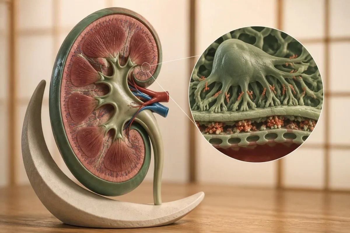
A positive antibody result can identify the immune process behind kidney protein leakage. It cannot, by itself, tell you how well your kidneys filter or whether a biopsy would add useful information.


- PLA2R antibodies support a diagnosis of PLA2R-associated membranous nephropathy, especially when nephrotic syndrome is present; they do not measure kidney filtration.
- A positive result may allow diagnosis without biopsy in selected patients, but unexplained kidney impairment, an unusual urine sediment, or suspected additional disease can change that decision.
- A negative result does not exclude membranous nephropathy: circulating antibodies are detectable in roughly 70–80% of untreated primary cases, depending on the population and assay.
- ELISA thresholds commonly include less than 14 RU/mL as negative, 14 to less than 20 RU/mL as borderline, and at least 20 RU/mL as positive; your laboratory's thresholds take priority.
- Antibody monitoring is often performed every 3–6 months, with additional testing around treatment decisions; use the same laboratory and method whenever possible.
- Immunologic improvement can precede a reduction in urine protein by several months, so a falling antibody level is encouraging even when proteinuria has not yet resolved.
- Kidney assessment still requires creatinine-based eGFR, urine protein measurement, serum albumin, blood pressure, and clinical review.
- Urgent symptoms such as sudden breathlessness, chest pain, one-sided leg swelling, or markedly reduced urine output need prompt assessment regardless of the antibody result.
What do PLA2R antibodies mean for your diagnosis?
PLA2R antibodies support a diagnosis of PLA2R-associated membranous nephropathy, particularly in someone with nephrotic syndrome. A negative result does not exclude it; selected positive patients can avoid biopsy, while atypical findings may require one. Antibody trends reflect immune activity—not kidney filtration, which needs separate assessment.
Membranous nephropathy is a disorder of the kidney's filtering structures that often causes substantial protein loss into urine; adult nephrotic-range proteinuria is conventionally above 3.5 g/day. A PLA2R antibody positive result becomes particularly persuasive when that protein loss accompanies low serum albumin and swelling, rather than appearing as an isolated finding on an unrelated screening panel.
Kantesti is an AI blood test analyzer that can help explain a PLA2R result alongside creatinine, albumin, and the laboratory's stated cutoff. Our organization and clinical purpose are separate from specialist diagnosis: even a result of 200 RU/mL cannot reveal how much permanent kidney scarring exists or establish the correct treatment on its own.
The clinical framework presented here by Thomas Klein, MD, separates 3 questions: what immune process is present, how much protein the kidney is losing, and how well filtration is preserved. Keeping those questions separate prevents a common misunderstanding—interpreting a falling antibody number as proof that every aspect of kidney health has already recovered.
How do anti-PLA2R antibodies affect kidney filters?
Anti-PLA2R antibodies bind the M-type phospholipase A2 receptor on podocytes, specialized cells surrounding the kidney's filtering capillaries. The resulting immune deposits and local complement activity can disrupt the filtration barrier, allowing albumin to enter urine even while overall filtration remains relatively well preserved.
PLA2R means phospholipase A2 receptor; the antibody is directed against the receptor, not against a routine digestive enzyme measurement. Beck and colleagues identified circulating antibodies in 26 of 37 patients with idiopathic membranous nephropathy in their landmark discovery study, establishing a disease-specific target rather than merely another nonspecific autoimmune signal (Beck et al., 2009).
The deposits in membranous nephropathy typically sit on the subepithelial side of the glomerular basement membrane, next to podocytes; this differs from several kidney disorders that produce more inflammatory urine findings. A person can therefore lose 6 g of protein per day without prominent visible blood in urine, although microscopic hematuria can occur and does not automatically invalidate the diagnosis.
An antibody result identifies a target-specific immune response, not simply a stronger or weaker version of a general autoantibody screen; 2 antibody tests with similar numerical values may have entirely different meanings. Our explanation of different positive antibody results helps distinguish autoimmunity from past infection, immunity, and other reasons a laboratory report says positive.
Who should have a PLA2R antibody test?
PLA2R testing is most useful when membranous nephropathy is suspected, especially in adults with substantial proteinuria, low albumin, or otherwise unexplained nephrotic syndrome. It is also useful after a biopsy diagnosis to establish an antibody baseline for subsequent monitoring.
Persistent foamy urine is a reason to check urine protein, not a reason to jump directly to autoimmune testing; bubbles alone cannot establish proteinuria of 3.5 g/day or any other amount. When foam persists alongside ankle swelling or puffy eyelids, a urine albumin or protein measurement provides a much more useful first step, as explained in our persistent foamy urine guide.
Consider an illustrative 52-year-old with ankle swelling, albumin of 2.4 g/dL, urine protein of 7 g/day, and an eGFR of 88 mL/min/1.73 m²: PLA2R testing addresses a plausible explanation for an established nephrotic pattern. Ordering the same test for a well person with a normal urinalysis changes its predictive value and creates more uncertainty around weakly positive or borderline findings.
Pretreatment testing is preferable when feasible because immunosuppression can reduce circulating antibodies before proteinuria improves; a negative sample obtained 3 months after treatment answers a different question from a negative sample obtained at presentation. The request should ideally record previous kidney findings and treatment dates, although medically necessary treatment should never be delayed simply to obtain a cleaner baseline.
How should you read anti-PLA2R antibody levels?
Anti-PLA2R antibody levels must be interpreted using the reporting laboratory's assay and thresholds. Some ELISA methods use less than 14 RU/mL as negative, 14 to less than 20 RU/mL as borderline, and at least 20 RU/mL as positive; these are not universal definitions.
RU/mL means relative units per milliliter, not a concentration that converts reliably into mg/dL or another laboratory's units. A result of 18 RU/mL may fall within a borderline category on one method, while a different method uses another decision threshold; the distinction between qualitative and quantitative testing explains why positive/negative reports and numerical results cannot always be substituted.
A borderline result deserves clinical correlation rather than an automatic diagnosis: 16 RU/mL in a patient with 8 g/day of proteinuria carries a different implication from 16 RU/mL with repeatedly normal urine protein. Depending on the circumstances, a nephrologist may request repeat testing or confirmation using indirect immunofluorescence, which detects antibody binding through a different testing approach.
Diagnostic negativity and complete immunologic remission are not necessarily the same threshold; KDIGO 2021 discusses an ELISA value below 2 RU/mL for complete immunologic remission with applicable assays, substantially below a common diagnostic cutoff of 14 RU/mL. That distinction matters after treatment: a report marked negative at 10 RU/mL may not mean the antibody is undetectable by a more sensitive method.
Why can membranous nephropathy be PLA2R negative?
PLA2R negative membranous nephropathy is a recognized diagnosis, not a contradiction. Circulating antibodies are detectable in roughly 70–80% of untreated primary cases, depending on the assay and population; other target antigens, low antibody concentrations, disease timing, and previous treatment explain some negative results.
Serum-negative, tissue-positive disease can occur because circulating antibody concentrations fall below detection while kidney tissue still demonstrates PLA2R-associated deposits. An early low circulating level may also reflect antibody binding within the kidney—the proposed kidney sink effect—so 1 negative measurement does not fully describe what is happening at the filtration barrier.
Other antigen-associated forms include THSD7A and NELL1, while EXT1/EXT2-associated deposits are often encountered in autoimmune-associated membranous disease; availability and interpretation of these tissue studies vary considerably. A negative PLA2R result therefore opens at least 2 questions: whether another antigen-associated form is present and whether a systemic condition is contributing, rather than proving that all autoimmune kidney disease has been excluded.
Low C3 or C4 can redirect the assessment toward lupus or another immune-complex process, although complement results alone cannot name the kidney disorder and normal values do not exclude it. If PLA2R is negative and there are 2 additional clues—such as an autoimmune rash and low complement—the broader complement and kidney assessment becomes particularly useful.
When is a kidney biopsy still needed?
A kidney biopsy may still be needed when PLA2R is negative, kidney function is unexpectedly impaired, or additional disease is suspected. KDIGO 2021 states that biopsy is not required to confirm membranous nephropathy in a patient with nephrotic syndrome and a positive anti-PLA2R result, but clinical circumstances can still justify tissue examination.
Avoiding biopsy is a selected-patient decision, not a rule that every positive result replaces tissue information; preserved eGFR, often above 60 mL/min/1.73 m² in studied diagnostic pathways, strengthens the case for a noninvasive approach. The decision also depends on assay certainty, the clinical pattern, and evaluation for associated conditions—not simply whether the laboratory prints the word positive (KDIGO, 2021).
An unexplained creatinine rise, red-cell casts, or another unusually active urine sediment can suggest an additional process that a PLA2R test will miss; 2 kidney disorders can coexist. Microscopic hematuria by itself is not the same as red-cell casts, which is why a detailed urine sediment interpretation is more useful than treating every positive urine blood flag as equally concerning.
Biopsy describes structure as well as diagnosis: it can reveal interstitial fibrosis, tubular atrophy, diabetes-related changes, or another glomerular disorder that affects prognosis and treatment. For an illustrative patient whose eGFR falls from 82 to 44 mL/min/1.73 m² despite declining antibodies, tissue information may change the plan; procedural risks, blood pressure, and anticoagulant use must be assessed individually.
Does a positive PLA2R result exclude secondary causes?
A positive PLA2R result does not remove the need to evaluate associated conditions. Infection, autoimmune disease, medication exposure, and malignancy can coexist with PLA2R-associated membranous nephropathy, and the relevance of each finding depends on the patient's history, examination, and other investigations.
Secondary evaluation should be targeted, not an indiscriminate search using dozens of unrelated tumor markers; a 35-year-old and a 75-year-old do not have identical screening priorities. KDIGO 2021 recommends assessment for associated conditions regardless of PLA2R or THSD7A antibody status, with age-appropriate cancer screening, infection testing, and medication review adapted to individual risk.
Autoimmune clues become more persuasive when they form a pattern: proteinuria, low C3/C4, and a compatible anti-dsDNA result are more informative together than 1 isolated ANA result. Our discussion of anti-dsDNA and lupus clues explains why antibody titers require symptom and organ context rather than serving as an independent diagnosis.
Complement tests assess a different immune pathway from PLA2R antibodies; a C3 result reported in mg/dL cannot be ranked numerically against a PLA2R result in RU/mL. The C3, C4 and ANA guide provides background for that distinction, but neither guide substitutes for checking hepatitis status, relevant drug exposures, or unexplained systemic symptoms when those findings could change kidney care.
Why do PLA2R antibodies not measure kidney function?
PLA2R antibodies measure a target-specific immune response, not the kidney's filtration rate. Creatinine-based eGFR estimates filtration, urine protein quantifies barrier leakage, and serum albumin reflects several influences including urinary protein loss; these measurements answer different clinical questions.
A normal eGFR does not exclude severe membranous nephropathy: an eGFR of 95 mL/min/1.73 m² can coexist with urine protein of 9 g/day and albumin of 2.1 g/dL. The kidney can still clear small waste molecules reasonably well while failing to retain proteins, so calling the entire kidney assessment normal because creatinine is normal misses an important part of the disease.
Kantesti is an AI biomarker interpretation platform that can organize anti-PLA2R, albumin, and creatinine results without treating them as interchangeable measures. An eGFR below 60 mL/min/1.73 m² for at least 3 months supports chronic kidney disease, while persistent kidney-damage markers can establish CKD even above that filtration threshold; our CKD staging guide explains the distinction.
Creatinine-based eGFR is an estimate, influenced by muscle mass, certain medications, and non-steady-state kidney changes; a single value of 58 does not prove irreversible decline. When the estimate seems inconsistent with the clinical picture, repeat measurements or cystatin C may help, although corticosteroid exposure and other nonfiltration influences also affect cystatin C and deserve consideration.
How can antibody trends guide monitoring?
Falling anti-PLA2R antibody levels generally suggest reduced immunologic activity, while persistent or rising levels can support concern about ongoing disease or relapse. Trends are most useful when measured with the same assay and interpreted alongside urine protein, albumin, kidney function, and treatment timing.
KDIGO 2021 suggests antibody monitoring every 3–6 months, with treatment-specific assessment commonly around months 3 and 6; patients with high baseline levels may need the shorter interval. A return from 120 to 12 RU/mL represents a 90% reduction on that assay, but it does not mean urine protein, swelling, or long-term kidney risk has fallen by exactly 90%.
Antibody disappearance can precede clinical improvement: Beck and colleagues linked rituximab-associated antibody depletion to subsequent improvement in proteinuria, demonstrating why immunologic and clinical responses should be tracked separately (Beck et al., 2011). At Kantesti, a useful trend explanation keeps the 2 timelines visible rather than labeling persistent proteinuria as treatment failure solely because the antibody level has already declined.
A small numerical fluctuation may not be a meaningful biological change; 18 versus 21 RU/mL is less convincing than a sustained rise from 20 to 160 RU/mL using the same method. Before interpreting a change, check assay consistency, the detection limit, and treatment dates—the same practical questions discussed in our guide to meaningful changes between tests.
What if antibodies and urine protein do not agree?
Discordant antibody and protein results often reflect different stages of recovery, but they can also indicate another source of proteinuria or renewed immune activity. Negative antibodies with ongoing protein loss do not automatically mean treatment failed, and rising antibodies do not by themselves prove a clinical relapse.
Proteinuria can persist for months after immunologic remission, because immune deposits and the damaged filtration barrier take time to resolve; improvement may continue over 6–12 months or longer. If antibodies disappear while albumin rises and urine protein falls gradually, that pattern differs from stable heavy proteinuria with falling eGFR, where scarring, another disease, or incomplete disease control may need investigation.
Urine collection differences can produce apparent disagreement: a protein-to-creatinine ratio of 4 g/g and a 24-hour collection showing 2 g/day are not automatically evidence of a sudden biological improvement. Collection completeness, urine creatinine excretion, and muscle mass affect interpretation; our urine creatinine and ratios guide explains why the denominator deserves attention.
A recurring antibody signal can precede proteinuria relapse, but the next step is confirmation and a full clinical assessment rather than immediate self-directed medication changes. If a 24-hour protein result looks inconsistent with symptoms or earlier ratios, review start and finish times and missed specimens using our 24-hour collection checklist before assuming the disease has abruptly changed.
Can a PLA2R level determine treatment by itself?
A PLA2R level alone cannot determine whether immunosuppressive treatment is needed. Nephrologists combine antibody activity with proteinuria, eGFR trajectory, albumin, nephrotic complications, and treatment risks; supportive kidney care remains relevant whether antibodies are high, low, or undetectable.
Higher antibody levels are associated with lower chances of spontaneous remission in several cohorts, but no single RU/mL value creates a universal treatment mandate. A patient with 150 RU/mL, stable eGFR, and improving proteinuria presents a different risk calculation from someone with the same antibody level, persistent 10 g/day proteinuria, and a sustained decline in filtration.
Supportive treatment may include blood-pressure control and antiproteinuric therapy, edema management, cardiovascular risk reduction, and individualized assessment of clot risk; immunosuppressive options depend on the overall risk category. For an illustrative patient with albumin of 2.0 g/dL and disabling edema, waiting for the next 6-month antibody measurement without clinical follow-up would be inappropriate even if the initial eGFR were preserved.
Calcineurin inhibitors can reduce proteinuria without equivalent elimination of the antibody response, and they require monitoring for kidney effects, drug interactions, and exposure. If tacrolimus is prescribed, a sample obtained 2 hours after a dose is not a trough result; our tacrolimus timing and monitoring guide explains why sampling details matter, without supplying a substitute dosing plan.
What should you bring to a follow-up appointment?
Bring the actual PLA2R reports, assay details, treatment dates, urine protein results, albumin, and creatinine/eGFR measurements. A follow-up record is most useful when immune activity, kidney leakage, and filtration can be viewed on the same timeline rather than as disconnected laboratory screenshots.
A practical monitoring record has at least 5 columns: collection date, anti-PLA2R result and method, urine protein measure, serum albumin, and creatinine/eGFR; medication changes belong alongside them. Home blood pressure, weight trends, swelling, and new symptoms add context, but a 1 kg weight change cannot be confidently labeled fluid gain without considering measurement conditions and the wider clinical picture.
Kantesti is an AI blood test interpretation platform that can help explain uploaded results and preserve the distinction between a report's reference range and a specialist treatment target. Our AI interpretation technology can assist with organization, but a roughly 60-second interpretation is not a nephrology assessment and should not decide whether biopsy or immunosuppression is appropriate.
Check extraction before interpreting a trend: confusing less than 2 RU/mL with 2 RU/mL, dropping a decimal, or missing a method change can distort the entire story. Before uploading sensitive reports, review our lab report privacy checklist and preserve the original files so the treating clinician can verify every clinically meaningful result.
Which symptoms need attention regardless of PLA2R levels?
Sudden breathlessness, chest pain, one-sided leg swelling, or markedly reduced urine output need prompt medical assessment, regardless of the latest PLA2R result. Membranous nephropathy with nephrotic syndrome can increase clot risk, and antibody negativity does not immediately remove complications from ongoing protein loss.
Low albumin is one contributor to clot-risk assessment, but there is no universal albumin threshold that automatically mandates preventive anticoagulation; KDIGO discusses decision ranges around 2.0–2.5 g/dL depending partly on the albumin assay. Urinary loss of anticoagulant proteins, increased clotting-factor production, immobility, and additional personal risks help explain why a falling antibody result is not enough reassurance.
A previous clot or an inherited thrombophilia can change the discussion, although a Factor V Leiden result alone does not calculate the risk from nephrotic syndrome or dictate anticoagulation. Someone with albumin of 2.2 g/dL and a prior pulmonary embolism deserves a different assessment from someone with the same albumin and no such history; our inherited clot risk guide provides background.
Albumin commonly measures about 3.5–5.0 g/dL in adults, with laboratory-specific variation, and low values can reflect urinary loss, liver disease, tissue responses, or other factors rather than kidney disease alone. The albumin and serum proteins guide explains those alternatives; rapid swelling or reduced urine output still warrants clinical contact rather than waiting for an antibody retest.
Which evidence supports safe PLA2R interpretation?
The core evidence comes from the discovery of PLA2R as a target antigen, studies connecting antibody depletion with response, and KDIGO's membranous nephropathy guidance. As of October 10, 2026, this article cites identifiable publications rather than presenting an unverified new cutoff as a universal standard.
Three external publications anchor this explanation: Beck et al. (2009) established the target antigen, Beck et al. (2011) examined antibody depletion and treatment response, and KDIGO (2021) addressed diagnosis, biopsy decisions, risk assessment, and monitoring. Their findings support clinically useful interpretation, but they do not make every low-positive result diagnostic or establish one antibody threshold that guarantees remission.
Kantesti's 2 listed Zenodo publications provide background on serum proteins and complement testing, not primary evidence validating PLA2R diagnosis or treatment thresholds. The formal citations and DOI records appear below; ResearchGate and Academia.edu links are discovery searches, not claims that an authenticated copy is hosted there, and our clinical standards and limitations should be read separately from any company publication.
Named authorship is not a substitute for individual medical review: the educational framework attributed here to Thomas Klein, MD, cannot establish what a particular patient's kidney tissue would show or whether a treatment's benefits exceed its risks. Kantesti's medical advisory structure describes oversight roles; decisions involving 1 patient's biopsy, anticoagulation, or immunosuppression belong with the treating clinical team.
Frequently Asked Questions
What does a PLA2R antibody positive result mean?
A PLA2R antibody positive result supports PLA2R-associated membranous nephropathy, especially when substantial urine protein loss and other nephrotic features are present. Some ELISA assays define positivity at 20 RU/mL or above, but the reporting laboratory's thresholds take priority. The result identifies a target-specific immune response rather than measuring kidney filtration. Creatinine/eGFR, urine protein, serum albumin, and clinical findings are still needed to assess kidney health and treatment urgency.
Can you have membranous nephropathy with negative PLA2R antibodies?
Yes, membranous nephropathy can occur with negative circulating PLA2R antibodies. Antibodies are detectable in roughly 70–80% of untreated primary cases, depending on the population and assay. Negative results may reflect another antigen-associated form, concentrations below detection, disease timing, or previous treatment. Kidney tissue can sometimes show PLA2R-associated deposits despite negative serum testing, so biopsy may remain useful when the clinical suspicion is strong.
Do positive PLA2R antibodies mean I can avoid a kidney biopsy?
Some patients with nephrotic syndrome and positive PLA2R antibodies can receive a diagnosis without kidney biopsy, as described in KDIGO 2021 guidance. Preserved kidney function, often an eGFR above 60 mL/min/1.73 m² in studied diagnostic pathways, supports that approach but is not the only consideration. Unexplained kidney impairment, an unusual urine sediment, suspected additional disease, or uncertainty about the antibody finding may justify biopsy. The decision should consider what tissue information would change and the individual's procedural risks.
What is a normal anti-PLA2R antibody level?
There is no single universal anti-PLA2R reference range because methods and decision thresholds differ. Some ELISA assays report less than 14 RU/mL as negative, 14 to less than 20 RU/mL as borderline, and at least 20 RU/mL as positive. A negative diagnostic result does not exclude membranous nephropathy. Treatment-response interpretation can use more stringent thresholds, including below 2 RU/mL on applicable assays, so follow the laboratory report and the nephrologist's monitoring plan.
How often should PLA2R antibodies be checked?
KDIGO 2021 suggests monitoring anti-PLA2R antibodies every 3–6 months, with timing individualized to disease activity and treatment decisions. Assessments around months 3 and 6 after treatment can help distinguish immunologic response from later changes in proteinuria. Repeat testing is most comparable when the same laboratory and assay are used. New symptoms, rising creatinine, or worsening nephrotic complications may require earlier clinical assessment rather than waiting for the next antibody measurement.
Why is urine protein still high after PLA2R antibodies become negative?
Urine protein can remain elevated after PLA2R antibodies become negative because immune deposits and filtration-barrier damage may take months to resolve. Clinical improvement can continue over 6–12 months or longer after immunologic improvement. Rising albumin, declining proteinuria, and stable eGFR can support recovery despite residual leakage. Persistent heavy proteinuria with worsening kidney function requires reassessment for ongoing disease, chronic damage, another kidney disorder, or measurement problems.
Get AI-Powered Blood Test Analysis Today
Join over 2 million users worldwide who trust Kantesti for instant, accurate lab test analysis. Upload your blood test results and receive comprehensive interpretation of 15,000+ biomarkers in seconds.
📚 Referenced Research Publications
Klein, T., Mitchell, S., & Weber, H. (2026). Serum Proteins Guide: Globulins, Albumin & A/G Ratio Blood Test. Kantesti AI Medical Research.
Klein, T., Mitchell, S., & Weber, H. (2026). C3 C4 Complement Blood Test & ANA Titer Guide. Kantesti AI Medical Research.
📖 External Medical References
Kidney Disease: Improving Global Outcomes (KDIGO) Glomerular Diseases Work Group (2021). KDIGO 2021 Clinical Practice Guideline for the Management of Glomerular Diseases. Kidney International.
Beck LH Jr et al. (2011). Rituximab-induced depletion of anti-PLA2R autoantibodies predicts response in membranous nephropathy. Journal of the American Society of Nephrology.
⚕️ Medical Disclaimer
This article is for educational purposes only and does not constitute medical advice. Always consult a qualified healthcare provider for diagnosis and treatment decisions.
E-E-A-T Trust Signals
Experience
Physician-led clinical review of lab interpretation workflows.
Expertise
Laboratory medicine focus on how biomarkers behave in clinical context.
Authoritativeness
Written by Dr. Thomas Klein with review by Dr. Sarah Mitchell and Prof. Dr. Hans Weber.
Trustworthiness
Evidence-based interpretation with clear follow-up pathways to reduce alarm.