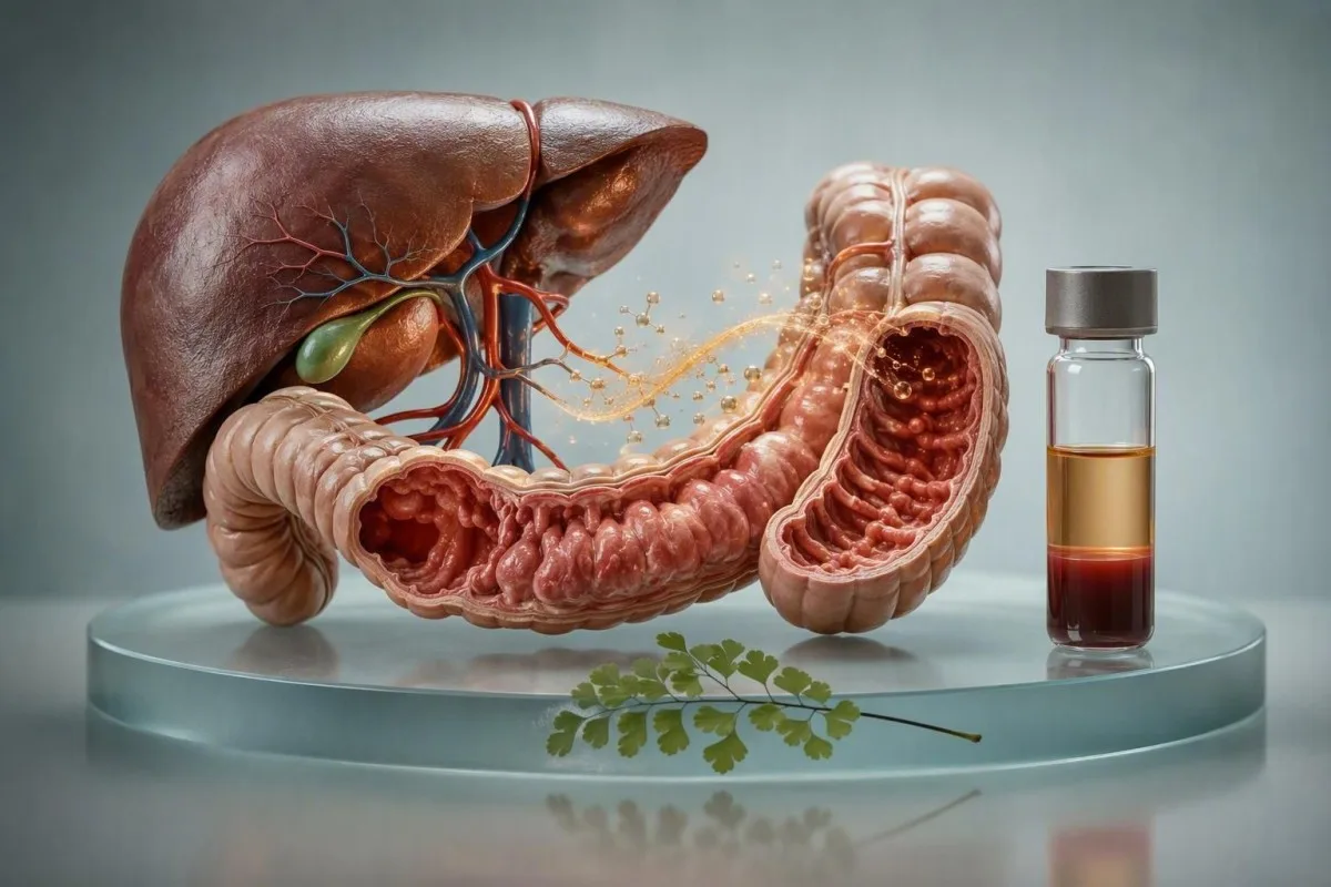
An elevated CEA does not itself cause symptoms; it is a protein marker, not a toxin or hormone. Symptoms, when present, usually come from the condition raising CEA—such as smoking-related airway disease, bowel inflammation, liver disease, or occasionally cancer—and the pattern matters far more than one result.



- CEA itself does not produce symptoms, even when results are above 10 ng/mL.
- Typical reference limits are below about 3 ng/mL in non-smokers and below about 5 ng/mL in smokers, although assays vary.
- A single CEA result cannot diagnose cancer and is not recommended as a population cancer screening test.
- Rising CEA trends on the same assay are usually more clinically useful than one isolated result.
- CEA above 10 ng/mL deserves timely clinical interpretation, especially if it is rising or accompanied by unexplained symptoms.
- Urgent symptoms include black or bloody stool, jaundice, persistent vomiting, new severe abdominal pain, or rapidly worsening weakness.
- Smoking cessation can lower a smoking-related CEA elevation, but repeat testing is usually delayed for several weeks.
- Kantesti AI reads CEA alongside liver tests, inflammatory markers, blood counts, symptoms, and prior results rather than treating it as a diagnosis.
Can high CEA cause symptoms directly?
No—high CEA does not directly cause symptoms. CEA is a glycoprotein measured in serum, and an elevated concentration does not make people feel unwell in the way low glucose, high calcium, or severe anaemia can.

People commonly notice fatigue, altered bowel habit, cough, appetite loss, or abdominal discomfort after seeing an abnormal result, then understandably connect the two. In my clinical experience, the result did not create those symptoms; either an underlying condition explains both, or the symptoms have a separate cause. A CEA concentration of 8 ng/mL is not inherently more symptomatic than 2 ng/mL.
Carcinoembryonic antigen is produced in fetal development and remains at low concentrations in many healthy adults. Adult tissues can release more CEA during irritation, impaired clearance by the liver, or some cancers, but it is a signal rather than an active disease process. This distinction is similar to the one patients ask about with elevated PSA: the marker is usually silent.
As of September 10, 2026, I would frame a high CEA result as a reason to ask, “What is causing this marker to rise?” rather than “What symptoms will this level give me?” Dr. Thomas Klein's practical rule is simple: assess the person first, then the trend, then the number.
Why the distinction changes what happens next
A symptom-causing disorder may need prompt treatment even when CEA is normal, while an incidental CEA elevation may only need confirmation and context. That is why clinicians do not use CEA alone to decide whether someone is well or unwell.
What CEA level is considered high?
Many laboratories use a CEA upper limit near 3 ng/mL for non-smokers and 5 ng/mL for smokers. The reference interval on your own report takes priority because assay calibration and local laboratory validation differ.
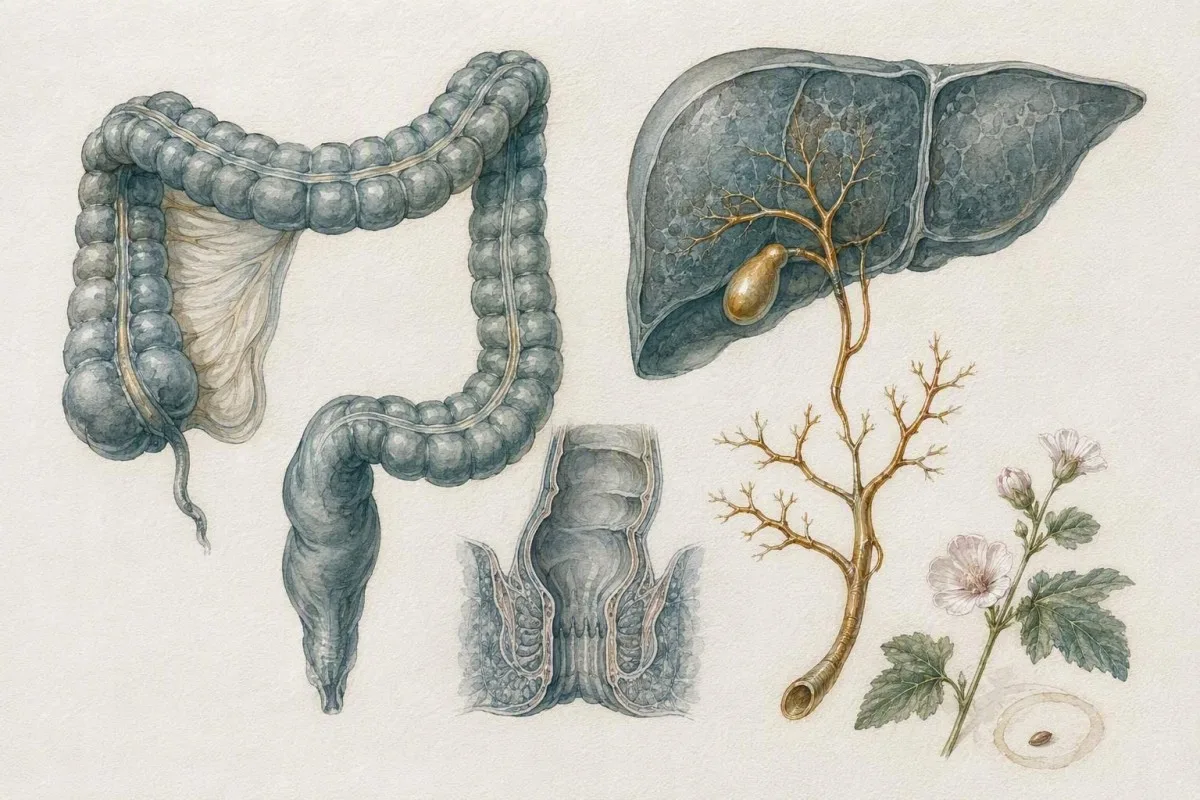
CEA is usually reported as ng/mL, which is numerically equivalent to micrograms per litre. A value of 4.2 ng/mL may be flagged in a non-smoker but fall within a smoker-adjusted interval; that does not mean smoking makes the result meaningless. It means interpretation starts with exposure history and repeatability.
CEA above 10 ng/mL is less commonly explained by uncomplicated smoking alone and usually warrants a clinician-led review, but it still does not establish cancer. Values above 20 ng/mL are more concerning when they rise on serial testing after known colorectal cancer treatment, particularly with abnormal imaging or liver tests. For perspective on how labs flag results, see what an out-of-range result means.
Kantesti is an AI blood test analyzer that places CEA beside the laboratory's own interval, smoking status, bilirubin, ALP, ALT, AST, full blood count, and earlier dates. A result moving from 2.1 to 5.8 to 11.4 ng/mL over 6 months deserves a different conversation from a stable 5.4 ng/mL measured three times.
Symptoms that may reflect the condition behind elevated CEA
Elevated CEA symptoms are really symptoms of the underlying illness, not symptoms of CEA. Persistent bowel change, rectal bleeding, unexplained weight loss, jaundice, chronic cough, or focal pain should be assessed on their own merits.
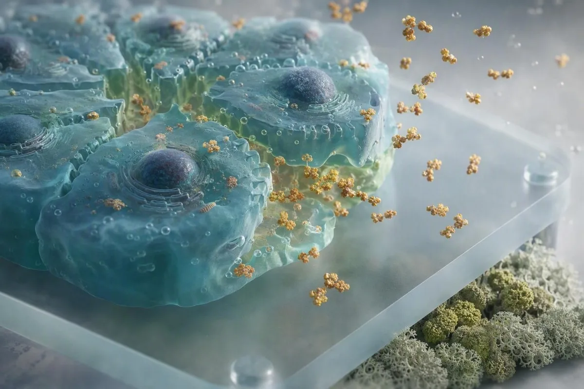
For bowel conditions, the more concerning pattern is a change in bowel habit lasting more than 3 weeks, iron-deficiency anaemia, blood mixed with stool, unexplained weight loss, or a new feeling of incomplete evacuation. A positive stool test needs formal follow-up regardless of CEA; our guide to a positive FIT result explains why colonoscopy is often the next diagnostic step.
Liver or bile-duct causes can bring itch, dark urine, pale stool, right-upper abdominal discomfort, nausea, or yellow eyes and skin. The reason CEA may rise here is partly reduced hepatic clearance and partly biliary epithelial release; CEA alongside raised ALP, GGT, and direct bilirubin is more informative than CEA alone. Review the cholestasis lab pattern if those tests are also abnormal.
Respiratory symptoms require a similarly broad view. A smoker with a persistent cough may have chronic airway disease, a recent viral illness, reflux, medication-related cough, or a more serious explanation; CEA cannot sort these possibilities by itself. In practice, duration, breathlessness, blood-streaked sputum, chest imaging, and smoking exposure matter much more.
Common non-cancer causes of a high CEA result
Smoking, liver disease, pancreatitis, inflammatory bowel disease, and some benign lung conditions can raise CEA. A mild elevation is therefore not synonymous with cancer, particularly when the result is stable over time.

Cigarette smoking is one of the best established non-malignant explanations. Smokers can have baseline CEA values around 5 ng/mL or modestly higher, and levels may decline after cessation, although I normally avoid testing too soon because the timing of normalization varies between individuals. This is not a reason to dismiss a steadily rising value.
Inflammatory bowel disease, diverticulitis, pancreatitis, cirrhosis, hepatitis, chronic obstructive pulmonary disease, and hypothyroidism have all been associated with elevations. When CEA is paired with a raised CRP and bowel symptoms, clinicians may prioritise stool inflammatory markers or imaging rather than jumping straight to a cancer conclusion; calprotectin versus lactoferrin can help clarify that branch of the work-up.
I once reviewed a patient with CEA 9.6 ng/mL, ALP 248 IU/L, GGT 410 IU/L, and new itch. The actionable finding was obstructive cholestasis from a gallstone pathway, not the CEA in isolation. Their CEA fell after the biliary issue resolved—an illustration of why clinicians read the liver panel as a pattern.
Which cancers can raise CEA, and what CEA cannot tell you
CEA can rise in colorectal cancer and also in pancreatic, gastric, lung, breast, ovarian, thyroid, and several other cancers, but it cannot identify the cancer site. Normal CEA also cannot exclude cancer.

CEA has its clearest established role in monitoring known colorectal cancer after treatment, not finding cancer in people without a diagnosis. The ASCO guidance led by Locker et al. recommends postoperative CEA monitoring in patients with resected stage II or III colorectal cancer who could be candidates for further treatment if recurrence is detected (Locker et al., 2006).
A CEA result cannot tell whether a growth is present, where it is, how advanced it may be, or whether symptoms are caused by cancer. In one useful practical contrast, a new CEA of 14 ng/mL with normal examination and a recent severe flare of colitis asks for a different sequence of tests than CEA 14 ng/mL plus anaemia, progressive weight loss, and a positive FIT.
For people already treated for cancer, a confirmed increase generally triggers clinical review and often imaging rather than treatment based on the marker alone. Our article on rising tumour markers after treatment explains why a repeat result, scan timing, and the original tumour's CEA behaviour all change the interpretation.
When high CEA symptoms need prompt or urgent medical review
Seek urgent medical care for black or maroon stool, vomiting blood, jaundice with fever, severe persistent abdominal pain, fainting, or new marked breathlessness—whether or not CEA is elevated. These are symptom-based red flags, not CEA thresholds.

Call emergency services or attend emergency care for collapse, confusion, severe chest pain, coughing up more than a trace of blood, or rapidly worsening abdominal swelling. A CEA of 30 ng/mL without acute symptoms is not automatically an emergency; black stool with a CEA of 2 ng/mL may be. That feels counterintuitive after a scary lab flag, but it is medically crucial.
Arrange a prompt appointment—usually within days to 2 weeks—for persistent rectal bleeding, unexplained 5% body-weight loss over 6 to 12 months, ongoing bowel change, recurrent vomiting, new jaundice, or pain that wakes you from sleep. Dark urine and pale stool deserve special attention; read our guide to bilirubin warning signs if these are present.
Kantesti AI is an AI blood test interpretation platform that can organise an abnormal CEA alongside related results and symptom timing, but it cannot replace a clinician's examination, imaging decision, or urgent-care assessment. Dr. Thomas Klein recommends bringing the original report, prior CEA values, medicines, smoking history, and a short symptom timeline to the appointment.
Why the CEA trend matters more than one number
A confirmed upward CEA trend is usually more meaningful than one mildly high result. Results should ideally be compared using the same laboratory method because switching assays can create an apparent change that is analytical rather than biological.

CEA assays differ enough that a 20% change across laboratories may not represent a true physiological rise. Clinicians look for direction across at least 2 measurements, the interval between them, intercurrent illness, and assay consistency. If the first unexpected result is only mildly high, repeating it in roughly 4 to 8 weeks is often reasonable when symptoms and examination do not suggest urgency.
A rise from 3.2 to 3.9 ng/mL can be ordinary variation, especially near a laboratory cutoff. A progression from 4 to 8 to 16 ng/mL over several months is more persuasive and normally merits expedited investigation. This is why comparing blood tests between visits is safer than interpreting each report in a vacuum.
Kantesti's neural network maps dates, units, laboratory intervals, and co-moving markers to identify patterns worth clinician review. Our clinical validation approach emphasises that trend detection is decision support: it does not declare recurrence or diagnose cancer.
Tests clinicians often check alongside CEA
Doctors usually pair CEA with a history, examination, full blood count, liver panel, and targeted stool or imaging tests. The added tests are chosen to answer a clinical question, not to create a larger panel without purpose.

A full blood count may reveal iron-deficiency anaemia, while ferritin, transferrin saturation, and MCV can help distinguish iron depletion from inflammation. In adults without obvious menstrual blood loss, confirmed iron-deficiency anaemia often prompts gastrointestinal evaluation even when CEA is normal; see low ferritin without heavy periods for the reasoning.
Liver tests matter because bilirubin, ALP, GGT, albumin, ALT, and AST can point toward cholestasis, liver-cell injury, or reduced synthetic function. CEA with isolated ALT 52 IU/L has a different meaning from CEA with bilirubin 68 micromol/L and ALP 320 IU/L. There is no universal “CEA panel”; a good clinician narrows rather than sprays.
Stool FIT, faecal calprotectin, colonoscopy, CT, ultrasound, chest imaging, and endoscopy are selected according to symptoms, risk, prior cancer history, and initial findings. The 2020 ESMO colorectal cancer guideline similarly treats CEA as one component of follow-up, integrated with imaging and clinical assessment rather than used as a stand-alone recurrence test (Argilés et al., 2020).
Smoking, medicines, and practical reasons CEA can shift
Smoking is a frequent reason for a modestly high CEA, while medicines usually influence CEA indirectly through liver, thyroid, or inflammatory effects. Do not stop prescribed medication simply to “improve” a tumour-marker result.

The useful question is not merely “Do you smoke?” but how much, for how long, whether vaping or other inhaled exposures are involved, and whether smoking has recently changed. Heavy smoking can keep CEA above a non-smoker reference limit, but unexplained symptoms or a climbing trajectory still need assessment. Smoking-related airway symptoms should never be written off as a laboratory artefact.
Hypothyroidism can be associated with an elevated CEA, and treating documented thyroid underactivity may reduce it over time. Drugs that affect the liver or provoke pancreatitis can matter indirectly, including some anti-seizure medicines, immunotherapies, and less commonly supplements. Bring every prescription, over-the-counter product, injection, and herbal preparation—dosage included—to review.
If several results change after a new medicine, check timing before assigning cause. Medication-related lab trend analysis can make that discussion more orderly, but the prescriber should decide whether any medication needs adjustment.
High CEA after cancer treatment: what changes the urgency?
After treatment for a CEA-producing cancer, a newly rising CEA should be confirmed and discussed promptly with the oncology team. The urgency depends on the rate of rise, symptoms, treatment history, and whether potentially treatable recurrence is plausible.

Not every colorectal cancer produces CEA, and not every recurrence raises it. The most useful baseline is the patient's own pre-treatment and post-treatment pattern: someone whose CEA was 1.4 ng/mL before surgery may need attention for a persistent rise to 5 ng/mL even though another patient remains stable at 6 ng/mL.
A rise should not automatically trigger treatment. Imaging can be falsely reassuring when done too early, while a repeated marker can prevent a cascade after a laboratory anomaly; this is one reason oncology teams sometimes repeat CEA in 2 to 6 weeks. For a wider comparison of marker roles, review CEA versus CA 19-9.
New focal bone pain, persistent cough, jaundice, neurological symptoms, rapidly progressive fatigue, or unintended weight loss should be reported sooner rather than waiting for the next scheduled draw. Most patients find it helpful to ask their oncologist, “What degree of change would alter the scan plan in my case?”
Four misleading beliefs about high CEA symptoms
The commonest mistake is assuming a high CEA means cancer is present and causing every symptom. CEA has both false-positive and false-negative limitations, which is why experienced clinicians avoid interpreting it in isolation.
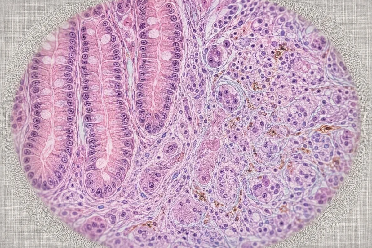
Misconception one: “A normal CEA means I do not have cancer.” That is false because many early cancers and some advanced cancers do not raise CEA. Screening decisions should follow age, family history, symptoms, and local programmes; FIT versus colonoscopy explains two tools with different purposes.
Misconception two: “A very high CEA proves where the cancer is.” It does not. A level of 25 ng/mL can occur in more than one malignancy and, rarely, severe benign hepatobiliary illness; tissue diagnosis and imaging establish location. Numbers guide probability, not certainty.
Misconception three: “Diet, detoxes, or supplements can safely lower CEA.” There is no evidence-based CEA-lowering regimen, and trying to suppress a marker can distract from finding its cause. Treat tobacco exposure, bowel disease, thyroid disease, liver disease, or cancer if identified—not the marker itself.
How to prepare for a CEA follow-up appointment
Bring your full CEA timeline, the laboratory name, symptom dates, smoking history, medicines, and any prior scans to a CEA appointment. This information often prevents both under-reaction and unnecessary testing.

Write down whether the sample was taken during a chest infection, bowel flare, liver illness, or shortly after hospital care. Also record appetite, weight, stool frequency, stool appearance, pain location, cough duration, and fevers; a 2-week symptom diary is often more useful than trying to remember details in the room. Our doctor-visit lab summary checklist can help structure this.
Useful questions include: Was this measured by the same assay as before? Could smoking, liver tests, thyroid status, or inflammation explain it? Should we repeat CEA, and when? Which symptom would make you want earlier imaging or referral? Those questions put the discussion on clinical ground.
Avoid extreme fasting, starting supplements, or making abrupt medication changes solely before a repeat test. A repeat result is most interpretable when the clinical circumstances are ordinary and documented. If a sample had an issue, a simple redraw may be enough; repeat testing after collection problems covers that separate issue.
Family history and screening: where CEA does not fit
CEA is not a recommended screening test for people who feel well, even with a family history of colorectal cancer. Screening uses validated approaches such as FIT, stool DNA testing in some settings, or colonoscopy according to individual risk.

A first-degree relative diagnosed with colorectal cancer before age 50, multiple affected relatives, known Lynch syndrome, or long-standing inflammatory bowel disease can alter screening age and interval. The marker may still be ordered in a specific clinical context, but a normal CEA should never reassure someone out of indicated screening. See our guide to family-history cancer testing priorities for a practical risk conversation.
Symptoms override screening schedules. Rectal bleeding, unexplained iron-deficiency anaemia, persistent bowel change, or weight loss should prompt diagnostic evaluation rather than waiting for the next routine FIT cycle. That distinction is especially relevant in younger adults, where “too young for screening” does not mean “too young for assessment.”
Kantesti is an AI-powered blood test analysis tool that helps users organise blood results in a privacy-focused record, but screening and referral decisions belong with qualified clinicians who know the family history and local pathway. If you need to understand how our medical review framework is governed, meet the medical advisory board.
A sensible next-step plan after an elevated CEA
A sensible plan is to verify the result, assess symptoms and smoking exposure, review companion tests, and arrange targeted follow-up. The timing ranges from emergency assessment for red-flag symptoms to a planned repeat in 4 to 8 weeks for a stable mild elevation without warning signs.
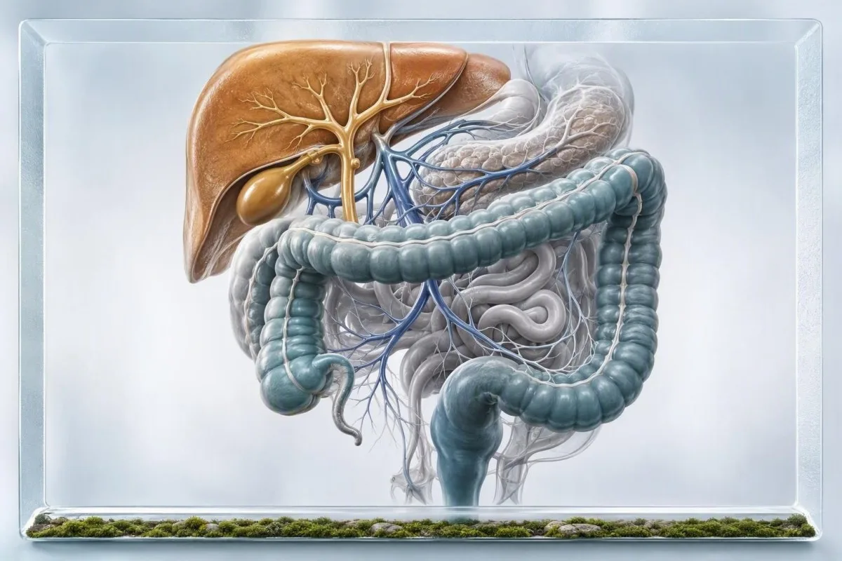
First, check the exact value, units, reference range, prior values, and whether you smoke. Second, identify red-flag symptoms and obtain a focused clinical examination. Third, use the pattern—CBC, ferritin, liver tests, CRP, thyroid studies, stool testing, or imaging—to answer the likeliest question rather than ordering every tumour marker available.
For stable values below 10 ng/mL without concerning symptoms, repeat testing and risk-factor review are often appropriate. For confirmed values above 10 ng/mL, a rising trend, or symptoms such as bleeding or weight loss, timely clinician assessment is prudent. This is a guide, not a substitute for personalised medical advice, because pregnancy status, cancer history, liver disease, and age alter the threshold for action.
Kantesti AI can help you review the chronology before your visit and prepare a concise question list. Our AI technology guide explains how the system extracts laboratory context while leaving diagnosis and treatment decisions to your healthcare team.
Research publications and clinical source notes
The evidence base supports CEA as a contextual monitoring marker, particularly in treated colorectal cancer, rather than a symptom-producing substance or a stand-alone screening test. The sources below separate clinical guidance from supplementary patient-education resources.

Locker et al. (2006) and Argilés et al. (2020) support serial CEA use within colorectal cancer surveillance, alongside examination and imaging rather than instead of them. Duffy (2001) remains a useful foundational review of CEA's sensitivity and specificity limitations. For plain-language digestion context, see the GI symptom research guide.
Kantesti Medical Editorial Team. (2026). Diarrhea After Fasting, Black Specks in Stool & GI Guide 2026. Figshare. https://doi.org/10.6084/m9.figshare.31438111. Supplementary access: ResearchGate and Academia.edu. This educational resource is not evidence that CEA causes gastrointestinal symptoms.
Kantesti Medical Editorial Team. (2026). Women's Health Guide: Ovulation, Menopause & Hormonal Symptoms. Figshare. https://doi.org/10.6084/m9.figshare.31830721. Supplementary access: ResearchGate and Academia.edu. About Kantesti Ltd: our clinical content is designed for informed follow-up, not self-diagnosis.
Frequently Asked Questions
Can high CEA cause symptoms?
High CEA does not directly cause symptoms because CEA is a measured protein marker rather than a substance that produces illness symptoms. A result above 5 ng/mL in a smoker or above 3 ng/mL in a non-smoker may reflect smoking, inflammation, liver disease, or cancer, but the symptoms come from the underlying condition. Persistent bleeding, bowel change lasting more than 3 weeks, jaundice, or unexplained weight loss need medical assessment regardless of the CEA value. A clinician should interpret the result with the trend, examination, and related tests.
What symptoms can happen with elevated CEA?
Elevated CEA itself causes no characteristic symptoms, but the condition raising it can cause bowel changes, rectal bleeding, abdominal discomfort, chronic cough, jaundice, fatigue, or weight loss. These symptoms are not specific for cancer and are commonly caused by non-cancer conditions such as inflammatory bowel disease, liver disease, smoking-related lung disease, or infection. New black stool, vomiting blood, severe abdominal pain, or jaundice with fever needs urgent assessment. A CEA level of 10 ng/mL without symptoms is not an emergency by number alone.
Is a CEA level of 10 high?
A CEA level of 10 ng/mL is above the usual adult reference limits of roughly 3 ng/mL for non-smokers and 5 ng/mL for smokers. It warrants timely clinical review, particularly if it is new, increasing on repeat testing, or accompanied by concerning symptoms or abnormal liver tests. It does not diagnose cancer, because smoking, liver disease, bowel inflammation, pancreatitis, and other conditions can raise CEA. Comparing results on the same assay over 4 to 8 weeks is often more useful than reacting to one isolated measurement.
Can smoking raise CEA levels?
Smoking can raise CEA and is a common reason for a mild elevation above a non-smoker reference range. Many laboratories use an upper limit near 5 ng/mL for smokers compared with about 3 ng/mL for non-smokers, although the reporting laboratory's interval should be used. A smoking-related elevation is usually modest and does not rule out other causes if CEA is rising or symptoms are present. Do not assume a high result is only smoking-related when CEA exceeds 10 ng/mL or shows a sustained upward pattern.
Should I worry if my CEA is high but I feel well?
Feeling well is reassuring, but an elevated CEA still deserves context-based review rather than dismissal or panic. A stable level of 4 to 8 ng/mL without symptoms may be handled differently from a rise from 3 to 12 ng/mL over several months, especially in someone with previous colorectal cancer. CEA is not a screening test and cannot confirm or exclude cancer by itself. Your clinician may repeat it, review smoking and liver tests, or choose targeted investigation based on your age, history, and risk factors.
How quickly should high CEA be rechecked?
For a mild, unexpected CEA elevation without red-flag symptoms, clinicians often repeat the test in about 4 to 8 weeks using the same laboratory method. A repeat may be sooner—often 2 to 6 weeks—after cancer treatment, when the value is clearly rising, or when the result would affect imaging plans. CEA should not be repeated as a substitute for urgent assessment when there is black stool, jaundice, progressive weight loss, persistent vomiting, or severe pain. The right interval depends on the clinical question, not a universal cutoff.
Get AI-Powered Blood Test Analysis Today
Join over 2 million users worldwide who trust Kantesti for instant, accurate lab test analysis. Upload your blood test results and receive comprehensive interpretation of 15,000+ biomarkers in seconds.
📚 Referenced Research Publications
Klein, T., Mitchell, S., & Weber, H. (2026). Diarrhea After Fasting, Black Specks in Stool & GI Guide 2026. Kantesti AI Medical Research.
Klein, T., Mitchell, S., & Weber, H. (2026). Women's Health Guide: Ovulation, Menopause & Hormonal Symptoms. Kantesti AI Medical Research.
📖 External Medical References
Duffy MJ (2001). Carcinoembryonic antigen as a marker for colorectal cancer: is it clinically useful?. Clinical Chemistry.




⚕️ Medical Disclaimer
This article is for educational purposes only and does not constitute medical advice. Always consult a qualified healthcare provider for diagnosis and treatment decisions.
E-E-A-T Trust Signals
Experience
Physician-led clinical review of lab interpretation workflows.
Expertise
Laboratory medicine focus on how biomarkers behave in clinical context.
Authoritativeness
Written by Dr. Thomas Klein with review by Dr. Sarah Mitchell and Prof. Dr. Hans Weber.
Trustworthiness
Evidence-based interpretation with clear follow-up pathways to reduce alarm.